Clustered Micro-Calcifications Extraction From
Mammogram Images Using Cellular Automata
Segmentation With Anisotropic Diffusion Filtering
N.N.Ganvir, D.M.Yadav
Abstract— Breast cancer is deadly disease amongst women in all over the world. Earliest symptoms of breast carcinomas and their detection plays vital role for improving early stage of cancer. Micro calcification is small calcium deposits in breast typically size of 0.1mm to 0.3mm and appears in a high frequency area. Many researchers worked on mammograms by developing efficient computer –aided diagnosis (CAD) using different algorithms. In this Paper, proposed system processed on real time images from Vinod hospital ,Jalgaon from India. Anisotropic diffusion filtering is used for preprocessing of mammogram images; this filter best known for enhances the low contrast mammogram and preserves the edges. Cancer detection refers to the extraction of region of interest ROI, typically represents the tumor, in the image. Here, cellular automata Moore neighborhood segmentation technique is used. This system extract clustered micro calcifications region that can process further for feature extraction. This method gives significally-improved results, which validated with Mini-Mias database.
Index Terms
—
Anisotropic, CAD, Cellular automata, DDSM, Mammograms, Micro calcifications, Mini-MIAS.—————————— ——————————
1
I
NTRODUCTIONBreast cancer is a kind of cancer in which abnormal cells starts forming with the normal cells in the region of the breasts. Like other types of the cancer, breast cancer diagnosed all over the world by observing and detecting early symptoms. Breast cancer observed in mostly in women. Breast cancer occurs when some breast cells begin to grow abnormally other than normal cells. These abnormal cells grow more rapidly than normal cells and continue to form a lump.Early detection of micro calcification is very much important as survival rate increases. Many computer aided diagnosis (CAD) system developed to detect micro calcifications from mammograms. In this paper real time images from hospital processed and it is validate with Mini-Mias database. Images are most effective way to see and analyzed any abnormalities. In medical imaging, segmentation is very much important for feature extraction and different analysis of images. Image segmentation is the process to divide a digital image into different sub-images of main image. These Image segmentation is the process in which sub-images created on the basis of image pixels of having same characterstics. In certain applications area, it may be useful to categorize the mammogram image pixels into anatomical region, such as bones, muscles and cancer, tissue deformities, multiple sclerosis lesions. Many researcher studied different preprocessing and segmentation methods .Biomedical images suffers from photon noise .Therefore it is very necessary to select proper filter to remove noise as well as preserves the edges. In this paper, non- linear approach of anisotropic diffusion is used. To get proper features for detecting micro calcification commonly used cellular automata segmentation is used.
2
MATERIALS
AND
METHODS
Wide varieties of filtering and segmentation techniques already proposed. Different assumptions about the type of the analyzed images direct to the use of different algorithms. [Bankman, 2009] [Lucas and Sinha,1996].Anisotropic diffusion, commonly called Perona–Malik diffusion, proposed the technique in which image noise reduces and preserves edges. [1,2].In bio-medical images it is very important to preserve the complete image contents with edges[2,3]. The space-variant filter is in isotropic in nature that relies on the image value that it generates and approximates an impulse function that are closed to edges and other characteristics that should be conserved in the image. Anisotropic diffusion concept is a commonly used to eliminate noise from mammogram images and preserving edges by selecting a proper constant diffusion coefficient. The anisotropic diffusion equations reduce to the heat equation that is equivalent to Gaussian blurring [1, 2, and 3]. This considered the best for noise filtering. When the diffusion coefficient selected as an edge level, such as in Perona- Malik [2], the resultant equations gives smoothing within regions and forbid it through tough edges. Most of the segmentation algorithms in image processing applications based on basic properties of gray-level values. Two types of discrimination of gray gray-level images mentioned. One is discontinuity and other is similarity. In discontinuity, image partitioning based on the sudden changes in intensity values of the pixels whereas similarity based on by selecting predefined criterion for image partitioning. The two broad categories of image segmentation based on the above properties are (1) edge-based segmentation techniques that work on edges between the regions and (2) region segmentation method work on determining regions. [Rosenfeld and Kak, 1982] [Sonka,1999]. The strategies for edge-based segmentation algorithms aim is to find out object boundaries [Sonka, 1999][Weska,1978].Thresholding is another region segmentation method; In this technique threshold values is selected to convert a gray-scale image into a binary image. Different mostly used methods are maximum entropy method, method, histogram etc. Some newly introduced methods suggest the use of fuzzy rule-based non-linear thresholds technique, which works on multi-dimensional. In this method, decision made to a segment to consider each
————————————————
N.N.Ganvir is currently pursuing Ph.D degree program in elctronics engineering in S.P.Pune University, India, PH-8007908391. E-mail: [email protected]
D.M.Yadav is Professor and Dean academics in S.P.Pune University, India, PH-9011063944. E-mail:
pixel's association based on multi-dimensional rules derived from fuzzy logic. In this paper, real time database is used. System validated with Mini- Mias. Mini –Mias database is freely available for researcher to compare and validate their results with their systems. Preprocessing of the image done by anisotropic diffusion filtering and segmentation carried out with cellular automata. Details of filtering and segmentation techniques mentioned below.
2.1 ENHANCEMENT OF MAMMOGRAPHIC IMAGES
Breast cancer considered deadly disease amongst women. Every pixel of the mammographic images is important. Preprocessing procedure always considered important part in CAD system. Precision of filtering will decide the success of the remaining process like segmentation and classification [4]. Overcome the adverse effects of linear filtering, as if blur the important edges of the bio-medical images. Anisotropic diffusion non-linear approach is very powerful tool for smoothing and edge detection. This filter can smooth noises while preserving the region boundaries within the image [6]. Anisotropic diffusion has been mostly used in biomedical imaging where non-linear approach of filtering is used [7, 8]. Perona and Malik referred to the formulation as anisotropic diffusion even though the locally adapted filter is isotropic [2, 3], but it has also been referred to as inhomogeneous and Perona-Malik diffusion. Perona, Malik proposed two approaches. Linear and non-linear filtering. Linear approach treats every pixel with the exact same convolution.
I(x,y) = Io(x,y)*G(x,y,t) (1) Non-Linear approach treats a pixel with varying intensity, depending on its neighborhood qualities. We can consider (x, y) image part of an edge then apply small amount of smoothing. If particular pixel is not a edge then apply full smoothing. Isotropic diffusion in continuous time domain given as follows [1]
, ,
M x y t
div
M
t
(2)Where, M is an input image and div is a mathematical diversion operation. Then, image smoothing using isotropic diffusion is equivalent to the Gaussian filtering is mentioned below,
, , 1
, ,
M x y t M x y t div M
(3)
As we know, Gaussian filter is symmetric and rotation invariant which turns into the image blurring. Thus, to avoid this problem, the anisotropic diffusion can be modified as given below,
, ,
M x y tdiv G M M
t
(4)
Thus, modified anisotropic diffusion processes only homogeneous regions (non-edge region). Image smoothing In terms of modified anisotropic diffusion is defined as,
, , 1
, ,
M x y t M x y t div G M M
(5)
The modified anisotropic diffusion process defined as given below,
, , , , , , ,, , 1 , ,
x y
x y x y i j i j i j
x y
M x y N M x y N
G M M
(6)Where, mammogram image is used as an initial
conditionM x y
, , 0
,
x y, , 0
denotes the pixel location, N denotes the number of iterations, is a stability constant <=0.25,
x y, represents the neighbourhoods of the centre pixel
x y,and x y,
shows the number of neighbourhoods.
,,
x y i j
M
represents the pixel difference located at
x y, and
i j,i.e. gradient. Selecting proper filters with statistics consideration used in [1]. The same algorithm is applied on real time images and remarkable results are obtained.
.
2.2 IMAGESEGMENTATION
Image segmentation is a procedure in which image is divided into sub sets of segments so that it covers the entire image. Segment division based on characteristics of intensity of pixels, color or texture. The popular method is Thresholding. In Thresholding, gray scale image converted in to binary and specific value is assigned to pixels called threshold value. Multi-dimensional fuzzy rule concept based on decision over each pixel. Histogram-based segmentation methods are powerful compared to other image segmentation methods. In this method, histogram can compute from pixels of the image. Intensity of the pixels gives the different peaks in the histogram which use to trace the clusters in the image. Intensity of pixel can use Ulam [2] and Von Neumann [3] originally introduced the measure Cellular automata (CA) for obtaining models of biological self-reproduction. A Cellular Automata (CA) is a rule-based states concept of complex system. It divides the image into number of cells and each cell can be in one or different final states. Its neighbor is to the simple rule affects cells. It has two types (a) Von Neumann method and (b) Moore method. As shown in Fig 1. Von Neumann neighborhood has five cells, consisting of the cell and its four immediate non-diagonal neighbor’s .Moore neighborhood has nine cells that are neighborhoods to main center cell, consisting of the cell and its eight surrounding neighbors. Cellular automata works on bi-directional. A triplet A = (S; N; δ), where S is a non-empty state set, N is the neighborhood system, and δ: SN →S is the local transition function (rule).
(a) (b)
Fig 1.Cellular Automata segmentation Methods (a) Von Neumann (b) Moore method [6].
3
RESULTS
AND
DISCUSSION
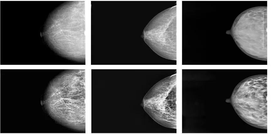
As per the mentioned methods, preprocessing work carried out with anisotropic diffusion filtering as per [1]. Here, work processed on Mini-Mias database and same work is validating on real time images those have been collected from from Vinod Hospital Jalgaon, India. Here, it has been observed that Mini-Mias and hospital images shows significant results, which gives proper segmentation. Figure 2 and 3 shows testing images from Jalgaon’s hospital, India. Figure 4 shows filtered output from Mini-Mias database. These images are filtered using anisotropic diffusion. A mammogram image suffers from photon noise and is of low contrast. Micro calcifications is a low calcium deposit in breast which significantly considered under stage 0 breast cancer and it appears on high frequency area. Hence, necessity occurs to filter the image and enhance the regions of mammogram to enlighten micro calcified area. Anisotropic filtering proved best for filtering and edge preservation. Figure 3 shows filtered output from Vinod hospital ,Jalgaon, India. Figure 4 shows filter output from Mini-Mias database. This filtered image further processed for segmentation. Segmentation plays vital role for classification of different categories of cancer as it divides the suspicious area with normal. Segmented results filled with colors to show suspected clustered micro calcifications.
Figure 5, 6 and 7 shows clustered micro calcification output using cellular automata segmentation technique. It is clearly visible the region of interest ROI of the image. This ROI used to extract a maximum feature that leads to classify different classes and stages of breast cancer.
4
C
ONCLUSIONBreast cancer is one of most deadly disease in women. Early detection plays vital role for chances of survival. Micro-calcifications is a small calcium deposition in breast of the size 0.1mm to 0.3mm.Detection of clustered of micro-calcification directly leads to stages of different classes of cancer. Since, mammogram images suffers from quantum noise and is of low contrast therefore non-linear approach of anisotropic diffusion filtering is used which enhance the image as well as preserves the edges. This image further processed to extract region of interest and to achieve this cellular automata segmentation technique is used. This technique gives noticeable result. This proposed system worked on real time database from Jalgaon mammogram images. To validate the result system processed on Mini-Mias Database.
5
F
UTURES
COPEThe proposed methodology can extend to extract suitable feature to learn and train best machine to differentiate the different stages of cancer.
Acknowledgment
We are sincerely thankful to radiologist of Vinod hospital Jalgaon, India for allow us to access mammogram database and validate the results. We are also thankful to faculty members of research center AISSMS, IOIT Pune.
Note: Upper images are testing images and bottom images are filtered output.
Fig. 3 Sample right breast images filtering results from Jalgaon’s Hospital database.
Fig. 4 Sample breast images filtered output from Mini- Mias database.
Note: Upper images are testing images and bottom images are segmentation results.
Fig.6 Sample Right images segmented results from Jalgaon’s Hospital database.
Fig.7 Sample segmented results from Mini-Mias database.
R
EFERENCES[1] Neha N.Ganvir, D.M.Yadav,‖Filtering Method for Pre-processing
Mammogram Images for Breast Cancer Detection‖, International Journal of Engineering and Advanced Technology.ISSN: 2249 – 8958, Volume-9 Issue-1, (pp. 4222- 4229),Oct 2019.
[2] Pietro Perona and Jitendra Malik ,‖ Scalespace and edge detection using anisotropic diffusion‖, Proceedings of IEEE Computer Society Workshop on Computer Vision,( pp. 16–22), ,November 1987.
[3] Pietro Perona and Jitendra Malik , ― Scale-space and edge detection using anisotropic diffusion. IEEE Transactions on Pattern Analysis and Machine Intelligence.12 (7), (pp.629–639), July 1990.
[4] Kshema , M. Jayesh George, ―Preprocessing filters for mammogram images: A review‖ ,Conference on Emerging Devices and Smart Systems (ICEDSS), IEEE xplore,2017. [5] Chourmouzios Tsiotsios a,Maria Petrou, ―On the choice of the
parameters for anisotropic diffusion in image processing‖, Journal on Pattern Recognition elsevier,2012.
[6] Priyanka Shotrya, Sanjeev Bhardwaj, ―Image Segmentation Using Cellular Automata: A Technical Survey‖, International Journal Of Engineering And Computer Science, ISSN: 2319-7242. (pp.1268-1272),2013.
[7] Xueyan Liu, Hongwei Li , ―A review on the modeling and simulations of solid-state diffusional phase transformations in metals and alloys, Manufacturing Review‖,2018
[8] J. Weickert, ― A review of nonlinear diffusion filtering, Scale-space theory in computer vision‖, Lecture Notes in Computer Science,(pp.3–28),1997.
[9] G. Gerig, O. Kubler, R. Kikinis, F. Jolesz, ―Nonlinear anisotropic filtering of MRI data‖, IEEE Transactions on Medical Imaging, (pp. 221–232),1992.
![Fig 1.Cellular Automata segmentation Methods (a) Von Neumann (b) Moore method [6].](https://thumb-us.123doks.com/thumbv2/123dok_us/8633412.1423120/2.612.318.576.540.685/fig-cellular-automata-segmentation-methods-neumann-moore-method.webp)