I
I
n
n
t
t
e
e
r
r
n
n
a
a
t
t
i
i
o
o
n
n
a
a
l
l
J
J
o
o
u
u
r
r
n
n
a
a
l
l
o
o
f
f
B
B
i
i
o
o
l
l
o
o
g
g
i
i
c
c
a
a
l
l
S
S
c
c
i
i
e
e
n
n
c
c
e
e
s
s
2014; 10(1):43-52. doi: 10.7150/ijbs.6818 Research PaperImportant Functional Roles of Basigin in Thymocyte
Development and T cell Activation
Hui Yao 1*, Yan Teng 2*, Qian Sun 1, Jing Xu 1, Ya-Tong Chen 1, Ning Hou 2, Xuan Cheng 2, Xiao Yang 2,
Zhi-Nan Chen 1
1. Cell Engineering Research Centre & Department of Cell Biology, State Key Laboratory of Cancer Biology, Fourth Military Medical Uni-versity, 169 West Changle Street, Xi'an 710032, China.
2. Genetic Laboratory of Development and Disease, Institute of Biotechnology, 20 Dongdajie, Fengtai District, Beijing 100071, China. * These authors contributed equally to this work.
Corresponding authors: Zhi-Nan Chen, Cell Engineering Research Centre & Department of Cell Biology, State Key Laboratory of Cancer Biology, Fourth Military Medical University, 169 West Changle Street, Xi'an 710032, China. Tel: +86-29-84773243; Fax: +86-29-83293906; E-mail: [email protected]. Xiao Yang, Genetic Laboratory of Development and Disease, Institute of Biotechnology, 20 Dongdajie, Fengtai District, Beijing 100071, China. Tel: +86-10-63895937; Fax: +86-10-63895937; E-mail: [email protected].
© Ivyspring International Publisher. This is an open-access article distributed under the terms of the Creative Commons License (http://creativecommons.org/ licenses/by-nc-nd/3.0/). Reproduction is permitted for personal, noncommercial use, provided that the article is in whole, unmodified, and properly cited.
Received: 2013.06.02; Accepted: 2013.11.22; Published: 2013.12.13
Abstract
Basigin is a highly glycosylated transmembrane protein that is expressed in a broad range of tissues
and is involved in a number of physiological and pathological processes. However, the in vivo role of
basigin remains unknown. To better understand the physiological and pathological functions of
basigin in vivo, we generated aconditional nullallele by introducing two loxP sites flanking exons 2
and 7 of the basigin gene (Bsg). Bsgfl/fl mice were born at the expected Mendelian ratio and showed
a similar growth rate compared with wildtype mice. After crossing these mice with Lck-Cre
transgenic mice, basigin expression was specifically inactivated in T cells in the resulting Lck-Cre;
Bsgfl/fl mice. Although the birth and growth rate of Lck-Cre; Bsgfl/fl mice were similar to control mice,
thymus development was partially arrested in Lck-Cre; Bsgfl/fl mice, specifically at the CD4+CD8+
double-positive (DP) and CD4 single-positive (CD4+CD8-, CD4SP) stages. In addition, CD4+ T cell
activation was enhanced upon Concanavalin A (Con A) or anti-CD3/anti-CD28 stimulation but not upon PMA/Ionomycin stimulation in the absence of basigin. Overall, this study provided the
first in vivo evidence for the function of basigin in thymus development. Moreover, the successful
generation of the conditional null basigin allele provides a useful tool for the study of distinct physiological or pathological functions of basigin in different tissues at different development stages.
Key words: Basigin; Cre-Loxp; gene targeting; thymocyte development; T cell activation
Introduction
Basigin, which is also known as CD147 or EMMPRIN (extracellular matrix metalloproteinase inducer), encodes a transmembrane glycoprotein with two immunoglobulin-like domains [1, 2]. Basigin is expressed in various tissues and is involved in a number of physiological processes, including sper-matogenesis [3, 4], fertilization [5], embryo implanta-tion [3, 5], and neural funcimplanta-tions, such as vision, be-havior, memory, and olfaction [6-8]. Within the
thy-mus, the expression level of basigin correlates with various differentiation stages of thymocytes, with the highest expression level found in early immature thymocytes. Treatment of immature fetal thymocytes using an anti-basigin monoclonal antibody (mAb) blocked thymocyte maturation ex vivo [9]. In adults, basigin was identified as a lymphocyte activa-tion-associated antigen [10]. Moreover, its expression was also significantly upregulated in activated T
Ivyspring
lymphocytes. MAbs specific for basigin inhibited an-ti-CD3-induced T lymphocyte activation, presumably via the inhibition of the reorganization and clustering of GPI-anchored co-receptors within lipid rafts, which, in turn, altered downstream signaling events [11]. Our previous study showed that basigin was functionally linked to the formation of the immuno-logical synapse [12]. However, the precise function and mechanism of basigin in regulating thymus de-velopment and T cell functions in vivo remains to be clarified. Basigin is also expressed in a large number of pathological conditions, including inflammation [13, 14], tissue repair/remodeling [15], ischemic dis-ease [16, 17] and Alzheimer’s disdis-ease [18]. It has been well established that basigin is overexpressed on the surface of tumor cells, and plays significant roles in tumor growth, angiogenesis, invasion, and metastasis by inducing the secretion of various matrix metallo-proteinases [19], stimulating the production of vas-cular endothelial growth factor [20], activating cell signaling pathways [21, 22], preventing anoikis [23], and regulating metabolism [24], among others. Fur-thermore, overexpression of basigin in tumor tissues is correlated with the poor prognosis of patients with several types of solid tumors [25-28]. These results indicated that basigin is a widely expressed multi-functional protein and is implicated in a variety of physiological and pathological conditions.
Although basigin has been recognized to be a multifunctional protein, the molecular mechanisms underlyingits diverse functionshave yet to be eluci-dated. To examine the functions of basigin in vivo, Igakura et al. generated a basigin (Bsg) null mutant mouse (Bsg-/-) using a conventional gene targeting
strategy [3]. Using this mouse model, they revealed that basigin played a critical role in embryo implanta-tion, reproducimplanta-tion, physiological neural functions, and the mitogenic response of lymphocytes upon a mixed lymphocytic response [4, 5, 29]. However, be-cause most of the Bsg-/- embryos died around the time
of implantation and only a small number of Bsg
-/-embryos survived until birth and into adulthood, it was very difficult to obtain a sufficient number of
Bsg-/- mice for a more detailed phenotypic analysis.
Moreover, the Bsg-/- mice that did survive into
adult-hood were smaller and weaker compared to wildtype controls. In addition, both male and female Bsg-/- mice
were infertile, which prevented the establishment of several disease models for the study of the functions of basigin in a pathological context. Furthermore, knocking out the expression of basigin ubiquitously in all tissues prevented the ability to study its roles in specific tissues or at specific developmental stages, as well as the corresponding cellular and molecular mechanisms.
To overcome these disadvantages, we generated a conditional null allele for basigin using the Cre-loxP
system. We also ablated the basigin gene in thymo-cytes and T lymphothymo-cytes using Lck-Cre transgenic mice. We found that Lck-Cre; Bsgfl/fl mice exhibited a
significant reduction in the number of DP (double positive, CD4+CD8+) and CD4SP (single positive,
CD4+CD8-) thymocytes, and a high proliferation index
of CD4+ T cells upon Concanavalin A (Con A) or
an-ti-CD3/CD28 stimulation. To the best of our knowledge, this is the first study to provide insight into the in vivo role of basigin in thymocytic develop-ment. Moreover, basigin conditional null mice will be a valuable genetic tool used to uncover the in vivo role of basigin in specific tissues and at specific develop-mental stages.
Materials and Methods
Construction of the Targeting Vector
The basigin conditional gene targeting vector was generated based on a PCR-based cloning strategy as previously described [30]. Basigin genomic DNA iso-lated from R1 ES cells (on 129/SV background) was used. To generate the targeting vector, the basigin
fragment containing exons 2-7, a 1.5-kb fragment of the 5’ arm and a 3.7-kb fragment of the 3’ arm were amplified using PCR. The following PCR primers were synthesized by Invitrogen and used in this study: Basigin fragment with exons 2-7 primers: 5’-GTCGACCTTGTAGTAACGGGTACTAACCCTT-3’ and 5’-ATCGATGACACACACATTGAGTCCCAG AGCA-3’; 5’ arm primers: 5’-AAGGAAAAAAGCGG CCGCAGGCTGAATTTGATATTAGGGTCTC-3’ and 5’-CCGCTCGAGCTCCATTTCTTTTCTGCTTGCGG GG-3’. 3’ arm primers: 5’-CCCATCGATAGATCTAT AACTTCGTATAATGTATGCTATACGAAGTTATA GGTGGATGGCTGCTGTTGAAATAA-3’ and 5’-CGGGGTACCCAGTTAATCAATGGTTGATCAA TCG-3’. The forward primer of the 3’ arm was de-signed to introduce a Bgl II site and a loxP site into the PCR product. High-fidelity Taq polymerase (TaKaRa) was used for PCR amplification and all of the PCR products were confirmed by sequencing. These frag-ments were sequentially subcloned into the pfrtI vec-tor (developed in our lab). A schematic of the condi-tional basigin targeting vector is shown in Fig. 1A.
Generation of Mice Carrying a conditional null allele for basigin
generate chimeric mice, which were further bred to C57BL/6 mice to generate F1 offspring. The condi-tional basigin allele (Bsgfn) mice were bred with Flp
transgenic mice to delete the FRT flanked Neo cassette to produce Bsgfl mice, which were then bred with
Lck-Cre transgenic mice to generate Lck-Cre; Bsgfl/fl
mice. All of the mice used in this study were housed in a specific pathogen-free facility. Moreover, all of the animal work was approved by the Fourth Military Medical University's institutional animal care and use committee and handled in strict accordance with good animal practice as defined by the relevant national animal welfare bodies.
Genotypic analyses
Mouse genotypes were analyzed using Southern blot and PCR. Genomic DNA from ES cells and tis-sues were prepared according to standard protocols. A Southern blot was performed using a digoxigen-in-based labeling and detection kit (Roche) according to the manufacturer's instructions. The 5’ arm ho-mologous recombination was identified by PCR using a Neo-specific primer pair, P1: 5’-ACAGACCCATAG CCTACAGC-3’ and P2: 5’-GCTACTTCCATTTGTCA CGT-3’, expected to amplify a fragment of 2.1 kb in
Bsgfn alleles. The loxP site flanked exon 7 was
identi-fied using a primer pair, P3: 5’-CTCTGGGACTCAATGTGTGT-3’ and P4: 5’-AGGTGGGTTTTCTGTAAGGT-3’, which was ex-pected to amplify a 371-bp fragment in the wildtype allele and a 405-bp fragment in the targeted allele. The Flp-mediated deletion of the Neo cassette was identi-fied using the primer pair, P5: 5’-CTGGAACTCCTAGCAATC-3’ and P6: 5’-TGGGAAAGGGTTAGTAC-3’, which was ex-pected to amplify a 445-bp fragment in the
Neo-deleted allele (Bsgfl) and a 377-bp fragment in the
wildtype allele. The Cre-mediated deletion of the floxed exons 2 to 7 was identified using the primer pair, P7: 5’-CTGGAACTCCTAGCAATC-3’ and P4, which was expected to amplify a 614-bp fragment (referred to as the deleted band). All of the PCR reac-tions were performed for 30 cycles of 94°C for 1 min, 55°C for 1 min, and 72°C for 0.5 or 2 minutes.
Cell isolation
Spleens and thymuses were harvested from five-week-old mice and mechanically disrupted through a 70-µm strainer. After treatment with red blood cell lysis buffer (0.14 M NH4Cl in HEPES, pH
7.0), the cells were washed with a sterile phosphate buffer solution (PBS) and resuspended as a single cell suspension. The absolute cell number was quantified. The splenocytes were further isolated using a mouse CD4+ T cell-specific and B cell-specific isolation kit
according to the manufacturer’s instructions (Miltenyi Biotec Inc.). The purity of the obtained cells was fur-ther detected using flow cytometry with CD4 or CD19 mAbs (BD-Biosciences). Only cells with a purity greater than 95% were used for further analysis.
Real-time quantitative PCR
Total RNA obtained from isolated CD4+ T and B
cells was extracted using Trizol reagent (Invitrogen).
Single strand cDNA was synthesized from 1 μg of
total RNA using the RNA reverse transcriptase kit (Takara). Real-time quantitative PCR was performed using the ABI PRISM 7000 Sequence Detection System (Applied Biosystems), and the TAKARA SYBR Pre-mix Ex Taq was used for amplification according to the manufacturer’s instructions. The mRNA level of
basigin was detected using the primer pair, Bsg-1: 5’-TGGACCGTGTTCACATCCAT-3’ and Bsg-2:
5’-ACCTCTTTAGCATAGTAGTCCGC-3’. β-actin
was detected using the primer pair: Actin-1: 5’-GGCTGTATTCCCCTCCATCG-3’ and Actin-2: 5’-CCAGTTGG TAACAA TGCCATGT-3’.
Western blotting analyses
Freshly isolated CD4+ T cells and B cells were
immediately homogenized in protein lysis buffer. Thirty-micrograms of protein were isolated on 10% SDS-PAGE, transferred onto a PVDF membrane, and blotted using a goat-anti-basigin antibody (R&D Sys-tems) or rabbit-anti-β-actin antibody (R&D Systems). After several washes, the membranes were incubated with HRP-conjugated anti-goat or anti-rabbit IgG (R&D Systems) and then visualized using ECL rea-gents (Amersham Bioscience).
Flow cytometry
Flow cytometry was performed according to previously described methods [9]. Freshly prepared single-cell suspensions of thymuses and spleens from at least eight animals per genotype were incubated using anti-CD16/32 to block FcRγ II/III, followed by staining with fluorochrome-conjugated mAbs. All of the antibodies were purchased from BD-Biosciences. Stained cells were analyzed using a FACSCalibur flow cytometer (BD-Biosciences) and CellQuest software (BD-Biosciences). The absolute cell number of each cell subset was calculated by the percentage of stained cells × total cell number.
T cell proliferation assay
Purified CD4+ T cells (5×10⁴ cells/well) were
incubated with either medium alone or with ConA (1
µg/mL, Sigma-Aldrich), plate-bound anti-CD3 and
96-well plates. Next, 20 µL of 20 µg/mL WST-1 (Roche) were added into each well, and the cultures were incubated at 37°C for an additional 4 hours. Next, the absorbance was measured at 450 nm and 630 nm. The proliferation index was calculated as (A450 nm - A630 nm) of the T cells cultured with different mitogens divided by (A450 nm - A630 nm) of the T cells cultured with medium alone.
Statistical analysis
The Graphpad prism software was used to manage the data. The results are presented as the means ± SD, and two-tailed Student’s t-test was used to compare the significance between two groups. *P < 0.05 was considered significant.
Results
Generation of a conditional null allele for
basigin in mice
The murine Bsg gene spans over 7.6 kb on
chromosome 10 and contains 8 exons, including the noncoding exon 1 [31]. The full-length transcript of
basigin is approximately 3.1 kb and encodes two isoforms of the basigin proteins 1 and 2, depending on the alternative splicing of exon 2.
Because the gene fragment from exon 2 to exon 7 encodes over 97% of
basigin amino acids, and deletion of this fragment will introduce a frame shift mutation in exon 8, a deletion of the gene fragment from exon 2 to exon 7 using Cre recombinase is ex-pected to result in a null allele of basigin for both isoforms. To gener-ate a conditional null allele for
basigin, we flanked exon 2 and exon 7 with two loxP sites. The pfrtI vector, which contains a Neo cassette flanked by two FRT sites, a loxP site and an
Hsv-tk cassette, was used to construct
the conditional gene targeting vector. First, we in-serted a 1.5-kb fragment located in intron 1 of basigin
into the pfrtI vector for the 5’ homology recombination arm, and then inserted a 3.2-kb fragment containing exons 2 to 7 downstream of the loxP site into the pfrtI
vector, followed by a 3.7-kb fragment containing a second loxP site and exon 8 inserted into the pfrtI
vector to form the 3’ homology recombination arm (Fig. 1A). After linearization by Not I, the targeting vector was electroporated into R1 embryonic stem cells (ES). Three out of 192 (1.6%) ES clones with G418- and ganciclovir- resistance showed correct re-combination as detected by Southern blot using an external 3’ probe, which exhibited a 4.1-kb targeted allele (referred to as Bsgfn, fn denotes floxP-neo) band
in addition to an 8.1-kb wildtype allele (referred to as
Bsg+) band (Fig. 1A and 1B). The 5’ arm homologous
recombination event of the targeted ES clones was also confirmed by PCR using a Neo specific primer pair, P1 and P2, which amplified a unique 2.1-kb fragment of the Bsgfn allele,but not the Bsg+ allele (Fig.
1A and 1C). These results demonstrated that three targeted ES clones with the floxed basigin allele (Bsgfn)
had been successfully generated.
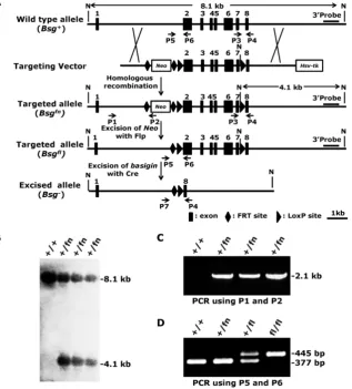
Figure 1. Generation of the conditional null allele for basigin. A. Strategy for generating the condi-tional targeting vector, targeted Bsgfn allele, targeted
Bsgfl allele and excised allele Bsg-. Flp-mediated
excision of Neo cassette from the Bsgfn allele
re-sulted in the Bsgfl allele, and Cre-mediated excision
of the loxP sites resulted in the Bsg- allele. The 3’
external probe for the Southern blot is indicated by the thick lines. The predicted lengths of the Southern fragments are indicated by the double arrow lines. PCR primers used to identify the targeted and deleted alleles are indicated by the arrows. N, Not I. B. Correctly targeted ES cells were confirmed by Southern blot analysis. Genomic DNA was isolated from ES cells, digested with Not I
and subjected to Southern blot analysis using a 3’
The correctly targeted ES cells were then mi-croinjected into blastocysts obtained from 129/SV mice to generate chimeric mice according to standard protocols [32]. F1 mice were analyzed by PCR using primers P3 and P4 to validate germline transmission of the Bsgfn allele (data not shown). Because the Neo
cassette located in intron 1 could potentially disrupt basigin mRNA transcription, splicing, or stability, we removed it by crossing the Bsgfn/+ mice to Flp
trans-genic mice [33], which resulted in Bsgfl/+ mice (Fig. 1A,
f denotes floxP). The deletion of the Neo cassettewas confirmed by PCR with primers P5 and P6, which amplified a 445-bp fragment from the Bsgfl allele but
only a 377-bp fragment fromthewildtype allele (Fig. 1A and 1D). Heterozygous Bsgfl/+ mice were
inter-crossed to generate homozygous Bsgfl/fl mice, which
were born at the expected Mendelian ratio (Bsg+/+:
Bsgfl/+: Bsgfl/fl = 54: 117: 61). Both male and female Bsgfl/fl
mice were viable, fertile, and presented no geno-type-dependent differences in gross morphological abnormalities, indicating that the floxed basigin allele (Bsgfl) was a functional basigin allele.
Conditional deletion of basigin allele using
Lck-Cre transgenic mice
To demonstrate that the Bsgfl allele could be
de-leted using Cre recombinase in vivo and toobtain ad-ditional information regarding the role of basigin in thymocyte development and T lymphocyte activation,
Bsgfl/fl mice were mated to Lck-Cre transgenic mice,
which have been successfully used to delete floxed genes in thymocytes and T lymphocytes [34]. We de-signed PCR primers (Fig. 1A) to amplify the deleted allele (using primers P7 and P4;
614-bp product in length) and undeleted allele (using primers P3 and P4; 405-bp product in length).
The PCR results showed no deleted band in tissues obtained from control mice, but a strong deleted band in genomic DNA from the thymus, a moderate band in the spleen, and no deleted allele band in the ge-nomic DNA from liver of the Lck-Cre; Bsgfl/fl mice (Fig.
2A) was observed, which was consistent with the lev-el of T lymphocytes in these tissues. However, a very weak undeleted band in the PCR results of the thy-mus was still present, which might have been ampli-fied from the genomic DNA of cells lacking Cre ex-pression in the thymus, such as thymic epithelial cells. To further investigate whether Lck-Cre-mediated ex-cision of the Bsgfl allele was T lymphocyte-specific, we
isolated CD4+ T lymphocytes and CD19+ B
lympho-cytes from the spleen and measured the basigin ex-pression both at the mRNA and protein level. Re-al-time quantitative PCR data showed that basigin
mRNA was normal in CD4+ T lymphocytes from
control Bsgfl/flmice, but was undetectable in the CD4+
T lymphocytes from Lck-Cre; Bsgfl/fl mice. However,
the basigin mRNA level in B lymphocytes showed no difference between mice of the two different geno-types (Fig. 2B). Using a western blotting analysis, the specific deletion of basigin protein in CD4+ T
lym-phocytes of Lck-Cre; Bsgfl/fl mice was also verified
us-ing an anti-basigin antibody (Fig. 2C). These results demonstrated that the Bsgfl allele could be specifically
deleted in thymocytes and subsequent T lymphocytes in Lck-Cre; Bsgfl/fl mice. In addition, the Bsgfl/fl mice
could be used for tissue-specific deletion of basigin
using previously developed Cre transgenic mouse models.
Figure 2. Deletion of the Bsgflallele in Lck-Cre;
Bsgfl/fl mice. A. PCR analysis of the basigin
deletion on genomic DNA obtained from the thymus, spleen and liver. Primers P3, P4 and P7 were added in a same tube for PCR reaction in this experiment. B. Real-time quantitative PCR. The relative mRNA level of basigin in CD4+ T cells isolated from Bsgfl/fl mice was presented as
1, and the mRNA levels of basigin in other cells were expressed as a relative ratio. n = 4. **, P <
0.01. C. Western blot analysis. Total protein from the CD4+ T cells and B cells were im-munoblotted using an anti-basigin antibody.
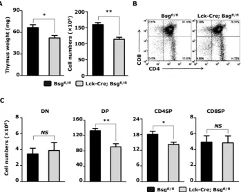
Figure 3. Targeted disruption of the basigin resulted in a decrease in thymic cellularity. Thymocytes were obtained from 5-week-old mice. A. Thymus weight (left) and absolute cell number of total thymocytes (right) were assessed. n = 8 per group. B. Analysis of CD4+ and CD8+ thymocytes from Lck-Cre;
Bsgfl/fl mice and their control littermates using flow cytometry. C. The absolute cell number of the DN, DP, CD4SP and CD8SP thymocytes. n = 8. NS, no
significance. *, P < 0.05; **, P < 0.01.
Analysis of thymocytesfrom Lck-Cre; Bsgfl/fl
mice
General observation and physical examination apparently showed that the Lck-Cre; Bsgfl/fl mice
showed no obvious difference in development and growth compared to the control mice (Bsgfl/fl). No
de-fects were found in the size and tissue structure of major organs in adult mice, including heart, lung, liver, kidney, and other organs. Previous reports have shown the involvement of basigin in thymus devel-opment ex vivo; thus, we examined whether the de-velopment of thymus was impaired in the absence of basigin in Lck-Cre; Bsgfl/fl mice. We isolated the thymus
from 5-week-old mice and found a significant reduc-tion in thymus weight in Lck-Cre; Bsgfl/fl mice
com-pared to that in control mice (Fig. 3A left). The total number of thymocytes in Lck-Cre; Bsgfl/fl mice was
ap-proximately 29% lower compared to control mice (Fig. 3A right). Quantification of individual thymocyte subsets using flow cytometry with CD4 and CD8 mAbs showed that the number of DP and CD4SP thymocytes from Lck-Cre; Bsgfl/fl mice was reduced by
32% and 22%, respectively, compared to control mice (Fig. 3B and 3C). However, the absolute number of
DN (double-negative, CD4-CD8-) and CD8SP
(CD4-CD8+) thymocytes showed no significant
dif-ference between mice of the two different genotypes
(Fig. 3C). Similar results were also found in the thy-mus from 2-week-old Lck-Cre; Bsgfl/fl mice and their
control mice (Supplementary Material: Fig. S1). The above results indicated that basigin expression in T lymphocytes is necessary for thymocyte development, particularly at the DP and CD4SP stage. Since the ob-served drop of cell number in thymus mainly came from DP subset, we assessed freshly isolated thymo-cytes by Annexin V versus PI staining to investigate whether increased apoptosis of DP thymocytes ac-counted for the reduced thymocyte numbers. How-ever, there were no significant difference of apoptotic DP thymocytes between Lck-Cre; Bsgfl/fl mice and the
control mice (Supplementary Material: Fig. S2).
Analysis of peripheral T cellsfrom Lck-Cre; Bsgfl/fl mice
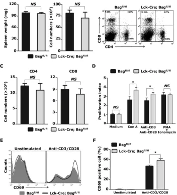
We also analyzed the number of peripheral T cells isolated from the Lck-Cre; Bsgfl/fl mice. In contrast
to the population variation found in thymocytes, the total number of splenocytes was slightly, but not sig-nificantly, reduced in Lck-Cre; Bsgfl/fl mice compared to
control mice (Fig. 4A). An analysis of splenocytes us-ing CD4 and CD8 mAbs also did not show significant changes in the percentage and total number of CD4+ T
lymphocytes and CD8+ T lymphocytes (Fig. 4B and
next analyzed whether T lymphocyte activation was affected in the absence of basigin expression. CD4+ T
lymphocytes were isolated from the spleen, and stimulated with ConA, anti-CD3/CD28 antibodies, PMA/Ionomycin and control medium. In the control medium treatment group, no difference in the prolif-erative index was observed between the Lck-Cre; Bsgfl/fl
mice and control mice. However, after stimulation with ConA and anti-CD3/CD28 antibodies, the pro-liferation index of the CD4+ T lymphocytes isolated
from Lck-Cre; Bsgfl/fl mice was significantly increased
compared to that of the CD4+ T lymphocytes from the
control mice (Fig. 4D). In contrast, the proliferation index of CD4+ T lymphocytes from Lck-Cre; Bsgfl/fl
mice stimulated with PMA/Ionomycin was the same as that of the control CD4+ T lymphocytes. To examine
whether deletion of basigin enhanced T cell activation
via T cell receptor (TCR), we measured the expression
of CD69 on CD4+ T cells stimulated with
an-ti-CD3/CD28 antibodies. As shown in Figure 4E, ex-pression of CD69 was nearly undetectable in unstim-ulated CD4+ T cells from both Lck-Cre; Bsgfl/fl mice and
control mice. However, expression level of CD69 on the CD4+ T cells from Lck-Cre; Bsgfl/fl mice was higher
than that from control mice after stimulation. Con-sistent with the proliferation index result, the per-centages of CD69+ cells were also significantly
in-creased in stimulated CD4+ T cells from Lck-Cre; Bsgfl/fl
mice compared to that from control mice (Figure 4F). Taken together, the above results indicated that basigin may regulate T cell activation via T cell re-ceptor activation and may not directly regulate Ca2+
storage release and protein kinase C signaling path-way.
Figure 4. Analysis of peripheral T cells from Lck-Cre; Bsgfl/fl mice and their control mice. T lymphocytes were freshly isolated from the spleens of 5-week-old
Discussion
In this study, we generated a conditional null allele for basigin (Bsgfl) in mice using the Cre-loxP
sys-tem. Bsgfl/fl mice were born at the expected Mendelian
ratio and exhibited a growth rate similar to wildtype mice. Next, we generated Lck-Cre; Bsgfl/flmice by
in-tercrossing the Bsgfl/fl mice with Lck-Cre transgenic
mice. In Lck-Cre; Bsgfl/fl mice, the floxed basigin gene
was specifically inactivated in thymocytes and T lymphocytes but not in B lymphocytes or other so-matic cells. A phenotypic analysis revealed that
Lck-Cre; Bsgfl/fl mice showed no obvious difference in
birth and growth rates compared to control mice, ex-cept for significant reduction in thymus weight and total number of thymocytes. Inactivation of basigin in T lymphocytes also significantly decreased the num-ber of DP and CD4SP cells in the adult thymus, and enhanced the proliferation of CD4+ T cells upon ConA
or anti-CD3/CD28 stimulation.
Basigin is a multifunctional protein that is thought to be involved in a variety of physiological and pathological activities, such as embryo develop-ment, reproduction, immune response and tumor metastasis [1, 3-5, 28]. However, the in vivo functions of basigin are still unclear, partly due to the shortage of appropriate experimental animal models. In addi-tion, basigin knockout mice have been previously generated using a conventional gene targeting strat-egy [3]; however, only a very small number of Bsg
-/-embryos survive to birth and develop into small, weak, and infertile adult mice. These developmental challenges prevented an elaborate phenotypic analy-sis and the establishment of some disease models. To overcome these disadvantages, Kadomatsu et al. generated a hybrid Bsg-/- mouse line known as
“re-verse F2,” by intercrossing F2 animals from the F1 hybrid offspring of 129/SV chimera and C57BL/6J animals with a C57BL/6J congenic mouse line [35]. The “reverse F2” Bsg-/- mice were born at an expected
frequency (~20%), possibly because of the specific genetic background and the composition of the flanking region of the Bsg gene, but not the normal function of basigin. Moreover, because basigin is ex-pressed in different cell types and may function via cell-cell interaction, knocking out basigin expression in all cells prevented the further study of its role in specific cell types and its corresponding cellular and molecular mechanisms. Here, we generated a condi-tional null allele for basigin using the Cre-loxP system. In contrast to the previously reported Bsg-/- mice, Bsgfl/fl
mice showed no fertility and growth defects, indicat-ing that the insertion of the loxP sites had no effect on
basigin gene activity. Intercrossing Bsgfl/fl mice with
Lck-Cre transgenic mice demonstrated an effective
knockout of the basigin gene in cells with lck promoter activity (thymocytes and T lymphocytes), which demonstrated that Bsgfl/fl mice could be a very useful
tool to study basigin’s in vivo physiological and pathological functions in specific cell types and at different development stages.
Lck-Cre; Bsgfl/fl mice also showed no difference in
birth and growth rate compared to control mice, which enabled further analysis of basigin’s function in thymus development. First, we found that the thy-muses of Lck-Cre; Bsgfl/fl mice were significantly
smaller compared to control Bsgfl/fl mice, which was
accompanied by a significant reduction in the number of thymocytes (Fig. 3A). Reno et al. previously re-ported that a basigin-specific mAb (RL73.2) could block the ex vivo development of fetal thymocytes into mature T cells by reducing the number of live cells in a fetal thymic organ culture, which predominantly occurred at the transition between the DN3 and DN4 stages; however, treatment with the mAb did not re-sult in an increase in cell death in adult thymocytes [9]. Because the expression of the Lck-Cre transgene used in our study might be initiated after the DN2 stage [34], the inactivation of basigin was expected to begin as early as the DN3 stage. This may have con-tributed to the weak undeleted band observed in the PCR test using thymus genomic DNA obtained from the Lck-Cre; Bsgfl/fl mice (Fig. 2A). However, Lck-Cre;
Bsgfl/fl mice did not show any significant change in the
number of DN cells in the adult thymus, and exhib-ited a significantly decreased cell number of DP and CD4SP cells in the adult thymus compared to con-trols, which were inconsistent with Reno’s findings. Our results showed an important in-vivo function of basigin in thymus development at the DP stage and CD4SP stage.
In contrast to the population variation found in the thymus, the relative proportions and absolute numbers of peripheral CD4 and CD8 T cell were sim-ilar between Lck-Cre; Bsgfl/fl mice and controls (Fig. 4B
and 4C). The possible reason was that maintenance of the peripheral T cell population does not only depend on the influx of thymic emigrants cells, but also de-pend on the homeostatic mechanism [36]. When the number of peripheral T cells is severely depleted in some pathological case, the remaining T cells sense the increased availability of “space” and undergo spontaneous proliferation to re-establish homeostasis. Although the molecular basis remains unclear, inter-action of TCR with self-peptide/MHC molecules seem to participate in the homeostatic proliferation of peripheral T cells [37]. We found that CD4+ T cells
from Lck-Cre; Bsgfl/fl mice expressed higher level of
compared with the control cells. These results sug-gested that deletion of basigin may plays an im-portant role in T cell activation and proliferation via elevating TCR signal strength. It is possible that dele-tion of basigin also augments the signals delivered by the complex of TCR and self-peptide/MHC mole-cules, and facilitates homeostatic proliferation of CD4+ T cells in Lck-Cre; Bsgfl/fl mice.
Igakura et al. previously found that the mito-genic response of lymphocytes upon a mixed lym-phocyte reaction was greater in Bsg-/- mice [3, 29]. Our
previous in vitro study showed that the engagement of basigin with a specific anti-basigin mAb in human T lymphocytes strongly inhibited T cell proliferation and disrupted the formation of the immunological synapse [12]. To the best of our knowledge, we are the first to show that the genetic ablation of basigin in T lymphocytes alone did not affect the proliferation or survival of CD4+ T cells isolated from the adult spleen
but, rather, resulted in a significantly enhanced
pro-liferation of CD4+ T cells upon ConA or
an-ti-CD3/CD28 stimulation. This finding suggested that basigin may not to be a general regulator of T cell survival or proliferation but may suppress the activa-tion or proliferaactiva-tion of T cells via TCR activaactiva-tion. Cy-tosolic Ca2+ dynamics is important for the precise
control of T cell activation, and relies on the extracel-lular Ca2+ influx following TCR activation and
intra-cellular storage release evoked by downstream sig-nals of TCR stimulation. Our previous study showed that anti-basigin mAb could inhibit cytosolic Ca2+
concentrations when added together with an-ti-CD3/CD28 antibodies. However, in the present study, no difference in the T cell proliferation index was observed between the Lck-Cre; Bsgfl/fl mice and
control mice upon PMA/Ionomycin stimulation, suggesting that the suppression of basigin on Ca2+
mobility may rely on TCR activation, or that basigin may only inhibit the TCR-mediated Ca2+ influx, but
not the release of intracellular Ca2+. Further
elucida-tion of the mechanistic funcelucida-tions of basigin in thymus development and T lymphocyte function are required and are currently ongoing in our lab. Moreover,
Lck-Cre; Bsgfl/fl mice will be a useful tool for the
ex-ploration of basigin function in T lymphocytes. To the best of our knowledge, this study pre-sented the first in vivo evidence for the function of basigin in thymus development. In addition, the suc-cessful generation of the conditional null allele for basigin provides a useful tool for the tissue- and de-velopmental stage-specific ablation of basigin using previously developed Cre transgenic mouse models.
Supplementary Material
Fig.S1 – Fig.S2.http://www.ijbs.com/v10p0043s1.pdf
Acknowledgements
We thank Dr. Hua Han for providing us with
Lck-Cre transgenic mice. This work is supported by China National Basic Research Program (NO. 2009CB521704); China National Key Project of Scien-tific and Technical Supporting Programs (NO. 2006BAI23B02); Key Project of China National Natu-ral Science Fund (NO. 81030058); China National Major Project (NO. 2013ZX09301301); the National Science and Technology Major Projects (NO. 2012AA020806) and the China National Natural Sci-ence Foundation (NO. 30800573).
Competing Interests
The authors have declared that no competing interest exists.
References
1. Muramatsu T, Miyauchi T. Basigin (CD147): a multifunctional transmembrane
protein involved in reproduction, neural function, inflammation and tumor invasion. Histol Histopathol. 2003; 18:981-7.
2. Biswas C, Zhang Y, Decastro R, et al. The human tumor cell-derived
colla-genase stimulatory factor (renamed EMMPRIN) is a member of the immuno-globulin superfamily. Cancer Res. 1995; 55:434-9.
3. Igakura T, Kadomatsu K, Kaname T, et al. A null mutation in basigin, an
immunoglobulin superfamily member, indicates its important roles in pe-ri-implantation development and spermatogenesis. Dev Biol. 1998; 194:152-65.
4. Toyama Y, Maekawa M, Kadomatsu K, et al. Histological characterization of
defective spermatogenesis in mice lacking the basigin gene. Anat Histol Em-bryol. 1999; 28:205-13.
5. Kuno N, Kadomatsu K, Fan QW, et al. Female sterility in mice lacking the
basigin gene, which encodes a transmembrane glycoprotein belonging to the immunoglobulin superfamily. FEBS Lett. 1998; 425:191-4.
6. Fan QW, Yuasa S, Kuno N, et al. Expression of basigin, a member of the
immunoglobulin superfamily, in the mouse central nervous system. Neurosci Res. 1998; 30:53-63.
7. Naruhashi K, Kadomatsu K, Igakura T, et al. Abnormalities of sensory and
memory functions in mice lacking Bsg gene. Biochem Biophys Res Commun. 1997; 236:733-7.
8. Philp NJ, Ochrietor JD, Rudoy C, et al. Loss of MCT1, MCT3, and MCT4
expression in the retinal pigment epithelium and neural retina of the 5A11/basigin-null mouse. Invest Ophthalmol Vis Sci. 2003; 44:1305-11.
9. Renno T, Wilson A, Dunkel C, et al. A role for CD147 in thymic development. J
Immunol. 2002; 168:4946-50.
10. Kasinrerk W, Fiebiger E, Stefanova I, et al. Human leukocyte activation
anti-gen M6, a member of the Ig superfamily, is the species homologue of rat OX-47, mouse basigin, and chicken HT7 molecule. J Immunol. 1992; 149:847-54.
11. Staffler G, Szekeres A, Schutz GJ, et al. Selective inhibition of T cell activation
via CD147 through novel modulation of lipid rafts. J Immunol. 2003; 171:1707-14.
12. Hu J, Dang N, Yao H, et al. Involvement of HAb18G/CD147 in T cell
activa-tion and immunological synapse formaactiva-tion. J Cell Mol Med. 2010; 14:2132-43.
13. Damsker JM, Okwumabua I, Pushkarsky T, et al. Targeting the chemotactic
function of CD147 reduces collagen-induced arthritis. Immunology. 2009; 126:55-62.
14. Zhu P, Lu N, Shi ZG, et al. CD147 overexpression on synoviocytes in
rheu-matoid arthritis enhances matrix metalloproteinase production and invasive-ness of synoviocytes. Arthritis Res Ther. 2006; 8:R44.
15. Gabison EE, Hoang XT, Mauviel A, et al. EMMPRIN/CD147, an MMP
mod-ulator in cancer, development and tissue repair. Biochimie. 2005; 87:361-8.
16. Boulos S, Meloni BP, Arthur PG, et al. Evidence that intracellular cyclophilin A
and cyclophilin A/CD147 receptor-mediated ERK1/2 signalling can protect neurons against in vitro oxidative and ischemic injury. Neurobiol Dis. 2007; 25:54-64.
17. Seko Y, Fujimura T, Taka H, et al. Hypoxia followed by reoxygenation induces
18. Zhou S, Zhou H, Walian PJ, et al. CD147 is a regulatory subunit of the gam-ma-secretase complex in Alzheimer's disease amyloid beta-peptide produc-tion. Proc Natl Acad Sci U S A. 2005; 102:7499-504.
19. Xu J, Xu HY, Zhang Q, et al. HAb18G/CD147 functions in invasion and
metastasis of hepatocellular carcinoma. Mol Cancer Res. 2007; 5:605-14.
20. Tang Y, Nakada MT, Kesavan P, et al. Extracellular matrix metalloproteinase
inducer stimulates tumor angiogenesis by elevating vascular endothelial cell growth factor and matrix metalloproteinases. Cancer Res. 2005; 65:3193-9.
21. Tang Y, Nakada MT, Rafferty P, et al. Regulation of vascular endothelial
growth factor expression by EMMPRIN via the PI3K-Akt signaling pathway. Mol Cancer Res. 2006; 4:371-7.
22. Dai JY, Dou KF, Wang CH, et al. The interaction of HAb18G/CD147 with
integrin alpha6beta1 and its implications for the invasion potential of human hepatoma cells. BMC Cancer. 2009; 9:337.
23. Yang JM, O'Neill P, Jin W, et al. Extracellular matrix metalloproteinase inducer
(CD147) confers resistance of breast cancer cells to Anoikis through inhibition of Bim. J Biol Chem. 2006; 281:9719-27.
24. Fanelli A, Grollman EF, Wang D, et al. MCT1 and its accessory protein CD147
are differentially regulated by TSH in rat thyroid cells. Am J Physiol Endo-crinol Metab. 2003; 285:E1223-9.
25. Ishibashi Y, Matsumoto T, Niwa M, et al. CD147 and matrix
metalloprotein-ase-2 protein expression as significant prognostic factors in esophageal squamous cell carcinoma. Cancer. 2004; 101:1994-2000.
26. Reimers N, Zafrakas K, Assmann V, et al. Expression of extracellular matrix
metalloproteases inducer on micrometastatic and primary mammary carci-noma cells. Clin Cancer Res. 2004; 10:3422-8.
27. Sienel W, Polzer B, Elshawi K, et al. Cellular localization of EMMPRIN
pre-dicts prognosis of patients with operable lung adenocarcinoma independent from MMP-2 and MMP-9. Mod Pathol. 2008; 21:1130-8.
28. Li Y, Xu J, Chen L, et al. HAb18G (CD147), a cancer-associated biomarker and
its role in cancer detection. Histopathology. 2009; 54:677-87.
29. Igakura T, Kadomatsu K, Taguchi O, et al. Roles of basigin, a member of the
immunoglobulin superfamily, in behavior as to an irritating odor, lymphocyte response, and blood-brain barrier. Biochem Biophys Res Commun. 1996; 224:33-6.
30. Wu X, Ding H. Generation of conditional knockout alleles for PDGF-C.
Gene-sis. 2007; 45:653-7.
31. Miyauchi T, Jimma F, Igakura T, et al. Structure of the mouse basigin gene, a
unique member of the immunoglobulin superfamily. J Biochem. 1995; 118:717-24.
32. Kühn R, Wurst W. Gene knockout protocols. 2nd ed. New York, USA:
Hu-mana Press; 2009.
33. Rodriguez CI, Buchholz F, Galloway J, et al. High-efficiency deleter mice show
that FLPe is an alternative to Cre-loxP. Nat Genet. 2000; 25:139-40.
34. Fu T, Zhang P, Feng L, et al. Accelerated acute allograft rejection accompanied
by enhanced T-cell proliferation and attenuated Treg function in RBP-J defi-cient mice. Mol Immunol. 2011; 48:751-9.
35. Chen S, Kadomatsu K, Kondo M, et al. Effects of flanking genes on the
phe-notypes of mice deficient in basigin/CD147. Biochem Biophys Res Commun. 2004; 324:147-53.
36. Surh CD, Sprent J. Regulation of mature T cell homeostasis. Semin Immunol.
2005; 17:183-91.
37. Surh CD, Sprent J. Homeostasis of naive and memory T cells. Immunity. 2008;