Available online on 15.06.2019 at http://jddtonline.info
Journal of Drug Delivery and Therapeutics
Open Access to Pharmaceutical and Medical Research
© 2011-18, publisher and licensee JDDT, This is an Open Access article which permits unrestricted non-commercial use, provided the original work is properly cited
Open Access
Research Article
Development and Validation of Stability indicating RP- HPLC method for
Simultaneous Estimation of Sofosbuvir and Ledipasvir in Bulk Tablet
Dosage Form
S.D. Mankar*, S.B. Bhawar, P.R. Dalavi
Department of Quality Assurance Technique, Pravara Rural College of Pharmacy, Pravaranagar, Tal-Rahata, Dist-Ahmednagar, India
ABSTRACT
The present research work describes a simple, accurate, precise, effective, Stability indicating RP-HPLC method for simultaneous estimation of Sofosbuvir and Ledipasvir in their tablet dosage form. A reverse phase high performance chromatographic method was developed for simultaneous estimation of Sofosbuvir and Ledipasvir their combined dosage. The separation was achieved by Inertsil ODS C18 column (150X4.6mm, 5µm) column, and ACN: 0.1% TFA in the proportion of 30:70 %v/v as mobile phase, at a flow rate of 1 ml/min. Detection was
carried out at 245 nm. For RP-HPLC method results of the validation indicate that the method was linear in the range of 100-600μg/ml for
Sofosbuvir and 22.5-135μg/ml for Ledipasvir. The % recoveries for Sofosbuvir and Ledipasvir obtained in the accuracy study were 99.92-100.31% and 99.84-100.55% respectively. The LOD for Sofosbuvir and Ledipasvir were found to be 0.395μg/ml and 0.132μg/ml respectively. LOQ for Sofosbuvir and Ledipasvir were found to be 1.197μg/ml and 0.401μg/ml respectively. Force degradation study also done and method
is stability indicating.Developed methods were found to be accurate, precise, rapid and stability indicating for simultaneous estimation of
Sofosbuvir and Ledipasvir.
Keywords: RP-HPLC, Sofosbuvir, Ledipasvir, ACN, TFA.
Article Info:Received 07 May 2019; Review Completed 06 June 2019; Accepted 09 June 2019; Available online 15 June 2019
Cite this article as:
Mankar SD, Bhawar SB, Dalavi PR, Development and Validation of Stability indicating RP- HPLC method for Simultaneous Estimation of Sofosbuvir and Ledipasvir in Bulk Tablet Dosage Form, Journal of Drug Delivery and Therapeutics. 2019; 9(3-s):500-509 http://dx.doi.org/10.22270/jddt.v9i3-s.2893
*Address for Correspondence:
S.D. Mankar, Department of Quality Assurance Technique, Pravara Rural College of Pharmacy, Pravaranagar, Tal-Rahata, Dist-Ahmednagar, India
INTRODUCTION
1.1 SOFOSBUVIR
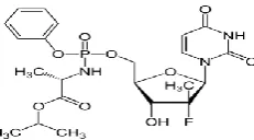
propan-2-yl (2S)-2-[[[(2R,3R,4R,5R)-5-(2,4-dioxopyrimidin- 1-yl)-4-fluoro-3-hydroxy-4-methyloxolan-2-yl]methoxy-phenoxyphosphoryl]amino]propanoate.[1] It is indicated for the treatment of chronic HCV genotypes 1, 4, 5, and 6 in adults and also indicated for the treatment of chronic HCV in patients co-infected with HIV. [2] Slightly soluble in water pH 1.7.7, freely soluble in ethanol and acetone, soluble in 2-propanol and insoluble in heptanes.[3]
Mol. formula: C22H29FN3O9P Mol. weight: 529.4525242
gm/mol . Structure:
Figure 1: Chemical structure of Sofosbuvir
Mechanism of action:Sofosbuvir is a direct-acting antiviral
agent against the hepatitis C virus. The HCV polymerase NS5B protein is an RNA-dependent RNA polymerase (RdRp). It is the essential initiating and catalytic subunit of this replication complex and is critical for the viral replication cycle. There is no human homolog for HCV NS5B RdRp. Sofosbuvir is a monophosphorylated pyrimidine nucleotide prodrug that undergoes intracellular metabolism to form the pharmacologically active uridine analog triphosphate (GS-461203). GS-461203 competes with natural nucleotides for incorporation (by HCV NS5B) into the nascent RNA strand during replication of the viral genome.GS-461203 differs from endogenous pyrimidine nucleotides in that it has been modified at the 2' position with the addition of a methyl and a fluoro functional group. Incorporation of GS-461203 into nascent RNA strongly reduces the efficiency of further RNA elongation by RdRp, resulting in premature termination of RNA synthesis. The stopping of viral replication leads to a rapid decline of HCV viral load and clearing of HCV levels in the body.[4,5]
1.2. LEDIPASVIR
[(2S)-1-{(6S)-6-[4-(9,9-
difluoro-7-{2-[(1R,3S,4S)-2-{(2S)-2- [(methoxycarbonyl)amino]-3-methylbutanoyl}-2- azabicyclo[2.2.1]hept-3-yl]-1H-benzimidazol-5-yl}-9H- fluoren-2-yl)-1H-imidazol-2-yl]-5-azaspiro[2.4]hept-5-yl}-3-methyl-1-oxobutan-2-yl]carbamate [6]. Ledipasvir is practically insoluble <0.1 mg/ml across the pH range of 3.0-7.5 and is slightly soluble below pH 2.3 1.1 mg/ml. The partition coefficient for Ledipasvir is 3.8 and the pKa1 is 4.0 and pKa2 is 5.0.[7]
Molecular formula: C49H54F2N8O6
Molecular weight: 888.999866 gm/mole.
Structure:
Figure 2: Chemical structure of Ledipasvir Mechanism of action:
Ledipasvir inhibits an important viral phosphoprotein, NS5A, which is involved in viral replication, assembly, and secretion. Ledipasvir is an inhibitor of the Hepatitis C Virus (HCV) NS5A protein required for viral RNA replication and assembly of HCV virions. Although its exact mechanism of action is unknown, it is postulated to prevent hyperphosphorylation of NS5A which is required for viral production. It is effective against genotypes 1a, 1b, 4a, and 5a and with a lesser activity against genotypes 2a and 3a of HCV [8,9]
According to the best of our knowledge, only four HPLC methods[10-13] have been published, during the preparation of the present work for publishing. The present study aimed to develop a simple, sensitive, short retention time and accurate RP-HPLC method for the simultaneous determination of both sofosbuvir and ledipasvir together in pure and tablet dosage forms with high sensitivity, selectivity that can be used for the routine analysis of production samples.
2. MATERIAL AND METHOD
2.1. Instruments and Apparatus : Ultrasonic Cleaner-5510
Glass wares – volumetric flask (10, 50 and 100 ml), pipettes beaker (500 ml), measuring cylinder(25, 50, 100 ml) (Borosil )
What man filter paper no.41
Thermo separation HPLC (Assemble), UV-200 detector (single wavelength), Rheodyne injector (20 µl). 0.45 µm Nylon 66 (Milipore, India)
Analytical Balance (Swisser)
All instruments and glass wares were calibrated.
2.2. Preparation of Solutions
Preparation of the Buffer solution
Diluted 1.0 ml of Trifluoro acetic acid in 1000ml of purified water and mixed.
Filter the resultant solution through 0.45μm Nylon membrane filter.
Preparation of the Mobile phase
Prepared a filtered and degassed mixture of buffer solution and acetonitrile in the ratio of (70:30 %v/v).
2.3. Preparation of Standard Solutions
Preparation of Standard Solution of Sofosbuvir
Accurately weighed quantity of Sofosbuvir 200.00 mg was transferred into 100 ml volumetric flask, sonicated to dissolve and diluted up to mark with diluent to give a stock solution 2000μg/ml. An aliquot 5.0 ml of the solution was transferred to 50 ml volumetric flask and diluted to the mark with diluent to obtain a working standard solution 200μg/ml of Sofosbuvir.
Preparation of Standard Solution of Ledipasvir
Accurately weighed quantity of Ledipasvir 45.00 mg was transferred into 100 ml volumetric flask, sonicated to dissolve and diluted up to mark with diluent to give a stock solution 450μg/ml. An aliquot 5.0 ml of the solution was transferred to a 50 ml volumetric flask and diluted to the mark with diluent to obtain a working standard solution 45μg/ml of Ledipasvir.
Preparation of Combined Standard Solution of
Sofosbuvir and Ledipasvir:
Accurately weighed Sofosbuvir 200.00 mg and Ledipasvir 45.00 mg were transferred into 100 ml volumetric flask, sonicated to dissolve and diluted up to mark with diluent to give a stock solution 2000μg/ml of Sofosbuvir and 450μg/ml Ledipasvir. Designate it as a combine stock solution
Stock solution 5.0 ml was transferred to 50 ml volumetric flask and diluted up to mark with diluent to obtain working standard solution 200μg/ml of Sofosbuvir and 45μg/ml Ledipasvir.
Preparation of Sample Solution of Sofosbuvir and
Ledipasvir
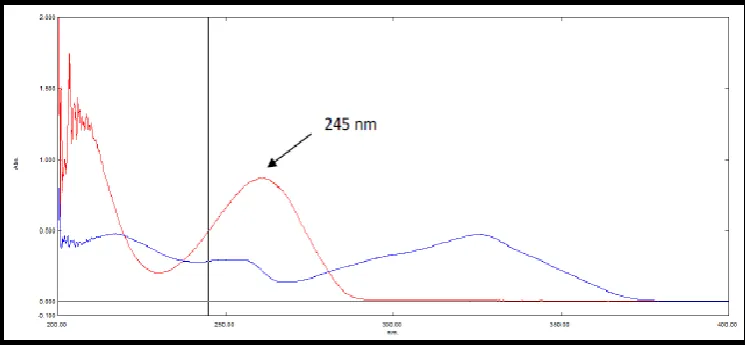
Twenty tablets were weighed and finely powdered. Powdered equivalent to 400 mg Sofosbuvir and 90 mg Ledipasvir was accurately weighed and transferred to 200 ml volumetric flask, and 140 ml of diluent was added and sonicated for 20 min finally volume was made up to the mark with diluent. The solution was filtered through whatmann filter paper (0.45µ). From this solution 5.0 ml was transferred to 50 ml volumetric flask and volume was made up to the mark with diluent to give a solution containing 200μg/ml Sofosbuvir and 45μg/ml Ledipasvir. 2.4. Selection of Wavelength for Estimation
Figure 3: Spectra of standard Sofosbuvir and Ledipasvir for wavelength selection
Table 1: Optimized chromatographic condition
Parameters Condition
Stationary phase Inertsil ODS C18 column (150mm X 4.6 mm, 5 µm particle size)s Mobile Phase 0.1% TFA : Acetonitrile (70:30%v/v)
Pump mode Isocratic
Flow rate (ml/min) 1.0
Run time (min) 7.0
Volume of Injection (µl) 20
Detection wavelength 245
Retention time (min) Sofosbuvir : 2.39 Ledipasvir : 5.51
Table 2: System suitability parameters for final optimized chromatographic conditions
Parameters Data obtained
Sofosbuvir Ledipasvir
Retention time (Rt) 2.39min 5.51 min
Resolution 7.22
Theoretical plates (N) 4025 8977
Tailing factor (Tf) 1.05 1.11
So, from all the above explained parameters, it was concluded that that the most efficient resolution and peak symmetry for Sofosbuvir and Ledipasvir were achieved with the above chromatographic conditions and with a mobile phase composed of 0.1%TFA :Acetonitrile (70 : 30 %v/v)
3. Method Validation
The described method has been validated for linerity, accuracy, limit of detection, precision, and robustness, as per ICH guidelines.[14]
3.1. Linearity and Range:
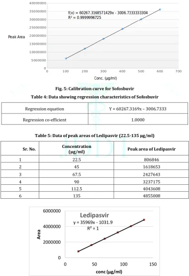
Linear relation was obtained between mean peak area and concentration of the drug in the range of 100-600 μg/ml for Sofosbuvir and 22.5-135 μg/ml for Ledipasvir. The data of the peak areas obtained with the respective concentration in μg/ml are shown in Table 3 and 5 for Sofosbuvir and Ledipasvir respectively. The linearity curves for Sofosbuvir and Ledipasvir are shown in Fig.5 and 6 respectively.
Table 3: Data of peak areas of Sofosbuvir(100-600μg/ml)
Sr.
No. Concentration (μg/ml) Peak area of Sofosbuvir
1 100 6018619
2 200 12053329
3 300 18074368
4 400 24115257
5 500 30130479
6 600 36151273
Fig. 5: Calibration curve for Sofosbuvir
Table 4: Data showing regression characteristics of Sofosbuvir
Regression equation Y = 60267.3169x – 3006.7333
Regression co-efficient 1.0000
Table 5: Data of peak areas of Ledipasvir (22.5-135 μg/ml)
Sr. No. Concentration (μg/ml) Peak area of Ledipasvir
1 22.5 806846
2 45 1618653
3 67.5 2427643
4 90 3237175
5 112.5 4043608
6 135 4855008
Fig. 6 : Calibration curve for Ledipasvir
y = 35969x - 1031.9
R² = 1
0
2000000
4000000
6000000
0
50
100
150
A
rea
conc (µg/ml)
Table 6: Data showing regression characteristics of Ledipasvir
Regression equation Y = 35968.52x-1031.8667
Regression co-efficient 1.0000
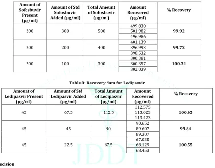
3.2. Accuracy
Accuracy refers to closeness of the test results obtained by the method to the true value. Accuracy was performed by the standard addition methods. To a fixed amount of the pre-analysed mixture add a 50%, 100% and 150% of the standard solution
and % recovery was calculated. The results are shown in Table 7
Table 7: Recovery data for Sofosbuvir Amount of
Sofosbuvir Present (µg/ml)
Amount of Std Sofosbuvir Added (µg/ml)
Total Amount of Sofosbuvir
(µg/ml)
Amount Recovered
(µg/ml)
% Recovery
200 300 500
499.830
99.92 501.982
496.986
200 200 400 401.139 396.993 99.72 398.532
200 100 300 300.381 300.357 100.31 302.039
Table 8: Recovery data for Ledipasvir Amount of
Ledipasvir Present (µg/ml)
Amount of Std Ledipasvir Added
(µg/ml)
Total Amount of Ledipasvir
(µg/ml)
Amount Recovered
(µg/ml)
% Recovery
45 67.5 112.5
112.575
100.45 113.023
113.423
45 45 90
90.652
99.84 89.607
89.307
45 22.5 67.5 67.035 68.129 100.55 68.453
3.3. Precision 3.3.1. Repeatability
It was performed by 100% test concentration level and % RSD was calculated. The data for repeatability for combined solution of Sofosbuvir and Ledipasvir is presented in Table 9
Table 9: Repeatability of Sofosbuvir and Ledipasvir
Sr. No. Sofosbuvir (200 μg/ml) Ledipasvir (45μg/ml).
1 12363864 1532054
2 12264518 1528040
3 12288474 1530889
4 12289526 1530928
5 12303266 1532196
6 12245790 1552151
Mean 12292573 1534376
SD 40493.37 8835.02
3.3.2. Interday and Intraday Precision of Sofosbuvir: The data for Intraday and Interday precision for Sofosbuvir are presented in Table 10 for Intraday Sample solutions containing 200µg/ml of Sofosbuvir and 45µg/ml Ledipasvir
were prepared six times and measured on the same day and % RSD was calculated. For Interday Sample solutions containing 200µg/ml of Sofosbuvir and 45µg/ml Ledipasvir were prepared six times and measured on the different days and % RSD was calculated.
Table 10: Intraday and Interday precision data for Sofosbuvir
Concentration (μg/ml)
Intraday Interday
Mean % Estimation ±
SD % RSD Mean % Estimation ± SD % RSD 200 100.10 ± 0.5462 0.55 100.24 ± 0.7162 0.71
3.3.3. Intraday and Interday Precision of Ledipasvir : The data for Intraday and Interday precision for Ledipasvir
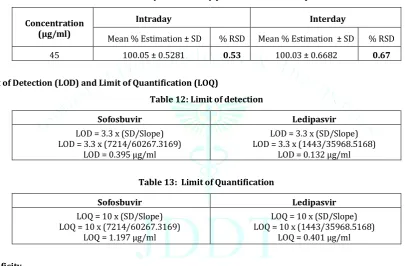
are presented in Table: 10 The % RSD for Intraday precision was found to be 0.53 for Ledipasvir. The % RSD for Interday precision was found to be 0.67 for Ledipasvir.
Table 11: Intraday and Interday precision data of Ledipasvir
Concentration (μg/ml)
Intraday Interday
Mean % Estimation ± SD % RSD Mean % Estimation ± SD % RSD 45 100.05 ± 0.5281 0.53 100.03 ± 0.6682 0.67
3.4. Limit of Detection (LOD) and Limit of Quantification (LOQ)
Table 12: Limit of detection
Sofosbuvir Ledipasvir
LOD = 3.3 x (SD/Slope) LOD = 3.3 x (7214/60267.3169)
LOD = 0.395 μg/ml
LOD = 3.3 x (SD/Slope) LOD = 3.3 x (1443/35968.5168)
LOD = 0.132 μg/ml
Table 13: Limit of Quantification
Sofosbuvir Ledipasvir
LOQ = 10 x (SD/Slope) LOQ = 10 x (7214/60267.3169)
LOQ = 1.197 μg/ml
LOQ = 10 x (SD/Slope) LOQ = 10 x (1443/35968.5168)
LOQ = 0.401 μg/ml
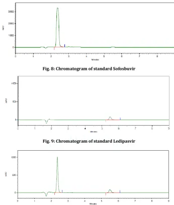
3.5. Specificity
The specificity was determined by the comparison of the chromatograms of:
Blank (mobile phase) (Fig 7)
Standard sample solutions of Sofosbuvir and Ledipasvir(Fig.8 and 9 for Sofosbuvir and Ledipasvir respectively),
Sample solution of Sofosbuvir and Ledipasvir (Fig. 10).
Fig. 8: Chromatogram of standard Sofosbuvir
Fig. 9: Chromatogram of standard Ledipasvir
Fig. 10: Chromatogram of sample solution containing Sofosbuvir(200μg/ml) and Ledipasvir (45μg/ml)
3.6. Robustness
Robustness study was performed in following altered chromatographic condition: Variation in the flow rate (± 0.2 mL/min)
Variation in mobile phase composition (± 2% )
Table 14: Robustness data for ± 0.2 mL/min variation in Flow rate Flow
rate SOFOS Peak area LEDI SOF Mean±SD LED SOF % RSD LED
0.8
mL/min 14313927 14386465 1757607 1756842 14350196 ± 51292.1117 1757224 ± 540.9367 0.36 0.03 1.2
mL/min
11094624 1380696 11096462 ±
2599.3245 1380890 ± 275.0645 0.02 0.02 11098300 1381085
Table 15: Robustness data for ± 2% variation in mobile phase Mobile
phase SOFOS Peak area LEDI SOF Mean±SD LED SOF % RSD LED
72:28 12441394 12460281 1540933 1545020 13355.1258 12450838± 1542977 ± 2889.9454 0.11 0.19
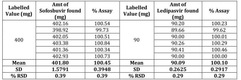
4. Assay Formulation analysis
Table 16: Estimation of Sofosbuvir and Ledipasvir in their Pharmaceutical Dosage Form (Tablet)
Labelled Value (mg)
Amt of Sofosbuvir found
(mg) % Assay
Labelled Value (mg)
Amt of Ledipasvir found
(mg) % Assay
400
402.16 100.54
90
90.20 100.23
398.92 99.73 89.66 99.62
402.05 100.51 90.00 100.01
403.38 100.84 90.26 100.29
401.36 100.34 90.41 100.46
402.93 100.73 90.00 100.00
Mean 401.80 100.45 Mean 90.09 100.10
SD 1.5791 0.3948 SD 0.2625 0.2917
% RSD 0.39 0.39 % RSD 0.29 0.29
5. Force degradation studies
5.1. Preparation of sample stock solution
Accurately weighed Sofosbuvir (200.00 mg) and Ledipasvir (45.00 mg) were transferred into 100 ml volumetric flask, sonicated to dissolve and diluted up to mark with diluent to give a stock solution (2000μg/ml) of Sofosbuvir and (450μg/ml) Ledipasvir. Designate it as a combine stock solution. Stock solution (5.0 ml) was transferred to 50 ml volumetric flask and diluted up to mark with diluent to obtain working standard solution (200μg/ml) of Sofosbuvir and (45μg/ml) Ledipasvir.
5.2. Acid degradation study (5NHCL)
From the test stock solution 1ml was taken in 10 ml volumetric flask, add 1ml of 5N HCL and heated at 60˚ for 30 min on a water bath. The flask was removed from the water bath and allows to cool at room temperature. Add 1ml of 5N NaoH to neutralize the solution and diluted to volume with diluents and mixed. 10µl solution were injected in to the system and the chromatograms were recorded to assess the stability of sample.
5.3. Alkali degradation studies (5N NaoH)
From the test stock solution 1ml was taken in 10ml volumetric flask, add 1ml of 5N NaoH and heated at 70˚c for 1h on a water bath. The flask was removed from the water bath and allowed to cool at room temperature. Add 1ml of 5 N HCL to neutralize the solution and diluted to volume with diluents and mixed.10µl solution were injected in to the systeam and chromatograms were recorded to asses the stability of sample.
5.4. peroxide (Oxidation) degradation studies (30%v/v of H2O2)
From the test stock solution 1ml Was taken in 10ml volumetric flask add 1ml of 30% H2O2 and heated at 70˚ c for 1hour on a water bath. The flask was removed from the water bath and allowed to cool at room temperature and diluted to volume with diluents and mixed. 10µl solutions were injected in to the systeam and the chromatograms were recorded to acess the stability of sample.
5.5. Thermal degradation studies (105˚c/ 48hr)
For the thermal degradation 200mg Sofosbuvir and 45mg lLedipasvir drug samples were weighed accurately and transfer to petridish heat the sample in oven for 48hr at 105˚c and transfer the sample into a 100ml volumetric flask dissolve and dilute to volume with diluents. Filter the solution using 0.45µ Nyllon filter. Transfer 5ml of above stock solution to 50ml volumetric flask and make up the volume with diluents to get the concentration of (200μg/ml)
of Sofosbuvir and (45μg/ml) Ledipasvir. 10 µl solutions was injected into the system and the chromatogram recorded to access the stability of sample.
5.6. Photolytic degradation (2600lux for 24hr)
For the thermal degradation 200mg Sofosbuvir and 45mg lLedipasvir drug samples were weighed accurately and transfer to petridish. The sample was exposed to UV light in a photolytic chamber at 1.2 millon lux hours for 24h and transfer the sample into a 100ml volumetric flask dissolve and dilute to volume with diluents. Filter the solution using 0.45µ Nyllon filter. Transfer 5ml of above stock solution to 50ml volumetric flask and make up the volume with diluents to get the concentration of (200μg/ml) of Sofosbuvir and (45μg/ml) Ledipasvir. . 10µl solutions was injected into the systeam and the chromatogram recorded to access the stability of sample.
6. RESULT AND DISCUSSION
6.1. Optimized chromatographic conditions
The optimized chromatographic conditions fig3. The best peak shape and maximum separation was achieved with mobile phase ACN : 0.1% TFA in the proportion of 30:70 %v/v, peak symmetry and reproducibility obtained on inetsil ODS C18 column (150X4.6mm, 5µm), optimum Wave length To detecting analyte was found to be 245 nm , a flow rate of 1ml/min yield optimum separation and peak symmetry. Chromatogram of Sofosbuvir and Ledipasvir fig 4 and optimized chromatographic condition is shown in table 1.
6.2. Linearity
Linear relation was obtained between mean peak area and concentration of the drug in the range of 100-600 μg/ml for Sofosbuvir and 22.5-135 μg/ml for Ledipasvir. Shown in the fig 5 and 6.
6.3. Accuracy
The percentage recovery for Sofosbuvir were 99.72-100.31% and for Ledipasvir 99.84-100.55%.shown in the Table 6 and 7.
6.4. Precision 6.4.1 Repeatability
6.4.2 Intraday and interday precision
The data for Intraday and Interday precision for Sofosbuvir are presented in Table: 9. The % RSD for Intraday precision was found to be 0.55 for Sofosbuvir. The % RSD for Interday precision was found to be 0.71 for Sofosbuvir.
The data for Intraday and Interday precision for Ledipasvir are presented in Table: 10. The % RSD for Intraday precision was found to be 0.53 for Ledipasvir. The % RSD for Interday precision was found to be 0.67 for Ledipasvir.
6.5. LOD and LOQ
The LOD for Sofosbuvir and Ledipasvir were found to be 0.395μg/ml and 0.132μg/ml respectively, shown in table no 11.
LOQ for Sofosbuvir and Ledipasvir were found to be 1.197μg/ml and 0.401μg/ml respectively, shown in table 12. 6.6. Specificity
The specificity of the method was evaluated with regard to interferance due to presence of any othe excipient. The figure
show that the selected drugs were clearly separated. Fig 7 and 10 show that the chromatogram of blank , standard. solution. There were no interfering peak at retention time of Sofosbuvir and Ledipasvir.
6.7.Robustness
Result of the robustness Table 13 & 14. The elution order and resolution for all components were not significantly affected. RSD of peak area were found to be well within the limit of 2.0%. 6.8. Degradation study
All the stability results were shown in table 16 and fig 11 (a-e) Table 17: stability results
Stress
Condition Sofosbuvir % Degradation Ledipasvir % Degradation
Acid 0.44 1.18
Basic 0.02 0.05
Oxidizing 0.41 1.73
Thermal 0.04 0.05
Photolysis 0.01 0.07
[a] [b]
[c] [d]
[e]
7. CONCLUSION
A simple, fast, accurate and precise stability- indicating HPLC analytical method has been developed and validated for the Quantitative analysis of Sofosbuvir and Ledipasvir in combined tablet dosage forms. The result of stress testing undertake according to the ICH guidelines reveal that the method is specific and stability indicating. The proposed method has the ability to separate these drugs from their degradation products in tablet dosage forms and hence can be applied to the analysis of routine quality control samples obtained from stability studies.
REFRENCES
1. Sofosbuvir Drug profile, January 2014,
http://www.drugbank.ca/drugs/DB08934
2. Gilead Files for U.S. Approval of Ledipasvir/Sofosbuvir
Fixed-Dose Combination Tablet for Genotype 1 Hepatitis C. Gilead Sciences, 2014.
3. Jeffery GH, Bassett J, Mandham J, Denny RC. Vogel’s. Text Book
of Quantitative Chemical Analysis. 5th Ed. Longman Scientific and Technical Publication, UK; 1994, 3-4.
4. Compound Summary for CID 45375808 “National Center for
Biotechnology Information” PubChem Compound Database; CID=45375808,
https://pubchem.ncbi.nlm.nih.gov/compound/Sofosbuvir#sec tion=Top
5. Vikas PM, Satyanarayana T, Kumar DV, Mounika E, Latha MS,
Anusha R , Sathish Y, Development and validation of new RP-HPLC method for the determination of Sofosbuvir in pure form, World Journal of Pharmacy and Pharmaceutical Science. 2016, 5(5):775-781.
6. Ledipasvir Drug profile, January 2014
http://www.drugbank.ca/drugs/DB09027
7. Devilal J, Durgaprasad B, Pal N, Rao AS, New method
development and validation for the determination of
Ledipasvir in bulk drug form by using reverse phase HPLC technique,World Journal of Pharmacy and Pharmaceutical Science. 2016, 5(8): 1312-1321.
8. Compound Summary for CID 67505836 “National Center for
Biotechnology Information” PubChem Compound Database; CID=67505836,
https://pubchem.ncbi.nlm.nih.gov/compound/67505836#sec tion=Top
9. Vikas PM, Satyanarayana T, Kumar DV, Mounika E, Latha MS,
Anusha R , Sathish Y, Development and validation of new RP-HPLC method for the determination of Sofosbuvir in pure form, World Journal of Pharmacy and Pharmaceutical Science. 2016, 5(5): 775-781.
10. Nagaraju T, Vardhan SVM, Kumar DR, Ramachandran D, A
New RP-HPLC Method for the Simultaneous Assay of SOFOSBUVIR and LEDIPASVIR in Combined Dosage Form, Int. J. of Chem Tech Research.2017,10(7): 761-768..
11. Mastanamma SK, Chandini SK, Reehana SK, Saidulu P,
Development and validation of stability indicating RP-HPLC method for the simultaneous estimation of Sofosbuvir and Ledipasvir in bulk and their combined dosage form, Future Journal of Pharmaceutical Sciences, 2017,4(2):116-123.
12. Rote AP, Alhat J, Kulkarni A, Development and validation of
RPHPLC method for the simultaneous estimation of Ledipasvir and Sofosbuvir in bulk and pharmaceutical dosage form, International Journal of pharmaceutical sciences and drug research. 2017; 9(6):291-298.
13. Rao BS, Reddy MV, Rao S, “Simulataneous analysis of
Sofosbuvir and Ledipasvir in bulk and tablet dosage Form by RPHPLC method, Global journal of research analysis.2017, 6 (4):505-509.
14. International Conference on Harmonization, ICH Guidelines,
![Fig 11:[e] (a) Acid degradation (b) alkali degradation (c) peroxide degradation (d) thermal degradation (e) photodegradation](https://thumb-us.123doks.com/thumbv2/123dok_us/1194597.1622400/9.595.55.525.228.753/degradation-alkali-degradation-peroxide-degradation-thermal-degradation-photodegradation.webp)