Abstract —Recent research has suggested that enhanced re-training for stroke patients using haptics (robotic forces) and graphics (visual display) to generate a practice environment that can artificially enhance error rather than reducing it, can stimulate new learning and foster accelerated recovery. We present an evaluation of early results of this novel post-stroke robotic-aided therapy trial that incorporates these ideas in a large VR system and simultaneously employs the patient, the therapist, and the technology to accomplish effective therapy.
I. INTRODUCTION
OBOT-APPLIED forces and sophisticated virtual reality feedback displays have already demonstrated promise of restoring function to several patient populations in rehabilitation training experiences [1-6]. In these early stages of exploration of how these technologies might be used, researchers have realized that the novel application of forces and feedback is full of limitless new possibilities, some which are not realizable combination of visualization and haptic technology. In the past few years the field has exploded with promising prospects in therapeutic robotics; most notably are the recent studies on adaptive training. Prolonged training in the presence of appropriately designed visual distortions [7-9] or mechanical distortions[10-13] is a novel way to use this technology to provide a beneficial change in movement ability.
One promising form of robotic training that leverages the knowledge of the central nervous system (CNS) and neuro-plasticity is error augmentation [11, 12, 14]. In this paradigm the computer singles out and magnifies the subject’s movement errors from a desired trajectory. . The presentation of this error in the visual and haptic systems used by the subject forces the subjects to strengthen their control as they counteract the error driven disturbance to their movements. This feedback is sometimes counterintuitive and differs greatly from the standard approach to treatment. However, such error-driven learning
Manuscript received April 21, 2009. This work was supported in part by the U.S. Department of Education NIDRR program under Grant H133E0700 13 and the NIH under grant H133E0700 13.
J. L. Patton is with the University of Illinois at Chicago, Illinois 60607 +01(312)413-7664 [email protected] and the Rehabilitation Institute of Chicago.
R. V. Kenyon is with the University of Illinois at Chicago, Illinois 60607 and the Rehabilitation Institute of Chicago.
S. Housman and M. Kovic are with the Rehabilitation Institute of Chicago.
S. V. Rozario is with the University of Illinois at Chicago, Illinois 60607
processes are believed to be central to the neuroplasticity and acquisition of skill in human movement [15, 16]. One concern is how to practically apply error augmentation, because it requires knowledge of the desired (or intended) movement, which is difficult to presume during complicated functional 3-dimensional (3D) activities. One solution is an adaptation of a current rehabilitation training where a therapist specifies the trajectory in real time. This technique also allows the expert therapist to customize their approach to therapy, focusing on what is critical for a particular patient’s recovery. For example, if a subject is having difficulty moving in a certain part of the workspace, the therapist can direct all practiced movements to that region.
Recent research points to intensive therapy, or “massed practice,” which appears to have a dramatic effect on recovery [17-19]. Even several years after a stroke, the potential for motor recovery is not lost. The question is not whether patients can improve their motor abilities after stroke, but to what extent and how meaningful is the rehabilitation to the patient. Research also supports “Task-specific” retraining, in which activities of daily living should be practiced [19-21]. Training on a variety of tasks provides better improvement in overall function than repetitions of the same task [22, 23].
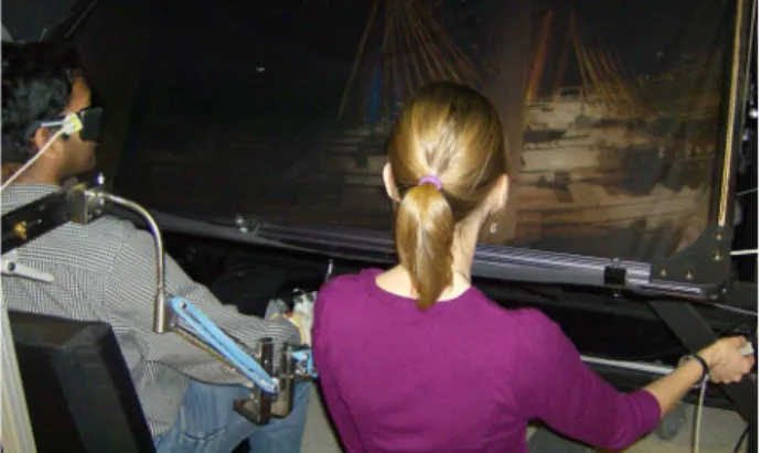
Fig. 1. Both therapist and patient were correctly positioned in front of the apparatus before performing simple reaching tasks.
In order to facilitate the repetitive practice of a variety of functional or pre-functional movements within a virtual reality interface, a large workspace is needed. A wide field of view that allows both the therapist and the patient to work side by side while undergoing treatment is optimal. Recent and unique developments in our lab using a state-of-the-art See http://smpp.northwestern.edu/Robotics for more information on this research.
Therapist-mediated post-stroke rehabilitation using haptic/graphic
error augmentation
Sylvester Vijay Rozario, Sarah Housman, Mark Kovic, Robert V. Kenyon, James L. Patton
R
display system interfaced with a haptic robot make this possible, and enable us to know the patients’ desired trajectory and amplify movement errors in real-time [24]. Here, we present data from five initial subjects.
II. PROCEDURE
A. Apparatus
The experiment used a haptics/graphics display presented previously [24], which combines a projected stereo, head-tracked rendering on a semi-silvered mirror overlay display with a robotic system that can record wrist position, track movements and generate force feedback (Fig. 1). H3D software provided haptics and graphics display. A cinema-quality digital projector (Christie Mirage 3000 DLP) displayed the images that span a five-foot-wide 1280x1024 pixel display, resulting in a 110º wide viewing angle. Stereo glasses were used with a field sequential stereo presentation of the images where Infra-red emitters synchronized separate left and right eye images through the LCD shutter glasses (Stereographics, Inc). Ascension Flock of BirdsTM magnetic elements tracked motion of the head so that the visual display was rendered with the appropriate head-centered perspective. A 6-degree of freedom PHANTOM Premium 3.0 robot (SensAble Technologies), capable of generating 3 Newtons (N) with transient peaks of 22 N, provided a workspace measuring approximately 0.9x0.9x0.3m.
Since it is debatable that training while holding a handle generalizes well to free hand motion [25, 26], and since a large percentage of stroke patients have difficulty raising the arm against gravity and opening and closing the hand, we provided appropriate supports. The weight of the entire upper limb was reduced with the WREX gravity-balanced orthosis and the robot handle was clamped directly to the forearm instead of being held in the hand. In addition, an exotendon glove was utilized to assist in hand opening. The center of the robot handle attached to the forearm was adjacent to epicondyle of the wrist so that its forces acted at the wrist but allowed the hand to be free (Fig. 2).
B. Participants
Five patients to date (4 male and 1 female subject) were recruited and consented using approved IRB and university guidelines for protection of human subjects and confidentiality protection of personal health information. Subjects were aged 36 to 69 (mean age 55 ± 12.07). Participants’ characteristics are summarized in Table I. Control data was collected from five young (ages 19-27) healthy volunteers who were not subjected to the treatment Averaged data from the control subjects is plotted in Figure 4.
Inclusion criteria were the following: Adults (age 18-80) who survived a single cortical stroke at least 6 months previously and demonstrated the presence of some active
shoulder and elbow movement (characterized by Arm Motor Fugl-Meyer scores ≥25≤50). Exclusion criteria included diffuse or multiple lesion sites or multiple stroke events, bilateral paresis, severe spasticity, severe concurrent medical problems, severe sensory deficits or severe ataxia, significant shoulder pain, botox injection to the hemiparetic upper extremity within the previous three months, aphasia, cognitive impairment or affective dysfunction that would influence the ability to perform the experiment, visual field cut or severe inattention that would influence the ability to perform the experiment, participation in other skilled upper extremity rehabilitation in a clinical or research setting, and the inability to provide informed consent.
Fig. 2. The WREX arm support counterbalances the weight of the arm against gravity while the exotendon glove assists in hand opening. The robot handle is clamped directly at the wrist the wrist, allowing the hand to be free.
C. Protocol
We tested two experimental treatments in a crossover design: each subject received, in randomized order, a control treatment of repetitive practice with no error augmentation, and an error augmentation treatment with the same amount of practice but with combined visual and haptic error augmentation. We hypothesized that combined haptic and visual error augmentation would lead to the best functional recovery.
The treatment protocol consisted of two phases (error augmentation or control). Each phase consisted of two weeks of training, with subjects receiving three 40-minute treatment sessions per week (6 sessions per phase). Between the two phases of treatment subjects received a one week rest. During error augmentation or control treatment phases, subjects were seated on a chair with the hemiparetic arm supported by the WREX gravity-balanced orthosis. The hand was placed into an exotendon glove which assisted with hand opening, and the glove was mounted on the
ROBOT HANDLE ATTACHMENT EXOTENDON GLOVE WRIST CLAMP EXOTENDON WREX
WREX wrist support. The wrist support swivels with forearm movement, which allowed the practice of forearm pronation and supination during training. The PHANTOM robot was attached to the forearm to provide augmentation forces (see Fig. 2). Forces were only applied during the error augmentation phase, however the robot was attached during both phases to assist in blinding the subjects and treatment therapist.
TABLE 1 Clinical Scores (WFMT – Wolf Motor Function Test, FAS – Functional Ability Scale, F/U – Follow-up, Pre – Evaluated before initial treatment session, Post – Evaluated following final treatment session). Subject data is color coded similar to that used in Fig. 4 for easy reference.
Sub 1 Sub 2 Sub 3 Sub 4 Sub 5
Sex M M M M F Age 58 36 54 59 69 Week 1 Pre 36 18 43 31 8 Week 2 Post 29 21 47 31 12 Week 4 Pre 31 23 51 32 7 Week 5 Post 32 26 52 35 8 Week 6 F/U 28 22 49 32 9 Week 1 Pre 42 39 45 41 26 Week 2 Post 43 38 43 43 25 Week 4 Pre 39 40 46 44 25 Week 5 Post 42 45 47 43 25 Week 6 F/U 41 46 45 43 25 Week 1 Pre 3.521 5.29 3.203 5.2 48.39 Week 2 Post 3.57 4.16 2.734 4 34.06 Week 4 Pre 4.395 4.8 2.405 4.42 37.8 Week 5 Post 3.017 4.01 2.657 3.74 34.97 Week 6 F/U 3.207 4.16 2.21 4.8 36.49 Week 1 Pre 3.8 3.67 3.8 3.47 2.4 Week 2 Post 4 3.67 3.867 3.33 2.733 Week 4 Pre 4 3.67 4.067 3.4 2.467 Week 5 Post 4.267 3.8 3.733 3.4 2.667 Week 6 F/U 4.2 4 3.867 3.47 2.867 W M FT (Mea n FAS ) B o x an d B lo c ks ( # o f bloc ks ) Fugl-Mey er (Total Sc o re ) W M FT (O ve ral l Me an Time )
During training, subjects viewed two cursors on the stereo display. One cursor was manipulated by the treating therapist while the other was controlled by the subject.
Participants were instructed to follow the exact path of the therapist’s cursor as it moved throughout the workspace.
Error-augmentation was provided both visually and by subtle forces generated by the robot. When subjects erred from therapist’s cursor, the error vector e was established as the instantaneous difference the position between the therapist’s cursor and the subject’s hand. Error was visually magnified by a factor of 1.5e (m) as part of the error-augmentation. Additionally we applied an error augmenting force of 100e (N/m), but saturating at 4 N.
The treatment protocol included the practice of specific movements for all subjects, including forward reach, side reach, shoulder-elbow coupling, and diagonal reaching across the body. The protocol also included customized training for each subject that was targeted at specific areas of weakness. Subjects alternated five-minute blocks of movement training with two-minute rest periods throughout each 40 minute treatment session.
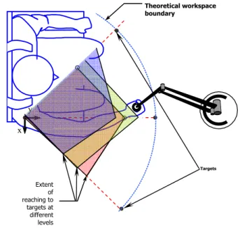
Figure 3: Schematic of the directions and layers of the functional workspace determined in the workspace parts of the experiment. Each color represents the reachable workspace of a separate subject.
D. Analysis
Subjects were tested immediately prior to the start of a treatment regime and again at the end of a treatment phase. Follow-up testing was also performed one week after each treatment phase ended. Effectively, follow-up to phase 1 and the pre-treatment before phase 2 represent the same data points in Fig. 4.
A single blinded rater performed the following clinical assessments. Manual dexterity was assessed with the Box and Blocks Test. The arm motor section of the Fugl-Meyer (AMFM) assessed arm movement ability outside of synergy patterns. Functional use of the arm was evaluated with the Wolf Motor Function Test (WMFT). Subtests of the WMFT include speed of movement and the Functional Ability Scale (FAS). A characterization of free reaching
x y Theoretical workspace boundary Targets Extent of reaching to targets at different levels
ability which requires subjects to reach the impaired hand to nine set targets throughout the workspace was assessed with the Flock of Birds 3D electromagnetic motion capture system and a Phantom robot set-up described earlier. Reaching range of motion (ROM) error was calculated as the linear distance between the position of the target and the final position of the cursor representing the subject’s maximum reach in the direction of the target.
We choose to measure the improvement in the arm motor Fugl-Meyer clinical score and the upper extremity reaching ROM test as main outcomes.
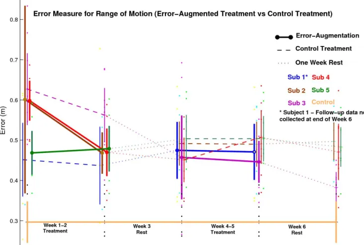
Fig. 4: Change in range of motion error over the weeks of training with different treatments. III. RESULTS
A. Range of Motion
As expected, all stroke subjects demonstrated higher errors than healthy controls (Fig. 4, lowest horizontal line) on the reaching Range of Motion (ROM) assessment. More interestingly, the ROM assessment exhibited a floor effect, where subjects that initially demonstrated fairly low reaching errors did not significantly improve their accuracy in reaching to targets. However, the reaching error on the ROM test did reduce for three subjects in the error-augmented and control treatment groups. It is notable that the error for two of the three subjects was significantly decreased following error augmentation treatment compared to control treatment.
Subjects provided with error augmentation during the first phase of treatment produced greater performance improvements. The two subjects given error-augmentation in phase two produced a smaller degree of improvement. An exception was Subject 5 who worsened during treatment. Overall, the improvement in performance gained over both treatment phases appear to be retained by three subjects (S2,S3,S4).
B. Correlation with Clinical Measures
No significant improvement, deterioration, or notable trends were demonstrated with the clinical measures. Subjects appeared to perform roughly the same on clinical evaluation measures over the six week period. The improvements seen from the error measures do not appear to correlate with any of the clinical measures. One thought is that the two week treatment blocks might not be sufficient to provide any
measurable change clinically. Longer treatment trials might serve to answer this question better.
IV. DISCUSSION
Overall the therapist-mediated training along with the error-augmented treatment produced improved ROM performance in the majority of subjects. Clinical measures however showed no correlating trend. Moreover the small number of subjects (five) is not sufficient to draw any definitive conclusion.
Subject 3 (and to a lesser extent subject 1) showed significant decrease in error over the course of all treatment, despite being put on the control treatment first, suggesting that the control treatment, while not providing error-augmentation, still improved functionality as a result of individualized repetitive therapist intervention. The benefits of patient specific treatment are intuitive and the data does allude to this possibility.
An exceptional case involves Subject 5 (S5) whose performance deteriorated despite being assigned to error-augmentation in Phase1 and whose error worsened slightly before the control treatment, leveled off and decreased during the week six follow-up. Some degree of both physical and mental fatigue with the task or the treatments themselves might have caused the observed result.
Another explanation for this might simply be that subjects unfamiliar with the task initially recorded larger errors and merely got better at executing the task at the end of week two. This ‘familiarity effect’ could explain why there is no significant performance change to either treatment during weeks 4-5.
The duration of training in this study is shorter when compared to the studies referenced in this paper. The CIMT trial [17] for instance utilized two week training periods with hours of intervention each day. This study however had six sessions roughly one hour in duration carried over two weeks for each phase. The lack of carryover observed when subjects are exposed to error-augmentation in Phase 1 might be caused by this shorter training period. While it might be expected from this discussion that a short training period may not provide significant improvement, the observed improvements might indicate that therapist-mediated therapy was indeed significantly beneficial. This assertion will be studied across a larger study population than at present.
In any case, this treatment modality at the very least did no harm to the subject, and feedback obtained from individuals on their experience was not negative and subjects were willing to return for each session. The error augmentation shows some significant improvements during the range of motion testing of being superior to simple repetitive practice (control treatment), and most importantly there appear to be benefits of this adaptive therapist-mediated training paradigm that accumulate with repeated visits to the lab.
ACKNOWLEDGMENT
We thank Santiago Acosta, Assaf Pressman, Felix Huang, Ross Bogey, Derek Kamper, Winsean Lin, Robert Kenyon, and Borko Javanovic for their excellent technical and clinical assistance and advice on this project.
REFERENCES
[1] E. Bisson, B. Contant, H. Sveistrup, and Y. Lajoie, "Functional balance and dual-task reaction times in older adults are improved by virtual reality and biofeedback training," Cyberpsychol
Behav, vol. 10, pp. 16-23, 2007.
[2] R. Boian, A. Sharma, C. Han, A. Merians, G. Burdea, S. Adamovich, M. Recce, M. Tremaine, and H. Poizner, "Virtual reality-based post-stroke hand rehabilitation," Studies in Health
Technology & Informatics, vol. 85, pp. 64-70, 2002.
[3] G. C. Burdea, "Virtual rehabilitation--benefits and challenges,"
Methods of Information in Medicine, vol. 42, pp. 519-23, 2003.
[4] W. S. Harwin, J. L. Patton, and V. R. Edgerton, "Challenges and Opportunities for Robot-Mediated Neurorehabilitation,"
Proceedings of the IEEE, vol. 94, pp. 1717 - 1726, 2006.
[5] R. J. Sanchez, J. Liu, S. Rao, P. Shah, R. Smith, T. Rahman, S. C. Cramer, J. E. Bobrow, and D. J. Reinkensmeyer, "Automating arm movement training following severe stroke: functional exercises with quantitative feedback in a gravity-reduced environment," IEEE Trans Neural Syst Rehabil Eng, vol. 14, pp. 378-89, 2006.
[6] B. T. Volpe, M. Ferraro, D. Lynch, P. Christos, J. Krol, C. Trudell, H. I. Krebs, and N. Hogan, "Robotics and other devices in the treatment of patients recovering from stroke," Curr Neurol
Neurosci Rep, vol. 5, pp. 465-70, 2005.
[7] B. Brewer, R. Klatky, and Y. Matsuoka, "Perceptual limits for a robotic rehabilitation environment using visual feedback distortion," IEEE Transactions on Neural Systems &
Rehabilitation Engineering, vol. 13, pp. 1-11, 2005.
[8] Y. Rossetti, G. Rode, L. Pisella, A. Farne, L. Li, D. Boisson, and M. T. Perenin, "Prism adaptation to a rightward optical deviation rehabilitates left hemispatial neglect," Nature, vol. 395, pp. 166-9, 1998.
[9] Y. Wei, P. Bajaj, R. A. Scheidt, and J. L. Patton, "Visual Error Augmentation for Enhancing Motor Learning and Rehabilitative Relearning," presented at IEEE-International Conference on Rehabilitation Robotics (ICORR), Chicago, IL, USA, 2005. [10] J. L. Emken and D. J. Reinkensmeyer, "Robot-Enhanced Motor
Learning: Accelerating Internal Model Formation During Locomotion by Transient Dynamic Amplification," IEEE
Transactions on Neural Systems & Rehabilitation Engineering,
vol. 13, pp. 33-39, 2005.
[11] J. L. Patton, M. Kovic, and F. A. Muss-Ivaldi, "Custom-designed haptic training for restoring reaching ability to individuals with stroke," Journal of Rehabilitation Research and Development, vol. 43, pp. 643-656, 2006.
[12] J. L. Patton, M. E. Stoykov, M. Kovic, and F. A. Mussa-Ivaldi, "Evaluation of robotic training forces that either enhance or reduce error in chronic hemiparetic stroke survivors,"
Experimental Brain Research, vol. 168, pp. 368-383, 2006.
[13] J. L. Patton, Y. Wei, J. Hitchens, C. Scharver, and R. V. Kenyon, "Exploiting the natural adaptive capacity of the nervous system for motor learning & rehabilitation," in From Basic Motor
control to Functional Recovery IV, N. Gantchev, Ed. Sofia,
Bulgaria: Martin Drinov Academic Publishing House, 2005, pp. 272-280.
[14] Y. Wei, P. Bajaj, R. A. Scheidt, and J. L. Patton, "A real-time haptic/graphic demonstration of how error augmentation can enhance learning," presented at IEEE International Conference on Robotics and Automation (ICRA), Barcelona, 2005.
[15] M. Kawato, "Feedback-error-learning neural network for supervised learning," in Advanced neural computers, R. Eckmiller, Ed. Amsterdam: North-Holland, 1990, pp. 365-372.
[16] D. M. Wolpert, Z. Ghahramani, and M. I. Jordan, "An internal model for sensorimotor integration," Science, vol. 269, pp. 1880-2, 1995.
[17] E. Taub, G. Uswatte, and R. Pidikiti, "Constraint-Induced Movement Therapy: a new family of techniques with broad application to physical rehabilitation--a clinical review.,"
Journal of Rehabilitation Research & Development, vol. 36, pp.
237-51, 1999.
[18] E. Taub, N. Miller, T. Novack, E. d. Cook, W. Fleming, C. Nepomuceno, J. Connell, and J. Crago, "Technique to improve chronic motor deficit after stroke," Archives of physical medicine
and rehabilitation, vol. 74, pp. 347-354, 1993.
[19] R. J. Nudo and K. M. Friel, "Cortical plasticity after stroke: implications for rehabilitation," Revue Neurologique, vol. 155, pp. 713-7, 1999.
[20] R. J. Nudo, "Recovery after damage to motor cortical areas,"
Current Opinion in Neurobiology, vol. 9, pp. 740-7, 1999.
[21] C. M. Dean and R. B. Shepherd, "Task Related Training improves performance of seated reaching tasks after stroke: A randomize controlled trial," Stroke, vol. 28, pp. 722-728, 1997. [22] R. E. Hanlon, "Motor learning following unilateral stroke,"
Archives of Physical Medicine & Rehabilitation, vol. 77, pp.
811-5, 1996.
[23] T. Jarus and T. Gutman, "Effects of cognitive processes and task complexity on acquisition, retention, and transfer of motor skills," Can J Occup Ther, vol. 68, pp. 280-9, 2001.
[24] J. L. Patton, G. Dawe, C. Scharver, F. A. Muss-Ivaldi, and R. Kenyon, "Robotics and virtual reality: A perfect marriage for motor control research and rehabilitation," Assistive Technology, vol. 18, pp. 181-195, 2006.
[25] N. Cothros, J. D. Wong, and P. L. Gribble, "Are there distinct neural representations of object and limb dynamics?," Exp Brain Res, vol. 173, pp. 689-97, 2006.
[26] M. Desmurget, M. Jordan, C. Prablanc, and M. Jeannerod, "Constrained and unconstrained movements involve different control strategies," Journal of Neurophysiology, vol. 77, pp. 1644-50, 1997.