Spine www.spinejournal.com 1721
A
NATOMY
ISSLS Prize Winner: Smudging the Motor Brain in
Young Adults With Recurrent Low Back Pain
Henry Tsao , PhD , * Lieven A. Danneels , PhD , † and Paul W. Hodges , PhD , DSc *
Study Design. Cross-sectional design.
Objective. To investigate whether recurrent low back pain (LBP) is associated with changes in motor cortical representation of different paraspinal muscle fascicles.
Summary of Background Data. Fascicles of the lumbar paraspinal muscles are differentially activated during function. Human studies indicate this may be associated with a spatially separate array of neuronal networks at the motor cortex. Loss of discrete control of paraspinal muscle fascicles in LBP may be because of changes in cortical organization.
Methods. Data were collected from 9 individuals with recurrent unilateral LBP and compared with 11 healthy participants from an earlier study. Fine-wire electrodes selectively recorded myoelectric activity from short/deep fascicles of deep multifi dus (DM ) and long/ superfi cial fascicles of longissimus erector spinae (LES), bilaterally. Motor cortical organization was investigated using transcranial magnetic stimulation at different scalp sites to evoke responses in paraspinal muscles. Location of cortical representation (center of gravity; CoG) and motor excitability (map volume) were compared between healthy and LBP groups.
Results. Individuals with LBP had a more posterior location of LES center of gravity, which overlapped with that for DM on both hemispheres. In healthy individuals, LES center of gravity was located separately at a more anterior location to that for DM. Map volume was reduced in LBP compared to healthy individual across muscles.
Conclusion. The fi ndings highlight that LBP is associated with a loss of discrete cortical organization of inputs to back muscles. Increased overlap in motor cortical representation of DM and LES may underpin loss of differential activation in this group. The results further unravel the neurophysiological mechanisms of motor
L
ow back pain (LBP) is associated with altered back muscle activation, 1 – 3 but the mechanisms are poorly understood.Although changes in muscle morphology, 4 – 6 denervation
from nerve root lesions, 7 and refl ex inhibition 6 are implicated,
changes in descending control by supraspinal centers may con-tribute. 8 – 11 Unraveling the mechanisms that underpin motor
changes with LBP has important implications for rehabilita-tion. For instance, interventions to reverse muscle atrophy 12
would differ from those to alter muscle recruitment. 13 Recent
insights into motor cortex organization using noninvasive magnetic brain stimulation provide a unique opportunity to explore neural mechanisms associated with movement chang-es in pain. 8 , 11
An observation that may provide insight into the mecha-nisms underlying motor control changes with pain is altered coordination between functionally distinct paraspinal muscle fascicles. Short/deep and long/superfi cial back muscle fascicles are differentially activated in healthy individuals in a range of tasks. 14 – 16 For instance, change in spinal curvature from fl at
to more lordosed postures increases activity of the medially positioned multifi dus muscle, whereas, this change has limited effect on the iliocostalis muscle. 15 , 16 Furthermore, predictable
challenges to the trunk are associated with earlier activation of the short/deep fascicles of deep multifi dus (DM) than the long/ superfi cial paraspinal muscles. 14 , 17 Such discrete activation
argues for fi ne control of the complex array of back muscle fascicles in healthy individuals.
Differential control of paraspinal muscles is compromised in LBP. In this group, paraspinal muscle fascicles tend to be recruited en masse with similar activation of iliocostalis and DM with changes in spinal curvature in sitting, 18 and
simulta-neous activation of deep and superfi cial multifi dus with trunk perturbation. 14 The mechanisms for loss of discrete control
are unclear, but one possibility can be gleaned from another painful condition associated with loss of discrete control—fo-cal hand dystonia. In that condition, loss of discrete corticontrol—fo-cal organization of somatosensory regions associated with each fi nger 19 ( i.e. , cortical map “smudging”) contributes to reduced From the * Centre for Clinical Research Excellence in Spinal Pain, Injury and
Health, School of Health and Rehabilitation Sciences, The University of Queensland, Brisbane, Australia ; and † Department of Rehabilitation Sciences and Physiotherapy, Ghent University, Ghent, Belgium .
Acknowledgment date: October 14, 2010. Acceptance date: March 24, 2011. The manuscript submitted does not contain information about medical device(s)/drug(s).
Institutional funds were received to support this work (P.H.). No benefi ts in any form have been or will be received from a commercial party related directly or indirectly to the subject of this manuscript.
Address correspondence and reprint requests to Paul Hodges, NHMRC Centre of Clinical Research Excellence in Spinal Pain, Injury and Health, School of Health and Rehabilitation Sciences, The University of Queensland, Brisbane, QLD 4072 Australia; E-mail: p.hodges@uq.edu.au
changes in recurrent LBP and suggest motor rehabilitation that includes training of differential activation of the paraspinal muscles may be required to restore optimal control in LBP.
Key words: low back pain , motor control , motor cortex organization , lumbar paraspinal muscles . Spine 2011 ; 36 : 1721 – 1727
1722 www.spinejournal.com October 2011
ability to isolate fi nger movements. Changes in brain organi-zation may underlie changes in paraspinal muscle activation.
Recent brain mapping studies using transcranial magnetic stimulation (TMS) suggest separate regions of the motor cor-tex underpin discrete activation in healthy individuals. 20 That
work showed a more posterior region of the motor cortex associated with corticospinal input to DM compared to that for the long/superfi cial fascicles of the longissimus erector spi-nae (LES). Although organization of these cortical networks has not been studied in LBP, responsiveness (excitability) of corticomotor pathways to paraspinal muscles using TMS is reduced 11 and the motor cortical map for other trunk muscles
is reorganized. 8 Those studies showed an association between
cortical changes and changes in pain intensity, 11 or motor
con-trol. 8 Central mechanisms may contribute to defi cits in
differ-ential control of paraspinal muscles.
This study aimed to investigate organization of areas of the motor cortex associated with inputs to different paraspinal muscle fascicles in individuals with recurring episodes of LBP. Identifi cation of smudging between discrete cortical networks with outputs to paraspinal muscle fascicles would provide novel and convincing evidence of supraspinal mechanisms underlying motor adaptation with pain.
MATERIALS AND METHODS
ParticipantsNine right-handed individuals with recurrent LBP were re-cruited and data were compared to that for 11 right-handed healthy participants reported previously. 20 Participants were
included in the LBP group if they had nonspecifi c episodic unilateral LBP ( ± buttock pain) lasting longer than 3 months with periods of symptom aggravation and remission. Each episode was to last at least 1 week with suffi cient intensity to limit function. LBP participants had minimal pain at time of testing and symptoms were not aggravated by experimental procedures. Pain-free participants were included in the earlier study if they had no history of back or lower limb pain or injury that limited their function and/or required treatment from a health profession. Participants were excluded from both groups if they had any major circulatory, neurological, respiratory disorders, recent or current pregnancies, previous spinal surgery, analgesic or anti-infl ammatory medication in the past month, or participation in trunk muscle exercise in the past 12 months. Subject demographics are shown in Table 1 . The institutional medical research ethics commit-tee approved the study and procedures conformed to the Declaration of Helsinki.
Electromyography
Fine-wire intramuscular electrodes recorded electromyograph-ic (EMG) activity from the paraspinal muscles (Tefl on-coated stainless steel wires, 75 um diameter with 1 mm Tefl on removed and tips bent back ∼ 1 and ∼ 2 mm to form hooks). Electrodes were inserted via hypodermic needles with ultrasound guid-ance into DM and LES bilaterally ( Figure 1 ), adjacent to the L4 spinous process. 14 A ground electrode was placed over the
ribcage. EMG data were amplifi ed 2000 times, band-pass fi l-tered between 20 and 1000 Hz and sampled at 2000 Hz using a Power1401 Data Acquisition System with Spike2 software (Cambridge Electronic Design, Cambridge, UK ).
Motor Cortex Mapping
TMS via a single-pulse monophasic Magstim 200 2 (Magstim
Company, UK) was used to map the motor cortex ( Figure 1 ). As motor-evoked potentials (MEPs) were diffi cult to elicit at rest, 21 – 23 motor cortex stimulation was conducted during
sub-maximal paraspinal muscle contractions. Subjects sat com-fortably with their arms crossed and feet on the fl oor. Three maximum voluntary contractions of the paraspinal muscles against resistance for approximately 3 seconds were used to determine submaximal contraction intensity. The highest root-mean-square EMG for 1 second was identifi ed from the DM bilaterally. The target DM activation during TMS was set at approximately 20% of this value (feedback provided on a monitor) and achieved by leaning forward with the back straight. 20 , 22
Motor cortex mapping was conducted using a 7-cm fi gure-of-eight coil to excite neuronal networks associated with paraspinal muscles. The coil handle was oriented along the sagittal plane to induce currents in an anterior direction. This coil orientation provides consistent paraspinal MEPs with minimal concurrent excitation of the opposite hemisphere. 22
Subjects wore a tight-fi tting bathing cap and the vertex was determined using the International 10/20 system. 24 Using a
stimulator intensity of 100%, stimuli were delivered over premarked scalp sites on a 5 × 7-cm grid over each hemisphere (0–5 cm lateral, and from 5-cm anterior to 2-cm posterior to the vertex; Figure 1 ). 8 Five stimuli were delivered at each cross
on the grid (interstimulus interval: ∼ 5 s). 25 Participants rested
for 1 minute after stimulation of each site to minimize fatigue.
Data Analysis
EMG was full-wave rectifi ed and MEPs were averaged at each scalp site. This was plotted with individual traces for visual
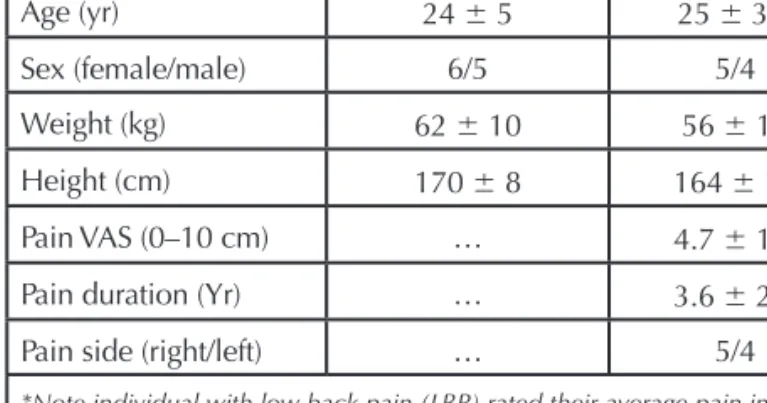
TABLE 1. Subject Demographics (Mean
±
SD)*
Healthy (n = 11) LBP (n = 9) Age (yr) 24 ± 5 25 ± 3.4 Sex (female/male) 6/5 5/4 Weight (kg) 62 ± 10 56 ± 15 Height (cm) 170 ± 8 164 ± 12 Pain VAS (0–10 cm) … 4.7 ± 1.1Pain duration (Yr) … 3.6 ± 2.4
Pain side (right/left) … 5/4
*Note individual with low back pain (LBP) rated their average pain intensity over the last 6 months on a visual analog scale (VAS). Note pain duration is the total time that participants had experienced recurrent LBP and including periods of aggravation and remission. No difference was detected between the two groups in age, weight, and height (The Student t test, P > 0.82).
Spine www.spinejournal.com 1723
identifi cation of MEP onset and offset. MEP amplitude was calculated as the root-mean-square EMG amplitude between the onset and offset, and background root-mean-square EMG from 55 to 5 m prior to stimulation was subtracted. MEP am-plitudes were averaged at each scalp site across subjects after normalization to the peak response to produce a topographi-cal map ( Figure 1 ). Normalized values less than 25% of peak values were removed and the remaining responses were res-caled from 0% to 100%. Removal of small amplitude MEPs minimally affects TMS maps. 26
Two parameters were calculated from normalized maps. Map volume (measure of total motor cortical excitability) was calculated as the sum of normalized MEP amplitudes across scalp sites with responses. Center of gravity (CoG) was calculated using the formula CoG = ∑z i x i /∑z i ; ∑z i y i /∑z i (x i – mediolateral location; y i – anteroposterior location;
z i –normalized amplitude). This measure provides an amplitude-weighted indication of map position. Although map volume is sensitive to changes in corticomotor excitabil-ity, the CoG is a valid and repeatable measure of a muscle’s motor cortex representation. 26 , 27
Statistical Analysis
Statistica 7 was used for analysis (Statsoft, USA). Map vol-ume, CoG location, and MEP onset at the optimal site were compared between muscles, group, and side using repeated measures analyses of variance. The Duncan test was used for
post hoc analyses. A separate analysis compared CoG loca-tion, map volume, and MEP onset between the painful and nonpainful sides (Pain) for the LBP group. The relationship between pain intensity/duration and cortical measures (CoG coordinates and map volume) were investigated using the Pearson correlation. Signifi cance was set at P < 0.05.
RESULTS
Data were rejected for left DM and right LES from one LBP participant each because of poor EMG quality. It was diffi cult to elicit MEPs ipsilateral to the stimulated hemisphere in all but one LBP participant. As MEPs contralateral to the stimu-lated cortex were consistently elicited, mapping was only un-dertaken using this data.
Figure 2 shows rescaled normalized TMS maps for the LBP group and the previous control data. 20 CoG location did
not differ between left and right sides ( P = 0.69), or between painful and nonpainful sides in the LBP group ( P = 0.15). In healthy individuals, DM CoG was located posteriorly to that for LES (interaction – pain × muscle: P < 0.001, post hoc : P
< 0.001; Figure 3 ). In contrast, for the LBP group, DM CoG was located similarly to that for LES ( Figures 3 , 4 ; post hoc: P
> 0.23). This is explained by a more posterior location of the LES CoG in the LBP group compared to controls (post hoc: P
< 0.001). There was no signifi cant association between CoG
Figure 2. Normalized maps of the left and right motor cortex for short/ deep fi bres of multifi dus (DM; left panel) and longissimus erector spi-nae (LES; right panel), for healthy (top panel) and low back pain (LBP) groups (bottom panel). Dotted lines denote sagittal and frontal planes, intersecting at the vertex. Note motor cortical maps for DM overlap that for LES in the LBP group, whereas DM is located posteriorly com-pared to LES in healthy group.
Figure 1. Mapping of the motor cortex using transcranial magnetic stimulation (TMS). A, Stimuli over the motor cortex excite intra-cortical neurons that provide synaptic input to corticospinal cells.
B, Recordings were made from short/deep fascicles of multifi dus (DM) and longissimus erector spinae (LES) at the L4 level. C, The descending volley excites the spinal motoneurons and results in a motor-evoked potential (MEP), mainly in the contralateral muscles. D, MEPs were recorded in both muscles from stimuli applied at each point on the grid placed over the scalp and aligned to the vertex (Cz). E , A three-dimensional map of MEP amplitude was created for each muscle from the individual and then group data.
1724 www.spinejournal.com October 2011
paraspinal muscle fascicles in LBP. Individuals with recurrent LBP had overlapped motor cortical areas for DM and LES, which contrasts recent evidence of discretely organized corti-cal inputs to these muscles in healthy individuals. 20 This
pro-vides novel evidence of a neural substrate that may underlie reduced potential to differentially activate parts of the para-spinal muscles during function in LBP. This work has impli-cations for motor rehabilitation that aims to restore optimal control of paraspinal muscles for management of LBP.
Smudging of Motor Cortical Organization and Reduced Motor Excitability in LBP
Paraspinal muscles receive descending input from the motor cortex and corticospinal pathways. 23 , 28 These pathways not
only contribute to voluntary activation, but also to postural control. For instance, motor cortical stimulation in cats elic-its fl exion of the contralateral forelimb and anticipatory pos-tural adjustments in the supporting forelimb. 29 Human data
demonstrate reduced trunk muscle postural activity with limb movements when motor cortex is inhibited with submotor-threshold TMS. 30 Furthermore, recent work confi rms a
re-lationship between organization of cortical neural networks and temporal parameters of postural activation of trunk mus-cles. 8 , 9 This suggests motor cortex contribution to a range of
motor functions including postural activity of trunk muscles. LBP is associated with adaptive changes in organization of cortical neuronal networks. 8 , 9 , 11 This shows these pain-related
cortical changes include smudging of cortical representation of functionally distinct paraspinal muscles fascicles. The pos-terior shift in LES representation refl ects motor cortex reor-ganization and cannot be accounted for by changes in excit-ability. 26 , 27
Smudging of motor cortical organization has functional implications for neural control of paraspinal muscles. It may reduce the potential to independently control these muscles. The degree of differential motor control is related to dis-crete organization of motor cortical networks; for example, discrete representation of individual fi ngers on the primary motor and sensory cortices are related to independent fi ne control of fi nger movements. 31 Reduced differentiation of
this discrete organization, such as smudging between areas location and pain intensity (all r 2 < 0.15; all P > 0.30) or
duration (all r 2 < 0.35; all P > 0.09).
Figure 5 shows map volume for healthy and LBP groups. Map volume was reduced in LBP compared to controls (main effect – pain: P < 0.001; post hoc : P < 0.021), although this did not differ between muscles ( P = 0.26) or sides ( P = 0.68). Reduced map volume suggests reduced corticomotor excit-ability in LBP, but this was not correlated with pain intensity ( r 2 < 0.18; P > 0.25) or duration ( r 2 < 0.23; P > 0.19).
The latency to onset of MEPs was shorter for DM com-pared to LES in both groups (muscle: P < 0.001; post hoc P
< 0.001), but no difference was observed between group ( P
> 0.35) and sides ( P > 0.39). There was no difference in MEP onset between painful and nonpainful side in the LBP group ( P > 0.19; Averaged across sides: healthy - DM = 15.8 ± 1.1 m, LES = 18.0 ± 3.0 m; LBP - DM = 15.2 ± 1.5 m, LES = 18.3 ± 4.0 m).
DISCUSSION
These results show for the fi rst time loss of discrete organiza-tion of neuronal networks that control funcorganiza-tionally distinct
Figure 4. Individual data for center of gravity (CoG) of short/deep
fi bres of multifi dus (DM; circles) and longissimus erector spinae (LES; squares) in healthy and low back pain (LBP). Data for left/right and pain/nonpainful sides were not different and are not separately
identi-fi ed in the fi gure. Cz indicates vertex.
Figure 3. Group data for center of gravity (CoG) of short/deep fi bres of multifi dus (DM) and longissimus erector spinae (LES) in healthy and low back pain (LBP). Mean and 95% confi dence interval (averaged across sides) are shown. * P < 0.05. Cz indicates vertex
Figure 5. Map volume of short/deep fi bres of multifi dus (DM) and
longissimus erector spinae (LES) for healthy (white) and low back pain (LBP; black) groups. Mean and 95% confi dence intervals are shown. Note reduced map volume in LBP group compared with healthy individuals. * P < 0.05. R indicates right; L, left.
Spine www.spinejournal.com 1725
after a disc lesion, but responsiveness of spinal pathways re-duced. 39 Thus, although reduced corticomotor excitability
would seem to contradict the suggestion of enhanced activa-tion of long/superfi cial fascicles (such as the LES) to protect the spine from further injury, the reduced responsiveness to TMS could simply refl ect the complex interaction of effects of pain along the motor pathway. The possibility that reduced map volume refl ects a more focal cortical network cannot be confi rmed as reduced excitability of spinal motoneurons can-not be excluded. Fortunately, changes of map organization, the key observation in this study, do not refl ect changes in excitability and interpretation is less problematic.
Although current data provide evidence of changes to the motor cortex and corticospinal tracts, the exact mechanisms of how pain induces such changes require further investiga-tion. Changes in other parts of the nervous system could con-tribute to altered motor coordination ( e.g. , basal ganglia, cer-ebellar, and spinal circuits). 41 Exactly how different networks
of the sensorimotor system contribute to changes in motor coordination requires further work.
Relationship Between Cortical Changes and Pain
Previous work has identifi ed a relationship between pain and cortical changes. Amplitudes of changes in the sensory rep-resentation of the back 42 and motor threshold 11 are related
to pain duration. No relationship was identifi ed in our data. This could be because of the small sample size or lesser pain intensity than previous studies. 11 However, pain report is
sub-jective and infl uenced by a complex array of biological and psychological ( e.g ., catastrophization) factors. 43 , 44 The lack of
association between pain and motor cortical parameters may refl ect this complex interaction.
Methodological Issues
The main limitation of the study is the small sample size. Yet, despite the small sample, fi ndings were consistent across jects and yielded signifi cant results. For practical reasons, sub-jects were only required to match DM EMG during TMS. We did not control LES EMG. Although this may increase inter-subject variability in motor cortical excitability for LES, this does not compromise our main fi nding as CoG measures are less affected by motor excitability. 26 Furthermore, this study
involved young adults and studies of older adult population should be considered.
Clinical Implications
A loss of differential activation of paraspinal muscle fascicles is likely to compromise fi ne-tuned segmental control and alter loading of spinal structures, 34 potentially increasing the risk of
LBP recurrence. 33 , 34 As our data show that such motor changes
may be mediated by loss of discrete organization at the motor cortex, it could be argued that a goal of LBP rehabilitation, at least initially, should include motor training to restore cortical organization. Although it is beyond this study to speculate on how best to restore cortical organization, studies in focal hand dystonia suggest redifferentiation of discrete fi nger representation is possible with training of isolated fi nger representing adjacent fi ngers in focal hand dystonia,
reduc-es the isolation of fi nger movements. 19 Smudging of motor
cortical representations of DM and LES may explain obser-vations of reduced discrete activation of different paraspinal muscle fascicles in LBP. 14 , 18 We did not investigate paraspinal
muscle activity in functional tasks. Thus, we cannot directly investigate the relationship between organization and func-tion. Nevertheless, this work provides the basis for studies into the relationship between cortical remodeling and altered trunk muscle coordination.
Motor cortex reorganization supports the notion that the nervous system adopts a new strategy for movement/stability with LBP. 32 It has been hypothesized that in the presence of
pain and/or injury, the nervous system implements new motor strategies to “protect the part” from further injury/pain. 33 , 34
This is often mediated by increased trunk muscle activity, par-ticularly large superfi cial muscles, to splint the spine. This is supported by several observations. First, when pain is induced in healthy individuals, trunk muscle EMG increases in keep-ing with the goal to protect the spine, although the pattern varies between individuals. 35 Second, biomechanical
model-ing suggests various patterns of muscle activity observed in LBP increase trunk stability. 36 In this case, fi ne coordination
between different fascicles of the paraspinal muscles may no longer be used in lieu of a simplifi ed protective strategy. That is, nervous system remodeling (including smudging of motor cortex) may simplify the motor strategy to a more en masse paraspinal muscle recruitment. Reorganization of LES but not DM appears consistent with the proposal that reorganization of inputs to long/superfi cial fascicles underpins the adaptive search for a new motor strategy. 34 , 37
Motor maps exhibited the same organization bilaterally in LBP, despite selection of patients on the basis of unilateral symptoms. The isolation of some motor control changes to the painful side 14 may question the association between
mo-tor control and symmetrical changes in cortical organization. However, many changes in motor control are not systemati-cally related to the side of pain. For instance, unilateral ex-perimental pain induces variation in motor coordination that is not exclusive to the pain side. 35 The nervous system appears
to select a protective solution that is generic, regardless of the location/side of pain. Further investigation is required to examine cortex reorganization in other LBP subgroups ( e.g. , bilateral LBP, specifi c spinal pathology).
DM and LES map volume was reduced in LBP. This sug-gests either reduced corticomotor excitability or that neural networks with input to these muscles occupy a smaller area of cortex. Reduced excitability has been interpreted from earlier data of increased paraspinal muscle motor threshold to TMS 11
( i.e. , greater stimulation intensity to evoke a response). How-ever, it is not possible to determine whether those changes were because of effects at the cortex or spinal cord as excitability of both may affect the responsiveness to TMS. 38 Recent animal 39
and human 40 studies highlight opposite changes at the cortex
and spinal cord, making interpretation impossible without techniques that isolate measures to a specifi c site. In animal studies, responsiveness to stimulation over the brain increased
1726 www.spinejournal.com October 2011
movements. 19 In addition, motor training with specifi c
activa-tion of deep abdominal muscles can reverse pain-associated reorganization at the motor cortex 9 and this is associated
with improved motor behavior. 9 , 13 , 45 A recent study of a small
group with LBP shows training that targets DM can improve coordination of the paraspinal muscle fascicles during func-tional tasks toward that observed in healthy individuals. 46
Al-though motor rehabilitation should not be exclusive to DM, changes in paraspinal muscle activation after motor training provide a marker for further investigations to study whether these improvements are associated with redifferentiation of muscle representation at the motor cortex (and improvement in clinical symptoms).
➢
Key Points
Fascicles of the paraspinal muscles are diff erentially activated during function.
Diff erential activation is associated with discrete or-ganization of neuronal networks at the motor cortex.
Loss of discrete organization of diff erent fascicles of the paraspinal muscles at the motor cortex is observed in individuals with recurrent low back pain.
Reorganization at the motor cortex may underpin this loss of diff erential activation in low back pain.
Acknowledgment
The authors thank Mr Ian Peeters for assistance with data analysis.
References
1. Danneels LA , Coorevits PL , Cools AM , et al. Differences in mul-tifi dus and iliocostalis lumborum activity between healthy subjects and patients with sub-acute and chronic low back pain . Eur Spine J 2002 ; 11 : 13 – 19 .
2. MacDonald DA , Moseley GL , Hodges PW . The lumbar mul-tifi dus: does the evidence support clinical beliefs ? Man Ther 2006 ; 11 : 254 – 63 .
3. Smith AJ , O’Sullivan PB , Campbell A , et al. The relationship between back muscle endurance and physical, lifestyle, and psy-chological factors in adolescents . J Orthop Sports Phys Ther 2010 ; 40 : 517 – 23 .
4. Danneels LA , Vanderstraeten GG , Cambier DC , et al. Clini-cal Science award 2000: CT imging of trunk muscles in chronic low back pain patients and healthy control subjects . Eur Spine J 2000 ; 9 : 266 – 72 .
5. Hides JA , Stokes MJ , Saide M , et al. Evidence of lumbar multifi -dus muscle wasting ipsilateral to symptoms in patients with acute/ subacute low back pain . Spine 1994 ; 19 : 165 – 72 .
6. Hodges PW , Holm AK , Hansson T , et al. Rapid atrophy of the lum-bar multifi dus follows experimental disc or nerve root injury . Spine 2006 ; 31 : 2926 – 33 .
7. Haig AJ . Paraspinal denervation and the spinal degenerative cas-cade . Spine J 2002 ; 2 : 372 – 80 .
8. Tsao H , Galea MP , Hodges PW . Reorganization of the motor cor-tex is associated with postural control defi cits in recurrent low back pain . Brain 2008 ; 131 : 2161 – 71 .
9. Tsao H , Galea MP , Hodges PW . Driving plasticity in the motor cor-tex in recurrent low back pain . Eur J Pain 2010 ; 14 : 832 – 9 . 10. Leinonen V , Kankaanpaa M , Luukkonen M , et al. Disc
herniation-related back pain impairs feed-forward control of paraspinal mus-cles . Spine 2001 ; 26 : E367 – 72 .
11. Strutton PH , Theodorou S , Catley M , et al. Corticospinal excit-ability in patients with chronic low back pain . J Spinal Disord Tech 2005 ; 18 : 420 – 4 .
12. Danneels LA , Vanderstraeten GG , Cambier DC , et al. Effects of three different training modalities on the cross sectional area of the lumbar multifi dus muscle in patients with chronic low back pain .
Br J Sports Med 2001 ; 35 : 186 – 91 .
13. Tsao H , Hodges PW . Immediate changes in feedforward postural adjustments following voluntary motor training . Exp Brain Res 2007 ; 181 : 537 – 46 .
14. MacDonald D , Moseley GL , Hodges PW . Why do some pa-tients keep hurting their back? Evidence of ongoing back muscle dysfunction during remission from recurrent back pain . Pain 2009 ; 142 : 183 – 8 .
15. O’Sullivan PB , Dankaerts W , Burnett AF , et al. Effect of different upright sitting postures on spinal-pelvic curvature and trunk muscle activation in a pain-free population . Spine 2006 ; 31 : E707 – 12 . 16. Claus AP , Hides JA , Moseley GL , et al. Different ways to balance
the spine: subtle changes in sagittal spinal curves affect regional muscle activity . Spine 2009 ; 34 : E208 – 14 .
17. Moseley GL , Hodges PW , Gandevia SC . Deep and superfi cial fi bers of the lumbar multifi dus muscle are differentially active during vol-untary arm movements . Spine 2002 ; 27 : E29 – 36 .
18. Claus A , Hidges J , Moseley GL , et al. How do we Sit and How Should we Sit? International Society of Posture and Gait Research . Bologna, Italy , 2009 .
19. Byl NN , Merzenich MM , Jenkins WM . A primate genesis model of focal dystonia and repetitive stain injury: I. Learning-induced dedif-ferentiation of the representation of the hand in the primary so-matosensory cortex in adult monkeys . Neurology 1996 ; 47 : 508 – 20 . 20. Tsao H , Danneels L , Hodges PW . Individual fascicles of the
paraspi-nal muscles are activated by discrete cortical networks in humans .
Clin Neurophysiol 2011 ; DOI:10.1016/j.clinph.2011.01.048. 21. Nowicky AV , McGregor AH , Davey NJ . Corticospinal control of
human erector spinae muscles . Motor Control 2001 ; 5 : 270 – 80 . 22. O’Connell NE , Maskill DW , Cossar J , et al. Mapping the
corti-cal representation of the lumbar paravertebral muscles . Clin Neurophysiol 2007 ; 118 : 2451 – 5 .
23. Taniguchi S , Tani T . Motor-evoked potentials elicited from human erector spinae muscles by transcranial magnetic stimulation . Spine 1999 ; 24 : 154 – 6 .
24. Jasper HH . Report of the committee on methods of clinical exami-nation in electroencephalography: 1957 . Electroencephalogr Clin Neurophysiol 1958 ; 10 : 370 – 5 .
25. Wilson SA , Thickbroom GW , Mastaglia FL . Transcranial mag-netic stimulation mapping of the motor cortex in normal subjects. The representation of two intrinsic hand muscles . J Neurol Sci 1993 ; 118 : 134 – 44 .
26. Uy J , Ridding MC , Miles TS . Stability of maps of human motor cortex made with transcranial magnetic stimulation . Brain Topogr 2002 ; 14 : 293 – 7 .
27. Boroojerdi B , Foltys H , Krings T , et al. Localization of the motor hand area using transcranial magnetic stimulation and functional magnetic resonance imaging . Clin Neurophysiol 1999 ; 110 : 699 – 704 .
28. Gandevia SC , Plassman BL . Responses in human intercostal and truncal muscles to motor cortical and spinal stimulation . Respir Physiol 1988 ; 73 : 325 – 38 .
29. Gahéry Y , Nieoullon A . Postural and kinetic co-ordination follow-ing cortical stimuli which induce fl exion movements in the cat’s limb . Brain Res 1978 ; 149 : 25 – 37 .
30. Hodges PW , Butler JE , Taylor JL , et al. Motor cortex may be in-volved in feedforward postural responses of the deep trunk muscles . In: Lord S , Menz HB eds. Proceedings of International Society for Posture and Gait Research . Sydney, Australia : International Society for Posture and Gait Research , 2003 : 53 – 4 .
31. Beisteiner R , Windischberger C , Lanzenberger R , et al. Finger so-matotopy in human motor cortex . Neuroimage 2001 ; 13 : 1016 – 26 . 32. Hodges PW . Changes in motor planning of feedforward postural responses of the trunk muscles in low back pain . Exp Brain Res 2001 ; 141 : 261 – 6 .
Spine www.spinejournal.com 1727 33. van Dieen JH , Selen LPJ , Cholewicki J . Trunk muscle activation in
low-back pain patients, an analysis of the literature . J Electromyogr Kinesiol 2003 ; 13 : 333 – 51 .
34. Hodges PW , Moseley GL . Pain and motor control of the lumbopel-vic region: effect and possible mechanisms . J Electromyogr Kinesiol 2003 ; 13 : 361 – 70 .
35. Hodges PW , Cholewicki J , Coppieters MW , et al. Trunk Muscle Activity is Increased During Experimental Back Pain, But the Pat-tern Varies Between Individuals . International Society for Electro-physiology and Kinesiology . Torino, Italy , 2006 .
36. Cholewicki J , Van Vliet IV JJ . Relative contribution of trunk muscles to the stability of the lumbar spine during isometric exertions . Clin Biomech 2002 ; 17 : 99 – 105 .
37. van Dieen JH , Cholewicki J , Radebold A . Trunk muscle recruit-ment patterns in patients with low back pain enhance the stability of the lumbar spine . Spine 2003 ; 28 : 834 – 41 .
38. Taylor JL , Gandevia SC . Noninvasive stimulation of the human corticospinal tract . J Appl Physiol 2004 ; 96 : 1496 – 503 .
39. Hodges PW , Galea MP , Holm S , et al. Corticomotor excitability of back muscles is affected by intervertebral disc lesion in pigs . Eur J Neurosci 2009 ; 29 : 1490 – 500 .
40. Martin PG , Gandevia SC , Taylor JL . Output of human motoneuron pools to corticospinal inputs during voluntary contractions .
J Neurophysiol 2006 ; 95 : 3512 – 18 .
41. Deliagina TG , Beloozerova IN , Zelenin PV , et al. Spinal and supraspinal postural networks . Brain Res Rev 2008 ; 57 : 212 – 21 .
42. Flor H , Braun C , Elbert T , et al. Extensive reorganization of pri-mary somatosensory cortex in chronic back pain patients . Neurosci Lett 1997 ; 224 : 5 – 8 .
43. Turk DC . Psychological aspects of chronic pain and disability . J Muscoskel Pain 1996 ; 4 : 145 – 53 .
44. Vlaeyen JWS , Kole-Snijders AMJ , Boeren RGB , et al. Fear of move-ment/(re)injury in chronic low back pain and its relation to behav-ioural performance . Pain 1995 ; 62 : 363 – 72 .
45. Tsao H , Hodges PW . Persistence of improvements in postural strat-egies following motor control training in people with recurrent low back pain . J Electromyogr Kinesiol 2008 ; 18 : 559 – 67 .
46. Tsao H , Druitt TR , Schollum TM , et al. Motor training of the lumbar paraspinal muscles induces immediate changes in motor coordination in patients with recurrent low back pain . J Pain 2010 ; 11 : 1120 – 8 .