Supporting Information
White et al. 10.1073/pnas.1119663109
SI Materials and MethodsRadiocarbon Materials and Methods.At the Oxford Radiocarbon Accelerator Unit (ORAU), Oxford, United Kingdom, each of the bones was sampled with an NSK Electer GX drill with a tungsten carbide drill bit. Then, 800–1,200 mg of bone was sampled for analysis. Samples were pretreated by using the manual Oxford method (1), which includes a decalcification with 0.5 M HCl, the removal of humates with 0.1 M NaOH, and afinal reacidification with 0.5 M HCl, with each step interspersed with distilled water rinses. Gelatinization was undertaken in water adjusted to pH 3 solution at 75 °C in an incubator (for 20 h). We recovered the supernatant with an EziFilter and ultrafiltered it with a Vivaspin 30-kDa molecular mass cut-off ultrafilter. We recovered the
>30-kDa fraction and freeze-dried it before accelerator mass spectrometry (AMS) dating.
Ultrafiltered gelatin from each of the dated bones was com-busted with a Europa Scientific ANCA-MS system consisting of a 20/20 IR mass spectrometer interfaced to a Roboprep CHN sample converter unit operating in continuous-flow mode using an He carrier gas. In Table 1, we report d13C values with respect to Vienna Pee Dee Belemnite (VPDB), nitrogen and carbon con-tents, and C:N ratios. CO2 from the sample combustion was graphitized by a reduction reaction over an iron catalyst in an excess H2atmosphere at 560 °C (2, 3) and then AMS-dated with the Oxford HVEE 2.5-MV accelerator. The radiocarbon dates in
this paper were corrected by using a unique background sub-traction model that sets the measurement limit at 49,900 y BP (4). The bone preparation at the Laboratoire des Sciences du Climat et de l’Environnement (Gif-sur-Yvette) was based on the specific reaction between collagen amino acids and ninhydrin (5, 6). Each dating required∼2,000 mg of cortical bone, which was crushed after being sand-blasted with carbon-free alumina. After a decalcification using 0.5 M HCl, the carbonate-free sample was treated with ninhydrin at 100 °C for 10 min to eliminate any “free”amino acids introduced from the archaeological sediment. After the collagen had been hydrolyzed with 6 M HCl at 100 °C overnight, the solution of amino acids wasfiltered and collected in a glass reactor where the filtrate was evaporated at 80 °C under nitrogen. Then the reactor was connected to a vacuum line. When the vacuum reached∼2.10−4mb, a second treatment with ninhydrin allowed extracting the CO2from the carboxylic groups of amino acids. The released CO2 is dried by “water traps,” trapped in liquid nitrogen, and quantified into a cali-brated volume. Finally, the extracted CO2 was reduced to graphite (7), which was submitted to the Tandétron AMS Fa-cility (UMS 2004, Gif-sur-Yvette). Bone“blank”specimens were prepared and measured alongside the archaeological samples. The13C/12C ratios measured during the AMS dating fell in the range of values obtained for bone; no other measurements were done on a mass spectrometer.
1. Brock F, Higham T, Ditchfield P, Bronk Ramsey C (2010) Current pretreatment methods for AMS radiocarbon dating at the Oxford Radiocarbon Accelerator Unit (ORAU).
Radiocarbon52:103–112.
2. Dee M, Bronk Ramsey C (2000) Refinement of graphite target production at ORAU.
Nucl Instrum Methods Phys Res B172:449–453.
3. Bronk Ramsey C, Higham T, Bowles A, Hedges R (2004) Improvements to the pre-treatment of bone at Oxford.Radiocarbon46:155–163.
4. Wood RE, Bronk Ramsey C, Higham T (2010) Refining background corrections for radiocarbon dating of bone collagen at ORAU.Radiocarbon52:600–611.
5. Nelson DE (1991) A new method for carbon isotopic analysis of protein.Science251: 552–554.
6. Tisnérat-Laborde N, Valladas H, Kaltnecker E, Arnold M (2003) AMS radiocarbon dating of bones at LSCE.Radiocarbon45:409–419.
7. Arnold M, Bard E, Maurice P, Duplessy JC (1987)14C dating with the Gif-sur-Yvette
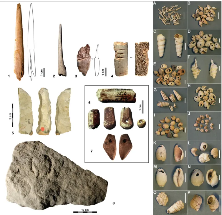
Fig. S1. Artifacts characterizing the Castanet-type Aurignacian. (Left) 1, Split-based antler point. 2, Bone awl. 3, Tongued piece in antler. 4, Decorated smoothing tool in herbivore rib. 5, Carinate scraper. 6, Basket-shaped beads and production stages. 7, Facsimile in ivory of a red deer vestigial canine, per-forated for suspension. 8, Limestone block engraved with“vulvar”images. (Right) Different species of Atlantic and Mediterranean gastropod species repre-sented in the ornament assemblage at Abri Castanet.
Fig. S2. Abri Castanet blocks from Peyrony excavations. (A) Vulvar engravings. (B) Juxtaposition of engraved“phallus”and an“anneau”gouged into the block’s surface. (C) Bichrome painting. (D) Vulvar engraving and cup marks.
Fig. S4. Abri Castanet, Southern sector. Multicomponentfire feature during excavation.
Fig. S6. Refitting of lithic artifacts shattered in place by impact of the ceiling collapse. At bottom,flint objects so pulverized by the roof collapse so as to be reduced to“gravel.”
Fig. S8. Bayesian model of the Castanet Northern sector results produced with OxCal 4.1 (1). The radiocarbon ages are compared against the IntCal09 dataset of Reimer et al. (2). The model is based on the assumption that the archaeological sequence consists of a single excavated phase. Individual radiocarbon likelihoods are shown by the light-shaded distributions, whereas the darker outlines represent posterior probability distributions. Note that the probability distribution function (PDF) for Castanet Aurignacian I is equivalent to the total date span for the occupation.
1. Bronk Ramsey C (2001) Development of the radiocarbon calibration program OxCal.Radiocarbon43:355–363.