Supporting Information
Structurally Colored Inks from Synthetic
Melanin-Based Crosslinked Supraparticles
Ziying Hu1, Hao Sun1,Matthew P. Thompson1,Ming Xiao4,#, Michael C. Allen5, Xuhao Zhou1,
Qing Zhe Ni6, Zhao Wang6, †, Weiyao Li4, Michael D. Burkart6,*, Dimitri D. Deheyn5,*, Ali
Dhinojwala4,*, Matthew D. Shawkey3,*, Nathan C. Gianneschi1,2,*
1Department of Chemistry, 2Department of Materials Science and Engineering, Department of
Biomedical Engineering, Department of Pharmacology, International Institute of
Nanotechnology, Simpson-Querrey Institute, Chemistry of Life Processes Institute, Lurie Cancer Center, Northwestern University, Evanston, Illinois 60208, United States
3Biology Department, University of Ghent, Ghent 9000, Belgium
4Department of Polymer Science, The University of Akron, Akron, Ohio 44325, United States 5Scripps Institution of Oceanography, 6Department of Chemistry & Biochemistry University of
California, San Diego, La Jolla, California 92093, United States
*N. C. G. Email: [email protected] *M. D. S. Email: [email protected]
*A. D. Email: [email protected] *D. D. D. Email: [email protected] *M. D. B. Email: [email protected]
Synthesis and Crosslinking Methods
SM@SiO2 NPs Synthesis. SM@SiO2 NPs were synthesized in two steps. SMNPs were first
synthesized by oxidative polymerization of dopamine hydrochloride (C8H11NO2·HCl, Alfa Aesar)
under basic conditions (pH = 10), and then coated SMNPs with a silica shell (SiO2) via a modified
Stöber method.1 To achieve this, typically, 1.1 mL of ammonium hydroxide (NH4OH, 28.0-30.0
% NH3 basis, Sigma-Aldrich) was mixed with a solution of 110 mL ethanol (200 proof, Decon
Labs, Inc.) and 20 mL Milli-Q water (resistivity of 18.20 MΩ·cm at 20oC, Thermo Scientific) in
5 min, and then 460 mg of dopamine hydrochloride was added with vigorous stirring. The size of SMNPs can be tuned by changing the amount of either dopamine hydrochloride or NH4OH. The
SMNPs were purified using a centrifuge (Fisher Scientific) under 10,000 rpm for 10 min and then dispersed in Milli-Q water. After repeating the purification procedure four times, the as-prepared SMNPs were dispersed in Milli-Q water at 3.7 mg/mL for the subsequent coating step. 8.75 mL of SMNPs solution was first mixed with 50 mL isopropanol (Sigma-Aldrich) followed by 1.2 mL NH4OH, and then 0.8 mL of tetraethyl orthosilicate (TEOS, Acros Organics), as the silica
precursor, was added dropwise into the mixture. Core shell particles with different shell thicknesses/diameters can be achieved by simply changing the reaction time. The core-shell particles were purified with water by centrifugation (5,000-10,000 rpm for 6 min). This was repeated three times before suspending the particles in water at 30 mg/mL.
Supraparticle Assembly. A reverse emulsion method was used to form supraparticles via a
modified published protocol.2 Briefly, 30 µL of the above aqueous core-shell particles solution
was added to 1 mL anhydrous 1-octanol (Sigma-Aldrich) in a 2.0 mL microtubes (Corning Inc.). The black aqueous solution and the clear 1-octanol form layered liquids at the beginning. A digital vortex (Fisher Scientific) was used to mix the solution to make water-in-oil emulsions. The shaking
speed was set at 1800 rpm/2.5 min (1,800 rpm holding for 2.5 min) and then reduced to 1,200 rpm/3 min. The whole solution became uniform and clear after vortexing. Larger particles disperse in the solution and then settle to the bottom within 10 min. We note that shaking speed and time depends on the size and shape of the containers which should be hydrophobic at the interior surface. For large scale supraparticle assembly (1 mL particle solution in 30 mL 1-octanol), a 50 mL centrifuge tube (Fisher Scientific) was used and the shaking speed was 2,000 rpm/2.5 min and then reduced to 1,800 rpm/3 min.
Supraparticle Crosslinking Procedure. Linear and 4-arm poly(ethylene glycol) (PEG) with
different molecular weights (Mw~2,000 g/mol; 5,000 g/mol; 10,000 g/mol) were used to crosslink supraparticles. These are: PEG2k Aldrich), PEG5k Aldrich), PEG10k (Sigma-Aldrich), 4-arm PEG2k (Nanosoft Polymers), 4-arm PEG5k (Nanosoft Polymers), and 4-arm PEG10k (Sigma-Aldrich). Stock solutions of the various types of PEGs were prepared by dissolving them in methanol (Fisher Scientific). After supraparticle assembly, the supernatant was removed and the PEG solutions were added to the supraparticles (mass ratio of PEG to supraparticles = 4:9). Free PEG was rinsed away with four washes of methanol after the supraparticle-PEG solutions were incubated overnight. Washes involved gentle shaking of the crosslinked supraparticles suspended in methanol, followed settling of the particles, removal of the supernatant and repeat.
Instruments and Characterization Methods
Transmission Electron Microscopy (TEM). Suspensions of SMNPs and core-shell
nanoparticles were loaded onto 400 mesh carbon grids (Ted Pella, INC.) and characterized using a Hitachi HT-7700 microscope. The images were captured at a voltage of 120 kV with an Orius SC 1000A camera. ROI (Region of Interest) manager in ImageJ was used to analyze the nanoparticle sizes. For each type of the nanoparticles, we measured the diameters of 50 nanoparticles and calculated the average values as well as standard deviations.
Scanning Electron Microscopy (SEM). Supraparticle suspensions were deposited on silica
wafers (Ted Pella, INC.) and dried under 40oC in air. The prepared samples were sputtered with a
layer of osmium to a thickness of 15 nm using an osmium coater (SPI Supplies). The morphology of the supraparticles was observed using a scanning electron microscope (SEM, Hitachi SU8030).
Optical Imaging and Overall Spectral Characterization. Optical images were acquired using an optical microscope (Nikon L-UEPI) equipped with ZEN software (Carl Zeiss Microscopy GmbH). Reflectance spectra of individual supraparticles were measured using a CRAIC AX10 UV-vis microspectrophotometer (CRAIC Technologies Inc.). A 75-W xenon short-arc lamp (Ushio UXL75XE) was used as a light source and a silver mirror standard was used as a reflectance standard. For each type of sample, we measured three supraparticles and averaged the spectra.
Nanoindentation Measurements. Red supraparticle suspensions were deposited on silica
wafers (Ted Pella, INC.) and dried under 40oC in air. Here, fewer supraparticles were deposited
on the silicon wafers because loading measurements were conducted supraparticle by supraparticle such that they needed to be separated in space. A nanoindenter (Hysitron TI 950 TriboIndenter,
Bruker) equipped with a cylindrical shaped probe with a flat end (probe diameter 200 µm) was used to quantify mechanical properties of noncrosslinked versus crosslinked supraparticles by measuring compressive force when compressing them down to 1,000 nm. The loading/unloading speed was set to 50 nm/s and held the probe for 2 s at maximum displacement. Measurements were made and repeated on the same supraparticle four times by changing the displacement (250 nm, 500 nm, 750 nm and 1,000 nm) each time. Displacement data of 1,000 nm are shown, providing direct information on the loading (compressive force) trends for each type of supraparticle and to make directly relevant comparisons. Further increases in displacement can break the supraparticles which resulted in difficulty obtaining the maximum compressive forces. Since the size of supraparticles would affect the loading results, measurements on supraparticles with an average diameter of 35 ± 7 µm were performed. For each type of sample, 10 to 15 supraparticles were measured, giving average compressive force values.
Hyperspectral imaging and spectral mapping. Using a PARISS® hyperspectral imaging
system (LightForm Inc.; http://lightforminc.com/index.html) that was mounted on a Nikon Eclipse 80i microscope, individual supraparticles were analyzed with a 50× objective, and their specular reflectance spectra collected for each pixel in the field of view when the top of the supraparticle was in focus. A silver mirror standard was used as a reflectance standard. We adopted a minimum correlation coefficient of 99.0% for the spectral analyses, indicating that pixels showed the same reference spectrum when sharing more than 99.0% similarity. Each reference spectrum (which shows the actual spectral color in wavelength) was then assigned a coded color that was mapped back to the area where it was acquired. Thus, the mapped images have a color-code that reflects the identity of corresponding spectra (and not the actual color of the sample). Wavelength
calibration was performed with an MIDL Hg+/Ar+ emission lamp (LightForm, Asheville, NC, USA), and accuracy was recorded and verified to be better than 2 nm.
1H Nuclear Magnetic Resonance (1H NMR) on The Amount of 4-arm PEG2k Bound in
Supraparticles. The supraparticles were assembled in a 2 mL microtube following the above
procedures (0.9 mg supraparticles were formed for each tube). The supraparticles were rinsed with deuterated methanol (CD3OD, Sigma-Aldrich) twice after removing the supernatant. Two stock
solutions of 4-arm PEG2k and Dimethylformamide (DMF, used as an internal standard, Fisher Scientific) in CD3OD were made separately. 0.4 mg 4-arm PEG2k was added into each tube of
supraparticles to mix overnight. Then, the supraparticles were rinsed with CD3OD three times with
the supernatant being collected each time (supraparticles would settle to the bottom). The supernatant sample was centrifuged and then 50 µL DMF, as an internal standard, was added (the volume of DMF to the remaining supernatant was 1:16). These samples were then ready for obtaining 1H NMR spectra (Figure S7-2). To test if there was still free 4-arm PEG2k left in the
supraparticle supernatant after three rinses, the supraparticles were washed one more time having the supernatant collected, and 50 µL DMF was added as an internal standard (Figure S7-3, same volume ratio). As a control experiment, 0.4 mg 4-arm PEG2k was dissolved in CD3OD and DMF
as an internal standard as above (Figure S7-1). 1H NMR spectra were recorded on a Bruker Avance
III HD system equipped with a TXO Prodigh probe (500 MHz) in CD3OD. For each type of
solution, the experiments were repeated three times. Integration was performed on each spectrum with the peak area of DMF set as 1.00. The average peak area of the standard solution was calculated to be 11.43 and that of sample supernatant was 8.84 (24.5 % loss in mass). No free 4-arm PEG2k was left in the rinsed supraparticles. The decrease of 4-4-arm PEG2k in solutions was ascribed to an amount of 4-arm PEG2k, now incorporated into the supraparticles, of 10 wt%.
Fourier Transform Infrared Spectroscopy (FTIR). FTIR (Nicolet iS50 FT-IR, Fisher Scientific) was used to verify hydrogen-bonds were present at ether oxygens of PEG when silica was present. We first mixed SiO2 NPs and PEG5k with a mass ratio of 1:4 in water and stayed
overnight before drying the solution to make a powder. Then, we ground the sample powder together with Potassium Bromide (KBr, International Crystal Laboratories) and made a thin film. The measurements were done under transmission mode. We selected PEG5k rather than other types of PEGs because it is a fine powder which is more suitable for this test. As control experiments, we characterized pure PEG5k and SiO2 NPs powders following the same procedure.
Supporting Information Figures
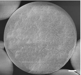
Figure S1. Morphology of synthetic melanin nanoparticles (SMNPs) and kinetic reaction of silica shell growth on the surface of SMNPs. a) Transmission electron microscopy (TEM) image of synthetic melanin nanoparticles (SMNPs). Scale bar: 100 nm. b) The diameter increases of synthetic melanin core silica shell nanoparticles (SM@SiO2 NPs) along with reaction time.
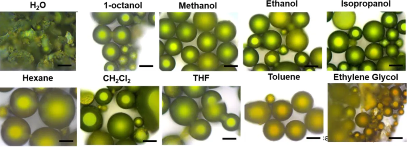
Figure S3. The stability of noncrosslinked green supraparticles in different commonly used solvents. Scale bar: 20 µm.
Figure S4. Here, supraparticles were suspended in methanol, without the addition of PEG. This control experiment was performed exactly as for the crosslinking experiment but excluding the PEG to check for the effect of methanol alone. SEM images show the resulting noncrosslinked red supraparticles that were suspended in methanol and then transferred to water without PEG present. a) low magnification; b) high magnification. Scale bar: a) 50 µm, b) 10 µm. We observed that the supraparticle surfaces roughened because some NPs dissociated. This confirms that PEG is needed to keep the SM@SiO2 NPs intact in water.
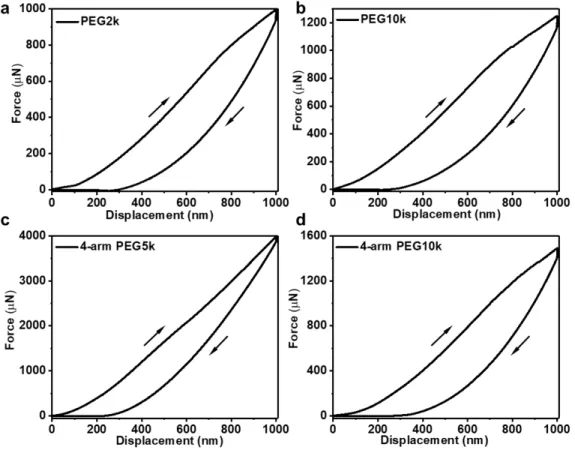
Figure S5. Typical Force-Displacement curves recorded during nanoindentation (the up right arrow-force loading, the down left arrow-force unloading). The indenter probe approached and retracted from red supraparticles crosslinked by different types of PEGs, a) PEG2k, b) PEG10k, c) 4-arm PEG5k, d) 4-arm PEG10k, at the speed of 50 nm/s and was held for 2s at 1000 nm.
Figure S6. Morphology of 4-arm PEG2k crosslinked supraparticles in full color exposed in water and then dried in air on silicon wafers. (a-d) SEM images of crosslinked a) blue, b) green, c) yellow and d) red SM@SiO2 supraparticles. Scale bars: a-d) 50 µm.
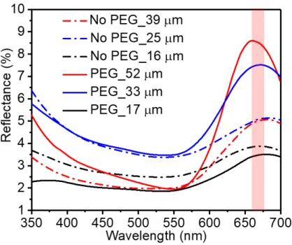
Figure S7. Reflectance spectra of noncrosslinked and 4-arm PEG2k crosslinked red supraparticles of different sizes. Dashed lines: average spectra from three noncrosslinked red supraparticles with average diameters of 40 µm, 25 µm and 15 µm as shown. Solid lines: average spectra of three 4-arm PEG2k crosslinked red supraparticles with average diameters of 50 µm, 30 µm and 15 µm respectively. All the reflectance peaks were concentrated around 670 nm with a slight peak shift within the red wavelengths, indicating that 4 arm-PEG2k and the supraparticle size had an imperceptible influence on the hue.
Figure S8. Hyperspectral imaging and spatially discriminated spectra of individual red supraparticles. a) 4-arm PEG2k crosslinked supraparticle; b) Noncrosslinked supraparticle. The color remained homogeneously red in all pixels. Note that the relative intensity of the color decreased gradually from the top of the supraparticle to the side edge (shown by the arrows), without significant change in the red hue. Scale car: 10 µm.
Figure S9. SEM images of SMNP supraparticles. a) SMNP supraparticles after assembly; b)
4-arm PEG2k crosslinked SMNP supraparticles exposed in water and dried on silicon wafer. Scale bar: a-b) 50 µm.
Figure S11. Fourier transform infrared spectroscopy (FT-IR) of PEG5k, SiO2, and SiO2 (20 wt%)
1. Stöber, W.; Fink, A.; Bohn, E. J. Controlled growth of monodisperse silica spheres in the micron size range. J. Colloid Interface Sci. 1968,26, 62−68.
2. Xiao, M.; Hu, Z. Y.; Wang, Z.; Li, Y. W.; Tormo, A. D.; Le Thomas, N.; Wang, B.; Gianneschi, N. C.; Shawkey, M. D.; Dhinojwala, A., Bioinspired bright noniridescent photonic melanin supraballs. Science Advances 2017,3, e1701151.