Miami Classification for Probe-Based Confocal Laser
Endomicroscopy
(Article begins on next page)
The Harvard community has made this article openly available.
Please share how this access benefits you. Your story matters.
Citation
Wallace, M., G. Lauwers, Y. Chen, E. Dekker, P. Fockens, P.
Sharma, and A. Meining. 2011. Miami classification for
probe-based confocal laser endomicroscopy. Endoscopy 43(10):
882–891.
Published Version
doi:10.1055/s-0030-1256632
Accessed
February 16, 2015 10:02:36 AM EST
Citable Link
http://nrs.harvard.edu/urn-3:HUL.InstRepos:12601541
Terms of Use
This article was downloaded from Harvard University's DASH
repository, and is made available under the terms and conditions
applicable to Other Posted Material, as set forth at
http://nrs.harvard.edu/urn-3:HUL.InstRepos:dash.current.terms-of-use#LAA
Miami classification for probe-based confocal laser
endomicroscopy
Authors M. Wallace1, G. Y. Lauwers2, Y. Chen3, E. Dekker4, P. Fockens4, P. Sharma5, A. Meining6 Institutions Institutions are listed at the end of article.
submitted 25. August 2010
accepted after revision
15. May 2011 Bibliography DOIhttp://dx.doi.org/ 10.1055/s-0030-1256632 Published online: 04.08.2011 Endoscopy 2011; 43: 882–891 © Georg Thieme Verlag KG Stuttgart · New York ISSN 0013-726X
Corresponding author M. B. Wallace, MD, MPH
Mayo Clinic Jacksonville 4500 San Pablo Road South Jacksonville FL 32224 USA Fax: +1-904-953-7260 [email protected]
Introduction
!Probe-based confocal laser endomicroscopy
(pCLE) and its related technology, endoscope-based CLE (eCLE), are rapidly emerging methods of imaging with the potential to fundamentally change the role of biopsy in gastrointestinal en-doscopy. Since the advent of endoscopic imaging, biopsy has been used to confirm or exclude pa-thology. Although highly valuable, biopsy is ex-pensive, and there can be delays between
endos-copy and a “confirmed” diagnosis and/or
treat-ment. In addition, although the risks associated with biopsy are small, the number of biopsies per-formed worldwide is enormous, and therefore risk becomes an important consideration. CLE methods offer the potential to decrease or possibly eliminate the need for some biopsies and to directly guide treatment of circumscribed lesions, such as colorectal polyps, in real time. CLE can also help to avoid biopsy, or if biopsy is necessary, to direct it much more efficiently in areas of very low diagnostic yield, such as in
sur-veillance of Barrett’s esophagus, ulcerative colitis,
and indeterminate bile duct strictures. Although it is still premature to determine the exact roles of pCLE in each gastrointestinal condition, it is es-sential to establish standards for image interpre-tation at an early stage of development.
We determined that a unique classification sys-tem was needed for pCLE due to the significant technical differences compared with eCLE (smal-ler field of view, fixed depth), as well as the fact that many of the eCLE images published in the lit-erature have used acriflavine (a nuclear stain) and
are not comparable to pCLE images, which are al-most exclusively obtained with fluorescein-only contrast.
Methods for development of criteria
!
A group of experts was assembled based on their early experience with pCLE imaging in a variety of gastrointestinal conditions. As pCLE technology is new, there was a limited number of individuals with significant expertise in each field prior to February 2009, and a limited number of clinical trials on which to establish standards. Thus the standards were largely based on expert opinion, and consensus development. In general, each group developed criteria based on the following algorithm:
▶ acquisition and review of unended images
with pathological confirmation
▶ description of features unique to each
histolo-gical state (e.g. metaplasia, neoplasia)
▶ pilot testing of accuracy in a small sample of
cases blinded to the histology
▶ where available validation, including
inter-ob-server agreement, in a large sample with mul-tiple users.
The Miami consensus conference involved pre-sentation of standard images and specific features of each condition, together with group discussion and consensus development. Following the con-ference, a report was drafted by the lead co-au-thor for each section, and the final criteria and manuscript were approved by all co-authors. An essential element for any new advanced
ima-ging technology is standardization of indications, terminology, categorization of images, and re-search priorities. In this review, we propose a state-of-the-art classification system for normal and pathological states in gastrointestinal disease
using probe-based confocal laser endomicroscopy (pCLE). The Miami classification system is based on a consensus of pCLE users reached during a meeting held in Miami, Florida, in February 2009.
General techniques for pCLE
!
Probe technology
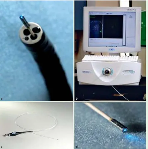
The confocal microscope used in pCLE captures microscopic ima-ges of untreated in vivo tissue. The microscope uses focused laser light of a defined wavelength and passes it through a confocal aperture. Images are then reconstructed in two dimensions. For pCLE (Cellvizio by Mauna Kea Technologies, Paris, France), both the laser scanning unit and light source are outside the body. The laser beam is transported via flexible confocal minip-robes and a distal lens sequentially scans it through a bundle of more than 10000 optical fibers. Confocal miniprobes are flexible, with diameters ranging from 0.9 mm to 2.5 mm. Therefore, this system may be introduced through the working channels of
en-doscopes (
●
" Fig. 1).Image acquisition, processing, and display
pCLE image data are collected at 12 frames per second, enabling video quality and direct visualization of blood on a single ery-throcyte scale. Depending on the probe used, the field of view
ranges from 240μm to 600μm (one-quarter to one-half of a
mil-limeter). The clinical system consists of a miniprobe connected to a laser scanning unit that is connected to a standard personal
computer for image data processing and display (
●
" Fig. 1).Ima-ges are reviewed with a specially designed software package (Cellvizio Viewer), allowing image correction and stabilization.
Contrast agents, dose, and safety
Different dyes, such as fluorescein or cresyl violet, have been used
for contrast enhancement [1–3]. Cresyl violet may only be
admi-nistered topically, whereas fluorescein may also be injected
in-travenously. Fluorescein is the current substance of choice be-cause it is relatively inexpensive, non-mutagenic, enables higher imaging depth than topically administered contrast agents, and it has been safely used for decades in ophthalmology [4]. Injection
of 1.0–5.0 mL of a 10% solution enables visualization of individual
cells with strong contrast of the capillary network. This is the cur-rent standard dosage in most settings. Hence, dynamic images of blood flow and supply are possible, making it a useful tool for de-tection of neo-angiogenesis [5].
The exact mechanism by which neoplastic cells appear dark is poorly understood. It is clear that fluorescence is not seen within these cells. Several mechanisms have been proposed including lack of fluorescein uptake, more rapid excretion from the cell [1], or greater leakage into the lamina propria, thus increasing the relative darkness of the epithelial cells [6].
Endoscopic techniques for high-quality image
acquisition of mucosal disease
The keys to obtaining high quality images are timing, positioning, and stability. Gradual changes in image quality occur in a time-dependant fashion after intravenous injection of fluorescein. Op-timal contrast is obtained within the first 10min after injection,
but good quality images can be acquired for a further 30–60 min
[3]. However, bleeding can impair image quality; therefore, the probe should be placed gently in contact with the tissue in order to avoid trauma, particularly if friability is expected, such as in neoplasms or severely inflamed tissue. As with other imaging methods, it is best to complete pCLE imaging in a region before biopsy or tissue removal is completed.
If possible, the probe should be perpendicular to the mucosa, rather than parallel to it. This can be difficult if the lumen is very
Fig. 1 The probe-based confocal laser endo-microscopy (pCLE) system.aProbe within standard endoscope.bLaser scanning unit and display. cFiber probe.dFiber probe with blue laser illuminated.
narrow, as in biliary strictures. In these circumstances, a cholan-gioscopical approach or biliary catheter with side hole or bend-able tip might help to maneuver the probe into the wall of the bile duct.
Movements caused by the examiner or the patient (breathing or peristalsis) can significantly impair positioning of the probe and cause artifacts. Using a clear 4-mm cap on the tip of the endo-scope and applying gentle suction helps to hold the probe in po-sition. This approach can be very helpful for the imaging of
Bar-rett’s esophagus or small colonic polyps.
Other methods to improve the match between pCLE imaging sites and biopsy sites include the use of a dual-channel endoscope (with pCLE probe in one channel and biopsy forceps in the other) or the use of cautery marking with argon plasma coagulation or
coagulating (“hot”) biopsy forceps. The most useful methods for
research studies require highly precise matching of image and biopsy locations.
Applications of pCLE
The main focus of pCLE has been the detection and classification
of neoplasia in Barrett’s esophagus and colonic polyps. However,
several other conditions have also been studied to a lesser extent
and others are just beginning to be studied.
●
" Table 1lists thesepotential applications in order of the current extent of evidence.
Comparison of eCLE and pCLE
Both endoscopy- and probe-based methods of CLE have advanta-ges and disadvantaadvanta-ges. eCLE offers a wider field of view, the
abil-ity to vary the depth of imaging from 0–250μm compared with a
fixed imaging plane (approximately 50μm below the surface of
GastroFlex), and slightly better lateral resolution (approximately
0.8μm vs. 1μm). Advantages of pCLE are the versatility of a probe
system that can be passed via any endoscope, or even needles,
and more rapid image acquisition (12 frames per second vs. 0.8–
1.6 frames/s), which allows imaging of in vivo blood flow. Both systems have inherent limitations compared with wide field en-doscopic methods, including the very small field of view, cost of the technology, learning curve, and extra time needed to view the images during endoscopy.
Barrett
’
s esophagus
!
Surveillance guidelines for the management of patients with
Bar-rett’s esophagus call for target biopsies from visible lesions,
fol-lowed by random four-quadrant biopsies every 2 cm [7]. This practice is time-consuming, costly, and challenging as sampling is not always accurate. Detailed analysis of esophagectomy
speci-mens from patients with Barrett’s esophagus has revealed that
areas of low-grade dysplasia (LGD), high-grade dysplasia (HGD), and cancer are extremely focal and occupy extremely small areas
within the Barrett’s segment [8]. To overcome these
shortcom-ings, advanced endoscopic techniques including pCLE have been developed to maximize the positive and negative predictive
val-ues of dysplasia detection in patients with Barrett’s esophagus.
CLE is a highly targeted technology that may be useful for lesion classification. However, other technologies, such as narrow band imaging (NBI), autofluorescence imaging (AFI), and
chromoen-doscopy are needed as“broad-base surface imaging”techniques
to initially detect and localize suspicious areas.
In patients with Barrett’s esophagus, CLE captures very high
reso-lution and detailed images including glandular architecture, crypts, columnar cells, goblet cells, and capillaries with red blood cells. In patients with a columnar-lined esophagus, the diagnosis
of Barrett’s esophagus can be confirmed by identifying and
con-firming the presence of intestinal metaplasia, which is character-ized by the presence of goblet cells. Furthermore, based on preli-minary data, CLE also has high accuracy in detecting HGD or can-cer.
Classification of CLE images as non-dysplastic or dysplastic has been evaluated in a few pilot studies using varying criteria [3,4]. An initial study by Kiesslich et al. evaluated criteria for gastric type, intestinal and neoplastic epithelium, based on cellular and vascular architecture details of CLE images [9]. Images that de-monstrate regular-shaped capillaries visible only in deeper mu-cosa, regular columnar-lined epithelium with round glandular openings, and typical cobblestone appearance, were suggestive of gastric-type foveolar epithelium; whereas, regular capillaries that were present in upper and deeper parts of the mucosal layer
along with identification of dark (“non-refractile”) mucin in
gob-let cells in columnar-lined mucosa, were diagnosed as intestinal
metaplasia (non-dysplastic Barrett’s esophagus). The diagnosis
of dysplasia/cancer was based on the identification of irregular capillaries in upper and deeper parts of the mucosal layer (sug-gestive of neo-angiogenesis) with black cells that had irregular apical and distal borders and shapes on confocal images. Using these criteria, Barretts-associated dysplasia could be predicted with a sensitivity of 93% and a specificity of 98%. Compared with
gastric metaplasia, Barrett’s metaplasia could be predicted with a
sensitivity of 98% and a specificity of 94%.
In another recent study, Pohl et al. tested the diagnostic
charac-teristics of pCLE for the detection of invisible Barrett’s-associated
dysplasia [10]. The investigators initially established pCLE criteria
for neoplastic Barrett’s esophagus including dark, irregularly
thickened epithelial borders, dilated irregular vessels, and in ade-nocarcinoma, disorganization of villiform structures and crypts and dark columnar cells. These criteria were established from 95 biopsies (15 patients) and then tested prospectively in 201 biop-sies (23 patients). pCLE videos were also assessed by two endos-copists and the inter-observer agreement was tested. The overall sensitivity, specificity, positive predictive value (PPV) and nega-tive predicnega-tive value (NPV) were 80%, 94%, 44%, and 99%, respec-tively, with good inter-observer agreement (Kappa = 0.6). A wide group of expert endoscopists evaluated these criteria and valida-ted them as reliable and reproducible.
Using a standardized set of 20 video images of dysplastic [11] and
non-dysplastic Barrett’s sites [10], Wallace et al. reported
preli-minary sensitivity of 88% and specificity of 96% and good inter-observer agreement (agreement rate 86%, Kappa = 0.72) [12]. These pCLE criteria have now been tested and validated in a large,
prospective, multi-center, randomized controlled trial (DON’T
Table 1 Potential applications of (probe-based) confocal laser endomicro-scopy.
Well-evaluated areas
Barrett’s esophagus guide to biopsy Colon polyp classification Areas of early exploration
Inflammatory bowel disease dysplasia Biliary strictures
Duodenal neoplasia Experimental areas
Solid and cystic tumor imaging Gastric neoplasia
BIOPCE trial) [13]. This trial used independent, blinded endosco-pists to perform tandem endoscopic procedures to evaluate the sensitivity and specificity of pCLE in addition to white light en-doscopy for the detection of HGD and early adenocarcinoma in
Barrett’s esophagus. Final data presented at Digestive Disease
Week 2010 (1–6 May, New Orleans, Louisiana, USA) included
874 sites (120 with high grade intra-epithelial neoplasia [HGIEN] or cancer) in 101 patients (31 with HGIEN or cancer). On a per pa-tient basis, the combination of pCLE with high-definition white
light endoscopy was significantly more accurate than endoscopy alone (sensitivity/specificity of pCLE + endoscopy 95%/67% vs. en-doscopy 85%/71%). The addition of pCLE to enen-doscopy and NBI was also more accurate than endoscopy + NBI alone (sensitivity/ specificity of pCLE + endoscopy + NBI 100%/56% vs. endoscopy + NBI 97%/56%). Using pCLE to guide biopsy in patients with
Bar-rett’s esophagus, where only suspicious sites are biopsied would
result in a 76% reduction in biopsies with no HGIEN patients mis-sed, although 24% of sites would have been missed. If pCLE was
Fig. 2 Esophagus.aGraphic of Barrett’s mucosa with villiform non-dysplastic epithelium (left side) progressing to higher grades of dysplasia and can-cer (right). The estimated image“slice”of probe-based confocal laser endomicroscopy (pCLE) is shown in blue.bpCLE images and histology of esophageal conditions including Barrett’s esopha-gus. In normal squamous epithelium, two pCLE images are shown as well as standard histology with hematoxylin and eosin (H&E). For Barrett’s esopha-gus, high-grade dysplasia, and adenocarcinoma, a pCLE image is shown on the left and a correspond-ing H&E image of the same condition is shown on the right.
Normal squamous epithelium
Non dysplastic Barrett‘s esophagus
– Flat cells without crypts or villi
– Bright vessels within papillae (intra papillary capillary loops)
– Uniform villiform architecture – Columnar cells (block arrow) – Dark „goblet“ cells (thin arrow)
High grade dysplasia
– Villiform structures – Dark, irregularly thickened epithelial borders (arrow) – Dilated irregular vessels (block arrow)
Adenocarcinoma
b
– Disorganized/loss of villiform structure and crypts
– Dark columnar cells (thin arrow)
– Dilated irregular vessels (block arrow)
used to guide biopsy, and four-quadrant random plus targeted biopsies were taken only in those patients with at least one ab-normal lesion on pCLE, then this would result in a 39% reduction in biopsies with no missed sites or patients. In summary, CLE
criteria for dysplastic and non-dysplastic Barrett’s esophagus
have been established and tested in pilot studies. Their validation in a large multicenter study is ongoing and eagerly awaited. It is likely that these criteria will continue to be refined as larger stud-ies are conducted.
●
" Fig. 2shows the current pCLE-based classification system with image and histological examples.Biliary disease
!
Accurate diagnosis and staging of pancreatobiliary cancers is im-portant for both prognosis and for guiding therapy; however, it remains challenging to differentiate benign from malignant stric-tures despite advanced imaging and tissue sampling methods. Up to 15% of all suspected cholangiocarcinomas originating from the liver hilum reveal benign histology following surgery [11]. Con-versely, only 20% of hilar cancers are resectable at the time of di-agnosis, and the rate of R0 resections is unfavorably low [14]. This diagnostic dilemma holds true despite the introduction of tissue sampling methods such as biopsy, brush cytology, or fine-needle aspiration (FNA) cytology through endoscopic retrograde cholan-giopancreatography (ERCP) or endoscopic ultrasound (EUS). The best outcomes are obtained when several sampling techniques are combined, but even they remain suboptimal. Rösch et al. re-ported that combining brush cytology with forceps biopsy yiel-ded a sensitivity of 54% and a specificity of 100% [15]. If
endo-scopic ultrasound-guided fine-needle aspiration (EUS–FNA) was
added to these methods, then the sensitivity increased to 71%, but specificity was still 100%. Ponchon et al. reported that com-bining brush cytology and forceps biopsy yielded a sensitivity of 63% and a specificity of 97% [16]. Schoefl et al. showed that the combination of brush cytology and biopsy yielded a sensitivity of 70.4% and a specificity of 100% [17]. Hence, there is a critical need to improve the diagnostic accuracy of pancreatobiliary pa-thology, thereby enabling a more tailored approach for patients and minimizing unnecessary operations.
pCLE that provides in vivo microscopic imaging of tissues in real time can be introduced through the working channel of an ERCP catheter or cholangioscope, in order to access and visualize the pancreatobiliary ductal system during the procedure. Meining et al. reported on a series of 14 patients with biliary strictures that were examined by pCLE via cholangioscopy, and compared the modality to standard endoscopic tissue sampling [18]. pCLE ima-ging of the biliary stricture was feasible in all cases. The presence
of irregular, dilated (“angiogenic”) vessels was the laser
micro-scopic hallmark for prediction of neoplasia, with an accuracy of 86%, sensitivity of 83%, and specificity of 88%. The respective numbers for standard histopathology were 79%, 50%, and 100%. The mean signal-to-noise-ratio of endomicroscopic images ac-quired from malignant strictures differed significantly from
those of benign origin (1.8 ± 0.8 vs. 2.6 ± 1.0;P= 0.005). As a
preli-minary conclusion, pCLE could considerably increase sensitivity for the detection of pancreatobiliary neoplasia, and therefore, re-presents a promising diagnostic approach. Large multicentric studies are ongoing.
To date, pCLE examination during ERCP procedures has been se-lectively applied to patients with signs, symptoms, or test results
that indicate malignancy or indeterminate pancreatobiliary stric-ture. The technique for pancreatobiliary pCLE is straightforward. ERCP is performed in accordance with the standard of care, and the stricture is accessed either with an ERCP catheter or a cholan-gioscope. The pCLE probe is introduced through the lumen of the catheter or the working channel of the cholangioscope, and the lesion of interest is targeted for microscopic imaging using fluoroscopy or endoscopic guidance. The pCLE probe is posi-tioned at the site(s) of suspected pathology immediately follow-ing intravenous injection of fluorescein sodium. Real-time in vivo sequences are acquired and stored for review, usually immedi-ately after a site of interest has been imaged.
The choice of the optimal access delivery system for pCLE during ERCP (catheter vs. cholangioscopic access and delivery) is cur-rently being investigated. In part, the decision depends on the endoscopy unit resources and operator preference. Initial experi-ence with pCLE during ERCP procedures shows that real-time se-quences can be obtained with either delivery method [19]. A vari-ety of catheter models have been used in conjunction with the
miniprobe (see
●
" Table 2). Access through the SpyGlass system(Boston Scientific, Natick, Massachusetts, USA), Olympus cholan-gioscopes (Olympus Corp., Tokyo, Japan), and Storz prototype cholangioscope (Karl Storz GmbH, Tuttlingen, Germany) are all also possible.
The Cellvizio CholangioFlex probe is dedicated to pCLE during ERCP procedures, and it has been miniaturized to an external di-ameter of 0.94 mm, in order to meet the constraints of intraduc-tal access devices. The probe fits inside lumens of 1.2 mm in di-ameter, has a field of view of 325 µm, a lateral resolution of 3.5
µm, and an imaging depth of 40–70 µm below the tissue surface.
Specific pCLE image interpretation criteria are under
develop-ment.
●
" Fig. 3shows typical pCLE findings in normal bile ductsand biliary cancer.
Colorectal disease
!
Several advanced imaging techniques may play a role in facilitat-ing both the detectionof dysplasia in the colon, as well as classifi-cationbetween neoplastic and non-neoplastic lesions during co-lonoscopy. CLE can provide imaging of single cells, making it un-suitable for the detection of lesions in the colon; however, it has
Table 2 List of endoscopic retrograde cholangiopancreatography (ERCP) devices that have been used successfully and can accommodate the Cholan-gioFlex probe (Cellvizio, Mauna Kea Technologies, Paris). The list is based on clinical experiences of current users of probe-based confocal laser endomi-croscopy and does not represent a recommendation from the manufacturers. Differences in size and diameter of the working channel might impair the compatibility in some cases.
Device name Manufacturera
Cotton®Graduated Dilation Catheter T7.0 Cook Medical
OASIS®One Action Stent Introduction
System
Cook Medical
Memory Dormia basket (Ref MSB_35_2X4 MemoryII)
Cook Medical
Howell Biliary Introducer (H-BIN) Cook Medical Geenen®Graduated Dilation Catheter Cook Medical
Swing Tip™ERCP cannula Olympus Medical
aCook Medical, Inc., Bloomington, Indiana, USA; Olympus Medical Systems, Tokyo,
Japan
the potential to differentiate dysplasia from non-neoplastic tis-sue in a targeted approach [2,20,21].
Chromoendoscopy, NBI, and a combination of NBI and AFI called endoscopic trimodal imaging (ETMI) may predict histology based on the Kudo pit pattern, vascular pattern intensity (VPI), and col-or. All of these techniques have a rather low specificity. CLE, how-ever, has the potential to provide real-time conventional histopa-thology with a very high specificity.
Ideally, a method with a high detection rate of neoplastic lesions (usually at the cost of lower specificity) should be combined with a method of optimal differentiation of all detected lesions. Such combinations can be made by pan-colonic chromoendoscopy or
autofluorescence as a“red flag technique”used in conjunction
with endoscopic pit pattern analysis and CLE for the differentia-tion of detected lesions.
Colorectal polyps
!
The detection of neoplastic lesions has been significantly im-proved by chromoendoscopy compared with conventional colo-noscopy, whereas NBI only showed an improvement during the learning phase [22,23]. In one randomized study ETMI (white light + NBI + AFI) did not result in an increased adenoma detec-tion rate [24].
Conventional endoscopy has limited ability to discriminate ade-nomatous from non-adeade-nomatous colorectal polyps. In daily practice, all identified lesions are routinely removed and sent for histopathology. This approach results in substantial increase of endoscopic workload, additional pathology costs, and increase in potential complications. If CLE could reliably predict histology, then it could increase cost-effectiveness and efficiency consider-ably. This is a concept suggested by several prominent colonosco-pists [25].
However, the decision to decide to leave a polyp in situ and not send it for pathology requires high accuracy and particularly high NPV of the imaging method. Chromoendoscopy, NBI, and AFI have shown unacceptable test characteristics for this pur-pose, with accuracies of 91%, 89%, and 79%, respectively [24]. CLE has shown high agreement with true histopathology. In ear-lier feasibility studies, Kiesslich et al. demonstrated an accuracy of 99% with the integrated system, whereas, feasibility studies
with the probe-based system reached an accuracy of 82–92%
[2,20,21].
A prospective study of CLE in polyp differentiation has been pub-lished. Sanduleanu et al. performed a prospective study using chromoendoscopy-guided CLE with the integrated system to evaluate differential features of adenomatous and non-adenoma-tous colorectal polyps, resulting in an accuracy of 96% [26]. Acri-flavine was used in conjunction with fluorescein, enabling discri-mination of HGD from LGD, with an accuracy of 97%.
Another large prospective double-blind trial compared pCLE with NBI and Fujinon Intelligence Color Enhancement (FICE, Fujinon, Fort Wayne, New Jersey, USA), for polyp classification. Buchner et al. demonstrated that pCLE was superior to digital chromoen-doscopy methods; however, it was primarily superior to FICE without significant improvement over NBI. The sensitivity of pCLE for neoplastic polyps was 91% and specificity was 76%, com-pared with virtual chromoendoscopy (NBI and FICE), which had a sensitivity of 77% and a specificity of 71% [27].
This same group evaluated the learning curve for pCLE for colo-rectal polyps and found that new users rapidly acquired the abil-ity to interpret pCLE images with accuracy similar to that of high-ly experienced experts. New users were able to interpret images with an accuracy of 93% after reading at least 35 cases. This study did not assess the accuracy of new users during live video endos-copies, which may require more experience [28].
Further prospective studies and data are eagerly awaited that ex-amine the cost-effectiveness of CLE for colorectal polyps with both the integrated as well as the probe-based system. Data on the assessment of degree of dysplasia, inter-observer variability, and learning curve are also anticipated. At the same time, other applications for the use of the CLE system (e.g. early detection of adenomatous tissue at a scar at the resection site of a polyp), should be explored.
●
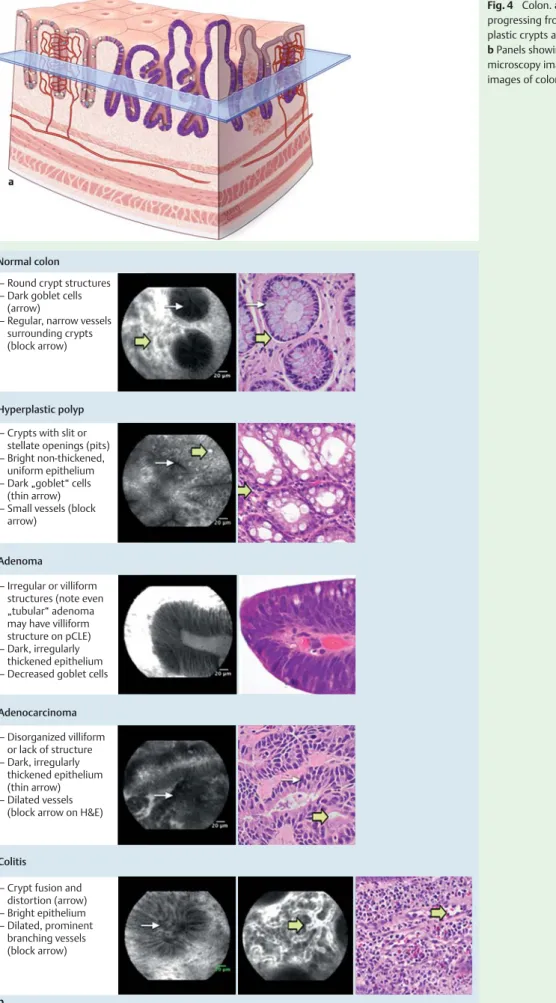
" Fig. 4shows the criteria for pCLE classification of colorectalpolyps.
Fig. 3 Bile duct.aNormal appearing bile duct with fine, reticular gray pattern (thin black arrow).bBiliary cancer. Note dark, irregular structures (thin black arrow) interspersed with bright areas of tortuous dilated blood vessel (thick white arrow).
Fig. 4 Colon.aGraphic showing colonic mucosa progressing from normal crypts (left side) to dys-plastic crypts and villiform structures (right side). bPanels showing probe-based confocal laser endo-microscopy images and hematoxylin and eosin images of colonic disease.
Normal colon
Hyperplastic polyp
– Round crypt structures – Dark goblet cells (arrow)
– Regular, narrow vessels surrounding crypts (block arrow)
– Crypts with slit or stellate openings (pits) – Bright non-thickened, uniform epithelium – Dark „goblet“ cells (thin arrow) – Small vessels (block arrow)
Adenoma
– Irregular or villiform structures (note even „tubular“ adenoma may have villiform structure on pCLE) – Dark, irregularly thickened epithelium – Decreased goblet cells
Adenocarcinoma – Disorganized villiform or lack of structure – Dark, irregularly thickened epithelium (thin arrow) – Dilated vessels (block arrow on H&E)
Colitis
b
– Crypt fusion and distortion (arrow) – Bright epithelium – Dilated, prominent branching vessels (block arrow)
Ulcerative colitis
!
As patients with ulcerative colitis have an increased risk of devel-oping dysplasia, guidelines recommend colonoscopic surveil-lance including targeted biopsies of suspicious lesions and multi-ple (40) random biopsies. However, the yield for dysplasia on random biopsy is very low, and standard endoscopic imaging cannot identify most neoplastic lesions. Several randomized studies have shown that targeting biopsies with chromoendosco-py significantly increases dysplasia detection rates in patients with longstanding ulcerative colitis [29,30]. In one randomized study, ETMI also increased dysplasia detection rates but NBI did not [31,32].
Using initial chromoscopy followed by directed eCLE combines the strengths of both techniques in ulcerative colitis. While pan-chromoscopy facilitates the detection of flat lesions in ulcerative colitis, subsequent targeted confocal endomicroscopy can be used to differentiate between neoplastic and non-neoplastic tis-sue, thus obviating the need for targeted biopsies of lesions that appeared non-neoplastic. This can also be used to possibly differ-entiate random biopsies in chromo-negative areas. By using this
diagnostic approach in a randomized study in 161 patients, Kies-slich et al. detected almost five times more dysplastic lesions than conventional colonoscopy with random biopsies [33]. Using the integrated CLE system, Hurlstone et al. demonstrated a high overall accuracy of 97% for in vivo differentiation of adeno-ma-like mass (ALM) and dysplasia-associated lesion or mass (DALM) in patients with chronic ulcerative colitis [34]. Case re-ports using the pCLE system suggest it may also be used to iden-tify DALM lesions [35].This technique could provide gastroenter-ologists with important information for selecting patients who are suitable for immediate endoscopic resection vs. referral for pan-proctocolectomy. Recent studies also suggest that CLE may be a valuable tool for the grading of colitis and detection of mi-croscopic colitis in regions of the colon without visible inflamma-tion [36].
All of these studies have been performed with the integrated CLE system. Studies with the probe-based CLE system are currently underway. Further data from non-tertiary referral centers and from investigation of the learning curve and inter-observer agreement in patients with chronic ulcerative colitis are neces-sary before implementation of chromoscopy-guided CLE can be recommended in daily practice.
●
" Fig. 4shows the criteria for pCLE classification of colitis andassociated dysplasia.
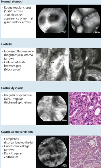
Normal stomach
Gastritis
– Round regular crypts (“pits“, arrow) – „Cobblestone“ appearence of normal glands (block arrow)
– Increased fluorescence (brightness) in stroma (arrow) – Cellular infiltrate between pits (block arrow) Gastric dysplasia
– Irregular crypt lumen – Dark, irregular, thickened epithelium Gastric adenocarcinoma – Completely disorganized epithelium – Fluorescein leakage (arrow) – Dark irregular epithelium
Fig. 5 Stomach. Panels showing probe-based confocal laser endomicros-copy images (normal, dysplasia, adenocarcinoma) and hematoxylin and eosin images of gastric disease. (Gastritis images kindly provided by Alex-ander Meining). Normal duodenum Duodenal adenoma – Villiform architecture – Bright, uniform epithelium – Villiform architecture – Dark, irregular epithelium (arrow)
Duodenum celiac disease
– Blunting or loss of villi (arrow)
– Expansion of lamina propia with chronic inflammatory cells (arrow head, H&E)
Fig. 6 Duodenum. Probe-based confocal laser endomicroscopy (pCLE) images and hematoxylin and eosin of normal and diseased duodenum. The top two panels show the high definition (UHD, Cellvizio, Mauna Kea Tech-nologies, Paris) probe of normal and adenomatous duodenum. The field of view was expanded using“mosaicing”software, which integrates multiple side-by-side frames into a single panoramic view. The bottom pCLE image (celiac disease) was obtained using the Z probe (Cellvizio, Mauna Kea), which provides a lower-resolution but wider field of view.
Gastric diseases
!
To date there are limited data exploring application of confocal imaging to the stomach. There is a potential role for CLE in the discrimination of indeterminate gastric lesions in countries where gastric cancer is more common and screening endosco-pies are performed. In two studies from Japan, CLE demonstrated typical features of dysplasia such as enlarged and increased
num-bers of nuclei [37]. CLE had an accuracy of 94–96% for diagnosis of
malignancy among 27 patients with early gastric cancer, when compared directly with histological biopsies [38]. Other potential but unexplored applications include distinction between
adeno-matous and fundic gland polyps.
●
" Fig. 5 shows typical pCLEimages of normal gastric mucosa, inflammation, and dysplasia.
Duodenal diseases
!
As with the stomach, there are limited data on duodenal applica-tions. Given the frequency of biopsies performed to detect celiac disease in patients with signs or symptoms of malabsorption, this would be a valuable role for CLE, especially given the relatively low yield of endoscopic biopsy. Two small case reports have sug-gested that confocal endomicroscopy can detect villous atrophy and increased intra-epithelial cellularity (presumed to be lym-phocytes) [39,40]. In a recent study of 17 patients with celiac dis-ease and 14 control individuals, CLE detected typical celiac changes of villous atrophy and crypt hypertrophy. The accuracy of CLE was excellent, with sensitivity of 94% and specificity of 92% [41].
Detection of duodenal adenomas is also a potentially valuable role for CLE. The risk of ampullary and non-ampullary duodenal cancer is substantially elevated in familial adenomatous polypo-sis, leading to recommendations for routine duodenal surveil-lance and ampullary biopsy [42]. A downside of biopsy of the am-pulla is the risk of inducing acute pancreatitis [43], such that non-biopsy methods would be preferable. Preliminary data from Sha-hid et al. at the Mayo Clinic (Florida, USA) suggest that pCLE may be highly accurate for the detection of both ampullary and non-ampullary adenomas, although more data are needed [44]. The features of dysplasia are typical of those in other gastrointestinal
sites including dark, irregular epithelium (
●
" Fig. 6).Research priorities
!
To determine the value of these potential roles and explore new ones, critical research is needed, including in the following areas.
▶ Determine precise estimates of accuracy compared with
cur-rent standard endoscopic methods in colon polyps, Barrett’s
esophagus, ulcerative colitis, biliary cancer, and gastric dys-plasia.
▶ Determine the reliability of pCLE in a community setting.
▶ Assess the ability to guide immediate endoscopic therapy in
Barretts-associated dysplasia, colorectal endoscopic mucosal resection, and polypectomy.
▶ Determine preliminary accuracy and reliability in bile duct
cancer.
▶ Assess the cost-effectiveness of pCLE compared with standard
competing technologies including biopsy.
▶ Develop improved methods for image stabilization.
▶ Develop improved methods for image interpretation systems
including computer-aided diagnosis.
▶ Assess the feasibility and accuracy of intratumoral and
intra-cystic CLE via needles (nCLE).
Summary and future directions
!
pCLE has the potential to directly guide endoscopic therapy of dysplasia and significantly reduce the number of non-targeted (random) biopsies. Application of this technology has the further advantage of visualizing a dynamic process on a microscopic level for monitoring and determination of blood flow in various condi-tions. Further miniaturization of the probe will imply new indi-cations, such as placing the miniprobe through an FNA needle for EUS-guided examinations of solid or cystic tumors and for in-terventions. In addition, integrating new biological fluorophores such as fluorescein-bound peptides will potentially enable the precise detection of dysplasia in vivo and guide subsequent inter-vention during the same procedure.
Competing interests:Dr Meining receives research funding from Mauna Kea Technologies and is co-patent holder of Cholangio-Flex probes. Dr Sharma receives funding from Barrx Inc., Cook Medical, Olympus, and Takeda. Dr Wallace receives research funding from Olympus, Cook Medical, Boston Scientific, Mauna Kea Technologies, and American BioOptics. Dr Dekker receives research funding from Olympus and has equipment on loan from Olympus and Mauna Kea Technologies. The other authors have nothing to declare.
Institutions
1Department of Gastroenterology, Mayo Clinic, Jacksonville, Florida, USA 2Division of Surgical Pathology and Gastrointestinal Pathology Service,
De-partment of Pathology, Massachusetts General Hospital and Harvard Medical School, Boston, Massachusetts, USA
3Division of Gastroenterology and Hepatology, University of Colorado Denver,
Denver, Colorado, USA
4Academic Medical Center, Amsterdam, The Netherlands
5Kansas University Medical Center and Department of Veterans Affairs Medical
Center, Kansas City, Kansas, USA
6Klinikum rechts der Isar, Technical University of Munich, Munich, Germany
Acknowledgment
!
We thank Ms Alice McKinney of the Mayo Clinic Jacksonville Gra-phic Department for creation of the graGra-phic representations of tissue histology, Ms Kelly Viola of the Mayo Clinic for editorial support, and Ms Anne Osdoit of Mauna Kea Technologies for pro-viding confocal images and confirming technical aspects of the pCLE system.
References
1Wang TD,Friedland S,Sahbaie P et al.Functional imaging of colonic mucosa with a fibered confocal microscope for real-time in vivo pa-thology. Clin Gastroenterol Hepatol 2007; 5: 1300–1305
2Meining A,Bajbouj M,von Delius S,Prinz C. Confocal laser scanning mi-croscopy for in vivo histopathology of the gastrointestinal tract. Arab J Gastroenterol 2007; 8: 14
3Becker V,von Delius S,Bajbouj M et al.Intravenous application of fluor-escein for confocal laser scanning microscopy: evaluation of contrast dynamics and image quality with increasing injection-to-imaging time. Gastrointest Endosc 2008; 68: 319–323
4Kwan AS,Barry C,McAllister IL,Constable I. Fluorescein angiography and adverse drug reactions revisited: the Lions Eye experience. Clin Experiment Ophthalmol 2006; 34: 33–38
5 Meining A,Wallace MB. Endoscopic imaging of angiogenesis in vivo. Gastroenterology 2008; 134: 915–918
6 Matysiak-Budnik T,Coron E,Mosnier J-F et al.780 Quantitative in vivo assessment of vascular permeability in human colonic mucosa using confocal endomicroscopy: clinical implications for colonic neoplasia. Gastroenterology 2009; 136: A-123
7 Wang KK,Sampliner RE. Updated guidelines 2008 for the diagnosis, surveillance and therapy of Barrett’s esophagus. Am J Gastroenterol 2008; 103: 788–797
8 Cameron AJ,Carpenter HA. Barrett’s esophagus, high-grade dysplasia, and early adenocarcinoma: a pathological study. Am J Gastroenterol 1997; 92: 586–591
9 Kiesslich R,Gossner L,Goetz M et al.In vivo histology of Barrett’s esoph-agus and associated neoplasia by confocal laser endomicroscopy. Clin Gastroenterol Hepatol 2006; 4: 979–987
10 Pohl H,Rosch T,Vieth M et al.Miniprobe confocal laser microscopy for the detection of invisible neoplasia in patients with Barrett’s oesoph-agus. Gut 2008; 57: 1648–1653
11 Uhlmann D,Wiedmann M,Schmidt F et al.Management and outcome in patients with Klatskin-mimicking lesions of the biliary tree. J Gas-trointest Surg 2006; 10: 1144–1150
12 Wallace M,Abrams J,Bajbouj M et al.Accuracy and inter-observer agreement of experts for probe-based confocal laser endomicroscopy detection of dysplasia in Barrett’s esophagus. Gastrointest Endosc 2009; 69: AB351
13 Sharma P,Meining A,Coron E et al.Detection of neoplastic tissue in Bar-rett’s esophagus with in vivo probe-based confocal endomicroscopy (DONT BIOPCE). Final results of a prospective international RCT: image guided versus 4 quadrant random biopsies?. Gastroenterology 2010; 138: S-155
14 Puhalla H,Schuell B,Pokorny H et al.Treatment and outcome of intra-hepatic cholangiocellular carcinoma. Am J Surg 2005; 189: 173–177 15 Rosch T,Hofrichter K,Frimberger E et al.ERCP or EUS for tissue
diagno-sis of biliary strictures? A prospective comparative study. Gastrointest Endosc 2004; 60: 390–396
16 Ponchon T,Gagnon P,Berger F et al.Value of endobiliary brush cytology and biopsies for the diagnosis of malignant bile duct stenosis: results of a prospective study. Gastrointest Endosc 1995; 42: 565–572 17 Schoefl R,Haefner M,Wrba F et al.Forceps biopsy and brush cytology
during endoscopic retrograde cholangiopancreatography for the diag-nosis of biliary stenoses. Scand J Gastroenterol 1997; 32: 363–368 18 Meining A,Saur D,Bajbouj M et al.In vivo histopathology for detection
of gastrointestinal neoplasia with a portable, confocal miniprobe: an examiner blinded analysis. Clin Gastroenterol Hepatol 2007; 5: 1261–1267
19 Stevens P,Chen Y,Shah R et al.Real-time intraductal confocal microsco-py during ERCP: feasibility and technical considerations. Gastrointest Endosc 2009; 69: AB267
20 Kiesslich R,Burg J,Vieth M et al.Confocal laser endoscopy for diagnos-ing intraepithelial neoplasias and colorectal cancer in vivo. Gastroen-terology 2004; 127: 706–713
21 Becker V,Vercauteren T,von Weyhern CH et al.High-resolution minip-robe-based confocal microscopy in combination with video mosaicing (with video). Gastrointest Endosc 2007; 66: 1001–1007
22 Adler A,Pohl H,Papanikolaou IS et al.A prospective randomised study on narrow-band imaging versus conventional colonoscopy for adeno-ma detection: does narrow-band iadeno-maging induce a learning effect?. Gut 2008; 57: 59–64
23 Adler A,Aschenbeck J,Yenerim T et al.Narrow-band versus white-light high definition television endoscopic imaging for screening colonosco-py: a prospective randomized trial. Gastroenterology 2009; 136: 410–
416
24 van den Broek FJ,van Soest EJ,Naber AH et al.Combining autofluores-cence imaging and narrow-band imaging for the differentiation of adenomas from non-neoplastic colonic polyps among experienced and non-experienced endoscopists. Am J Gastroenterol 2009; 104: 1498–1507
25 Ignjatovic A,East JE,Suzuki N et al.Optical diagnosis of small colorectal polyps at routine colonoscopy (Detect InSpect ChAracterise Resect and
Discard; DISCARD trial): a prospective cohort study. Lancet Oncol 2009; 10: 1171–1178
26 Sanduleanu S,Driessen A,Gomez-Garcia E et al.In vivo diagnosis and classification of colorectal neoplasia by chromoendoscopy-guided confocal laser endomicroscopy. Clin Gastroenterol Hepatol 2010; 8: 371–378
27 Buchner AM,Shahid MW,Heckman MG et al.Comparison of probe-based confocal laser endomicroscopy with virtual chromoendoscopy for classification of colon polyps. Gastroenterology 2010; 138: 834–
842
28 Buchner AM,Gomez V,Heckman MG et al.The learning curve of in vivo probe-based confocal laser endomicroscopy for prediction of colorec-tal neoplasia. Gastrointest Endosc 2011; 73: 556–560
29 Kiesslich R,Fritsch J,Holtmann M et al.Methylene blue-aided chromo-endoscopy for the detection of intraepithelial neoplasia and colon can-cer in ulcan-cerative colitis. Gastroenterology 2003; 124: 880–888 30 Rutter MD,Saunders BP,Schofield G et al.Pancolonic indigo carmine
dye spraying for the detection of dysplasia in ulcerative colitis. Gut 2004; 53: 256–260
31 van den Broek FJ,Fockens P,van Eeden S et al.Endoscopic tri-modal imaging for surveillance in ulcerative colitis: randomised comparison of high-resolution endoscopy and autofluorescence imaging for neo-plasia detection; and evaluation of narrow-band imaging for classifica-tion of lesions. Gut 2008; 57: 1083–1089
32 Dekker E,vandenBroek FJ,Reitsma JB et al.Narrow-band imaging com-pared with conventional colonoscopy for the detection of dysplasia in patients with longstanding ulcerative colitis. Endoscopy 2007; 39: 216–221
33 Kiesslich R,Goetz M,Lammersdorf K et al.Chromoscopy-guided endo-microscopy increases the diagnostic yield of intraepithelial neoplasia in ulcerative colitis. Gastroenterology 2007; 132: 874–882
34 Hurlstone DP,Thomson M,Brown S et al.Confocal endomicroscopy in ulcerative colitis: differentiating dysplasia-associated lesional mass and adenoma-like mass. Clin Gastroenterol Hepatol 2007; 5: 1235–
1241
35 Palma GD,Staibano S,Siciliano S et al.In-vivo characterization of DALM in ulcerative colitis with high-resolution probe-based confocal laser endomicroscopy. World J Gastroenterol 2011; 17: 677–680
36 Li CQ,Xie XJ,Yu T et al.Classification of inflammation activity in ulcera-tive colitis by confocal laser endomicroscopy. Am J gastroenterol 2010; 105: 1391–1396
37 Kakeji Y,Yamaguchi S,Yoshida D et al.Development and assessment of morphologic criteria for diagnosing gastric cancer using confocal en-domicroscopy: an ex vivo and in vivo study. Endoscopy 2006; 38: 886–890
38 Kitabatake S,Niwa Y,Miyahara R et al.Confocal endomicroscopy for the diagnosis of gastric cancer in vivo. Endoscopy 2006; 38: 1110–
1114
39 Trovato C,Sonzogni A,Ravizza D et al.Celiac disease: in vivo diagnosis by confocal endomicroscopy. Gastrointest Endosc 2007; 65: 1096–
1099
40 Zambelli A,Villanacci V,Buscarini E et al.Confocal laser endomicrosco-py in celiac disease: description of findings in two cases. Endoscoendomicrosco-py 2007; 39: 1018–1020
41 Leong RW,Nguyen NQ,Meredith CG et al.In vivo confocal endomicro-scopy in the diagnosis and evaluation of celiac disease. Gastroenterol-ogy 2008; 135: 1870–1876
42 Vasen HF,Moslein G,Alonso A et al.Guidelines for the clinical manage-ment of familial adenomatous polyposis (FAP). Gut 2008; 57: 704–713 43 Nugent KP,Spigelman AD,Williams CB,Phillips RK. Iatrogenic
pancrea-titis in familial adenomatous polyposis. Gut 1993; 34: 1269–1270 44 Shahid M,Buchner A,Hasan M et al.The role of probe-based confocal
laser endomicroscopy (pCLE) in detection of dysplasia in duodenal polyps. Gastrointest Endosc 2009; 69: AB369