Technology Assessment
Technology
Assessment Program
Agency for Healthcare Research and Quality
540 Gaither Road Rockville, Maryland 20850
Horizon Scan on Hip
Replacement Surgery
Horizon Scan on Hip Replacement
Surgery
Prepared by
ECRI Evidence-based Practice Center David Snyder, Ph.D.
Richard Chapell, Ph.D. Wendy Bruening, Ph.D. Karen Schoelles, M.D., S.M. Janice Kaczmarek, M.S. Evelyn Kuserk, M.L.S, M.A. Eileen Erinoff, B.A.
Horizon Scan on Hip Replacement
Surgery
This horizon scan is based on research conducted by the ECRI Evidence-based Practice Center (EPC) under contract to the Agency for Healthcare Research and Quality
(AHRQ), Rockville, MD (Contract No. 290-02-0019). The findings and conclusions in this document are those of the author(s) who are responsible for its contents; the findings and conclusions do not necessarily represent the views of AHRQ. Therefore, no
statement in this horizon scan should be construed as an official position of the Agency for Healthcare Research and Quality or of the U.S. Department of Health and Human Services.
The information in this horizon scan is intended to help health care decision-makers; patients and clinicians, health system leaders, and policymakers, make well-informed decisions and thereby improve the quality of health care services. This horizon scan is not intended to be a substitute for the application of clinical judgment. Decisions concerning the provision of clinical care should consider this horizon scan in the same way as any medical reference and in conjunction with all other pertinent information, i.e., in the context of available resources and circumstances presented by individual patients. This horizon scan may be used, in whole or in part, as the basis for development of clinical practice guidelines and other quality enhancement tools, or as a basis for
reimbursement and coverage policies. AHRQ or U.S. Department of Health and Human Services endorsement of such derivative products may not be stated or implied.
Date: December 22, 2006
Prepared for:
Agency for Healthcare Research and Quality 540 Gaither Road
Table of Contents
Tables... i
Background ...1
Total Hip Replacement ...1
Hemiarthroplasty...1
Hip Resurfacing ...2
Primary Arthroplasty ...4
Indications...4
Operative Approaches...4
Minimally Invasive Approaches ...5
Computer/Robotic Assisted Surgery...6
Short-term Outcomes ...7 Adverse Events ...8 Thrombosis ...9 Infection ...9 Fracture ...9 Heterotopic Ossification ...9 Dislocation ...10 Long-Term Outcomes ...11 Revision ...11 Dislocation ...11 Sepsis ...11 Wear...12 Loosening...15 Breakage ...15
Influence of Patient Factors ...16
Weight...16
Activity ...16
Bone Quality ...16
Age...16
Effects of the Clinical Environment ...18
Types of Replacement Hips ...19
Materials ...19
Metal-on-plastic ...19
Highly cross-linked polyethylene ...20
Metal-on-metal...20 Ceramic-on-ceramic...21 Ceramic-on-Plastic...22 Ceramic-on-Metal ...22 Cushion Bearings ...22 Surface Coatings ...22 Fixation Methods ...23 Coatings ...24
Effects of Femoral Head Size ...24
Bipolar Designs...24
Hip Replacement Revision ...25
Difficulties in Interpreting Studies of THR ...28
Ongoing or Planned Clinical Trials of THR ...29
References and Included Studies ...34
Appendix A. Inventory of Prosthetic Hips Currently Available...41
Tables
Table 1. Contraindications to Hip Replacement Surgery(26,27)...4
Table 2. Short-Term Outcomes...8
Table 3. Short-Term Complications...8
Table 4. Risk Factors for Hip Instability...10
Table 5. Reported Linear Wear Rates of Different Implant Materials ...14
Table 6. Reported Patient Ages at initial THA ...17
Table 7. Reported Hip Revision Rates Under various Conditions ...26
Disclaimer
This report is a horizon scan on hip replacement surgery and hip implant technology.
The purpose of a horizon scan is to compile a broad overview on existing and future technologies from reviews, guidelines, studies and news reports on the subject. A horizon scan is not a full technology assessment and does not provide a systematic review and critical synthesis of the clinical studies of the technology under consideration.
Background
Over the past 30 years, total hip replacement (THR) surgery, also known as total hip arthroplasty (THA), has become commonplace in the United States and throughout the world. It has been described as the greatest achievement in orthopedic surgery in the twentieth century.(1) Although no surgery is without risk, the utility of THR to relieve pain and restore function among patients with damaged or degenerated hips and chronic pain is well-accepted as indicated by the large number of procedures that take place in the United States each year.(2,3) In 2003 alone, 201,545 THR procedures and 34,688 revisions of THR were performed in the United States.(4) From 1990 through 2002, the number of THR procedures per 100,000 individuals in the United States increased by 46%, from approximately 45 to 66 per 100,000 individuals.(5) The same study reported a 60% increase in revisions of THR during the same time period. The rates of both primary and revision THR are expected to continue to increase. A recent report estimates that the annual number of THR revision surgeries will increase 137% by 2030.(6) This horizon scan looks at some of the important issues facing orthopedic surgeons and other healthcare providers as they plan for the increasing utilization of THR. Areas of concern include:
• The selection of operative approaches (standard vs. minimally invasive or computer/robotic assisted)
• The design of the replacement prosthesis (metal-on-plastic, metal-on-metal, ceramic-on-ceramic, or some other combination)
• The surface coating of the prosthesis (untreated vs. treated) • Cemented vs. uncemented fixation of the prosthesis.
Total Hip Replacement
The THR procedure generally involves removal of the head of the femur and its replacement with a metal or ceramic prosthesis that fits into the remaining bone. The ball end of the artificial femur then fits into a cuplike socket (acetabular cup) that is installed in the patient’s pelvis. Hip replacement can be unilateral (one hip) or bilateral (both hips). The longevity of currently available implants, the rate at which surgical revisions are needed to replace failed implants, and the ease with which implants can be replaced are primary concerns noted in the hip replacement literature. Artificial hips may work loose, break, wear out, or dislocate. Any of these occurrences may require revision surgery and replacement of part or all of the implant. Determining the primary reasons for revision surgery is difficult because some published studies report the reasons for revisions while others report only the number of revisions performed. Revision surgery tends to be more dangerous and less successful than primary surgery.(7-11)
The development of new implant materials has focused primarily on extending longevity in order to avoid revision. A secondary consideration is preserving the integrity of the remaining bone, to make future revision surgery easier.
Hemiarthroplasty
Hemiarthroplasty refers to the replacement of only the femoral head and is most often performed in elderly patients who have sustained a displaced femoral neck fracture.(12) In this situation, the femoral head is at risk for avascular necrosis. Patients undergoing hemiarthroplasty have shown
faster recovery of function and better function than patients treated only with internal
fixation.(13) Some orthopedic surgeons have expressed a concern that hemiarthroplasty may not be the best option for active individuals expected to have long life expectancies.(14) Acetabular cartilage erosion from hemiarthroplasty leading to persistent pain and discomfort will occur if the device remains in place for longer periods. Therefore, younger more active individuals with a displaced femoral neck fracture may benefit more from THR than hemiarthroplasty.
In hemiarthroplasty the femoral head is usually replaced with a bipolar prosthesis. The bipolar prosthesis has an additional joint that allows movement to occur both at the prothesis-acetabular interface and within the prothesis. A proposed advantage of the doubled-jointed bipolar prothesis is a reduced risk of dislocation.(15) If pre-existing arthritis has damaged the acetabular cartilage, THR should be performed instead of hemiarthroplasty.
Hemi-resurfacing is similar to hemiarthroplasty, but only the surface of the femoral head is removed and replaced. Resurfacing arthroplasty involves minimal femoral head removal rather than resection of the entire femoral head and neck. The prosthesis forms a cap over the
remaining femoral head and is secured with a stem inserted into the femoral head and neck bone. Hemi-resurfacing of the femoral head has a role in treating young patients with osteonecrosis of the hip in order to delay the need for THR in these patients.(16-18) The ideal candidate is less than 40 years old and has minimal acetabular cartilage damage. Patients receiving
hemi-resurfacing arthroplasty rather than THR achieve higher activity levels but may experience more groin pain related to wear of the acetabular cartilage.
Hip Resurfacing
Total hip resurfacing, involving resurfacing of both the femoral head and acetabular cup, is an alternative to THR. It is performed primarily on younger patients who would be expected to live long enough and remain active enough to wear out several THR devices.(19) Better stability and range of motion than THR has been cited as an advantage of this procedure.(20) With this procedure, the femoral head is preserved, reshaped, and capped with a metal shell. The socket is fitted with a prosthetic cup, as is the case with THR. This procedure can only be performed if the patient has sufficient healthy bone stock to support the resurfacing prosthetic. The ideal
candidate for this procedure is less than 60 years old, has normal proximal femoral bone geometry and bone quality, and is expected to outlive any current conventional prosthesis.(21) Sales in resurfacing implants are a fast-growing market worldwide, but the procedure has seen limited use in the United States.(22) In the United States, only one manufacturer has obtained FDA approval to market its hip resurfacing system. Smith & Nephew Orthopaedics received premarket approval from the FDA on May 9, 2006 for the Birmingham Hip Resurfacing
system.(23,24) Femoral head resurfacing systems are cleared for marketing in the U.S. for use in hemi-resurfacing arthroplasty as discussed above.
Clinical data on the efficacy of total hip resurfacing are limited. A systematic review of studies on hip resurfacing was conducted by the U.K. National Health Service in 2002. They found that the data describing the procedure were too limited for firm conclusions to be reached.(19) Two studies, both published in 2004, have been cited as indicating good success rates but data were only available at 4 years of follow-up.(17) The average patient age in both studies was 48 years. Periprosthetic fracture of the femoral neck is the most common complication with hip
resurfacing systems, but better patient selection and improved surgical technique have reduced the frequency of this complication.(21,22)
Primary Arthroplasty
Indications
Indications for hip replacement include radiological evidence of joint damage and persistent pain and/or disability that is not adequately relieved by nonsurgical treatment such as analgesics or physical therapy. Joint damage leading to hip replacement may be the result of inflammatory or degenerative disorders, or of trauma such as hip fracture.(25) Contraindications for THR include conditions that would limit or prevent the success of the procedure.(26) These are listed in Table 1. The main absolute contraindication is active infection.(27)
Generally, patients between 65 and 80 years of age have been considered candidates, but in recent years the age range has expanded at both ends. Patients as young as 19 and as old as 90 have undergone THR.(27) Rheumatoid arthritis and other inflammatory arthritides typically affect patients younger than age 65, and may lead to joint replacement when patients are in their 50s or younger.(27)
Table 1. Contraindications to Hip Replacement Surgery(26,27)
Absolute Contraindications Relative Contraindications Acute or active infection (includes localized septic
arthritis and osteomyelitis as well as regional and systemic infection elsewhere in the body) Skeletal immaturity
Bone stock inadequate to support the device due to severe osteoporosis or severe osteopenia
Patient inability to follow preoperative and postoperative instructions
Previous history of local infection such as septic arthritis, or thought to be at high risk of infection due to
co-morbidities Neuropathic arthritis
Vascular insufficiency, muscular atrophy, co-morbidities, or neuromuscular disease severe enough to compromise implant stability or postoperative recovery
Family history of severe osteoporosis or severe osteopenia
Operative Approaches
Several surgical approaches to THR are possible. The surgeon’s choice appears to be largely a matter of personal preference. Surgeons may choose an anterolateral, direct lateral,
transtrochanteric, or posterolateral approach. A posterolateral approach may be associated with a higher rate of postsurgical dislocation.(28)
Regardless of the approach, the acetabulum of the pelvis is reamed to the appropriate size and depth to accept the prosthetic cup. When a cemented cup is used, cement fixation holes are drilled before the cup is fitted and cemented in place. An osteotomy of the femoral neck is performed and the medullary canal within the femur is reamed to accept the stem of the prosthetic femoral head. The canal is plugged below the level into which the stem protrudes to prevent bone cement from spreading into the canal. The artificial hip is then assembled using trial components to test for range of motion and eliminate potential impingements that may interfere with hip function. When the test motions are satisfactory, the permanent components are screwed, cemented or otherwise fitted into place. Test motions are again performed and the surgical site is closed. Performing a capsular repair on the joint rather than allowing scar tissue to form a pseudocapsule may reduce the incidence of dislocation.(29)
Minimally Invasive Approaches
Many surgeons employ minimally-invasive techniques, and those who perform THR are no exception. In general, proponents of these minimally invasive techniques believe that they lead to faster recovery and less short-term morbidity than traditional techniques. Two basic approaches have been proposed; one involves a single incision and the other involves two incisions.(30) The incision length in minimally-invasive surgery is less than 10 centimeters, while the incision length in conventional surgery is 15-25 cm.(30,31) Surgeons can also work around muscles and soft tissue instead of cutting through them.(32) Minimally invasive surgery may be associated with less blood loss and a lower prevalence of gait disturbance at early followup. However, minimally invasive techniques, with the restricted field of view of the working area, can be difficult to perform. A recent review of the two-incision approach examines the complications that can occur with this approach even when performed by experienced
surgeons.(33) Femoral fracture is the most common complication with this approach, especially in the individual with osteoporotic bone. Such individuals are therefore not good candidates for this surgical technique. Excess fat and muscle can limit the minimally invasive approach to the hip and abnormal hip anatomy can complicate the placement of the prosthesis. The ideal patient is a thin individual with normal hip anatomy and thick femoral cortices.
Although minimally invasive surgery can lead to cost savings and more rapid recovery for many patients, a recent study of the two-incision approach found a higher rate of complications
compared to open surgery.(32) This retrospective study, which was presented at the 2005
meeting of the American Academy of Orthopedic Surgeons, found that 14% of patients receiving THR by the two-incision method experienced an early complication, compared to 3.75% in the open surgery group. We did not identify any information in the examined literature regarding the effect of patient age on the incidence of complications among patients undergoing minimally invasive surgery.
Minimally invasive surgery requires special training of the surgeon. A learning curve is associated with the procedure before surgeons may be considered adept.(30) Some clinicians believe that minimally invasive surgery is being marketed to patients faster than evidence supporting its use can be found.(34) This may lead to increased demand from patients who may pressure surgeons to adopt the technique whether or not the evidence supports its use.
Several recent reviews have cited the general lack of data on long-term outcomes in patients treated with minimally invasive THR.(22,35-37) Pour et al. (35) cites 16 separate studies of minimally invasive THR, including three randomized controlled trials, but only five of the studies (two RCTs) reported follow-up data more than six months after surgery. Hou and Gilbert (36) cited many of these same studies when they concluded that advocates of minimally invasive surgery should collect and publish long-term data to compare with the supposed short-term advantages of this procedure. Weng and Fitzgerald (37) cited data from two RCTs (also mentioned in Pour et al.) to conclude that “we do not yet know the long-term outcomes of a smaller incision.”
Both the National Institute for Health and Clinical Excellence (NICE) in their examination of two-incision THR(38) and the Canadian Coordinating Office for Health Technology Assessment in their examination of minimally invasive hip resurfacing(39) have cited the lack of evidence on the long-term safety and efficacy of minimally invasive procedures. NICE identified four case series studies (517 patients) of minimally invasive two-incision THR and has concluded that the
evidence is not sufficient to recommend the procedure without special arrangements.(38) NICE did find sufficient evidence to recommend single mini-incision THR.(40) Their recommendation was based on evidence from two randomized controlled trials (279 patients) and five
non-randomized comparative studies reporting significantly less intraoperative blood loss with the mini-incision than with the standard THR procedure.(41) The Cochrane collaboration has recently begun a systematic review of the subject, but the results of this review are not yet available.(42) The Canadian Coordinating Office for Health Technology Assessment report, searched in November 2004, found no trials, case reports, or abstracts that assessed the harm or benefit of minimally invasive procedures for hip resurfacing.
Computer/Robotic Assisted Surgery
Computerized surgery systems have been used in THR for two purposes.(43) The first is to position the acetabular cup in such a way that the prosthesis has maximal range of motion
without impingement. This may have the effect of decreasing wear and reducing the potential for dislocation. The second application for computer-assisted surgery is precision reaming of the medullary cavity of the femur so that it makes maximal contact with the stem of the prosthesis. This may increase initial stability and improve bonding of bone to prosthesis.
Newly marketed computer-assisted navigation systems are designed to aid in implant
positioning. They provide surgeons with both preoperative and intraoperative information by displaying three-dimensional computer images of patient anatomy.(44) Computer-assisted navigation involves three processes: data acquisition, registration, and tracking. Fluoroscopic, CT-guided, magnetic resonance imaging (MRI)-guided or imageless systems facilitate data acquisition. Image data are then used for registration and tracking. Registration is the means of establishing a spatial relationship between all image locations and the corresponding locations on the patient. Tracking occurs during the actual surgery, as sensors and measurement devices provide real-time feedback regarding the orientation and position of instruments and implants relative to bone anatomy. For THR, computer-assisted navigation systems use the registered landmarks to navigate the needed surgical tools (cup reamer, cup inserter, stem rasp, bone saw, and implant) to the planned position so that the prosthesis is properly placed.
Computer-assisted surgery has also contributed to the development of minimally invasive surgical techniques for THR.(45) Many surgeons believe that the standard, large incision approach, is necessary to adequately visualize the surgical area and properly align the implants. Computer-assisted surgical navigation overcomes the need for a large visual field by providing patient-specific anatomical data and proper instrument positioning without direct visual contact. Computer-assisted minimally invasive surgery may be able to provide the benefits associated with less invasive surgery while insuring proper implant alignment and better long-term outcomes. However, data available to support this claim are sparse.
ECRI has systematically reviewed and assessed the available literature on computer-assisted navigation for THR and can therefore comment on the totality of this evidence base.(44) ECRI did not identify any randomized, controlled trials (RCTs) comparing computer-assisted navigation to other alternatives, and no study reported long-term followup of patients who underwent THR using computer-assisted navigation. Most of the evidence came from meeting abstracts and uncontrolled case series performed in Europe. Due to the lack of RCTs, ECRI also considered single-arm studies (case series). In conditions that are not likely to improve without intervention, such as with degenerative hip disease, single-arm studies may provide useful
information about treatment efficacy. Only four reports met the inclusion criteria: two controlled studies and two prospective case series. The evidence base was limited by the lack of prospective RCTs, short follow-up periods, small patient numbers, and unique patient populations that may have results that are not generalizable to a broader population. In addition, the use of different navigation systems and various traditional and minimally invasive surgical approaches limited the interpretation of the results. ECRI concluded that insufficient evidence is available to determine whether using computer-assisted navigation systems for THR reduces postprocedure complications (e.g., increased wear, reduced range of motion, dislocation, and need for revision). A few nonrandomized European studies suggest that use of these systems improves the accuracy of prosthesis placement, but improvements in accuracy have not yet been shown to improve longevity of the devices.
Our examination of the literature did not identify any information regarding the effect of patient age on the incidence of complications among patients undergoing robotic or computer-assisted surgery.
Short-term Outcomes
Short-term outcomes typically assessed following hip replacement include pain and ability to walk unassisted and without a limp. Instruments commonly used to assess outcomes in hip replacement include those designed specifically for assessing THR, those designed to assess hip-related difficulties, and general health questionnaires. Occurrence of adverse events is also an important measure of short-term outcome. Some common outcome measures are listed in Table 2, and adverse events are listed in Table 3.
Many validated instruments are available for assessing the outcome of hip surgery. The earliest of these is the numerical grading system introduced by d’Aubigne and Postel in 1954.(46) This scale assesses pain, walking, range of motion and overall patient satisfaction. It has
subsequently been modified numerous times, and other instruments may be based in part on the d’Aubigne model.(1) The Harris Hip Score is the most commonly used hip scoring system.(47) It was developed to assess the results of hip surgery in general, not just THR. Harris scores range from 0 to 100, with higher scores indicating greater health. The Oxford Hip Score (OHS) was developed specifically to assess outcomes of THR.(48) This validated questionnaire consists of two subscales (pain and function) containing six questions each. Five response categories for each question are summed to yield scores of 6 to 30 for each subscale.(49) Higher scores indicate more pain and impaired function. The WOMAC (Western Ontario and McMaster Universities) Osteoarthritis Index for hips consists of 24 items grouped into three categories: pain
(five questions), stiffness (two questions) and physical function (17 questions). Lower scores indicate greater disability. The Charnley hip score relies on surgeon assessments of patients’ pain, mobility, and walking, with lower scores indicating greater disability.(50) A hip-rating questionnaire published by Johanson et al.(1992) scores patients in four domains: Pain, Walking, Function, and Overall impact of arthritis.(51) Final scores range from 16 (Worst) to 100 (Best). The outcome evaluation questionnaire developed by the American Academy of Orthopedic surgeons provides information on patient motivations and experiences, but does not provide a single score or a rapid method of comparing outcomes between patient or treatment groups.(52) Instruments designed to measure general health and health-related quality of life include the Medical Outcomes Study Short Form-36 (SF-36), the Nottingham Health Profile and the
Sickness Impact Profile. Such scales tend to be long, and may include factors or utilities of questionable relevance to THR patients.(50)
Table 2. Short-Term Outcomes
Pain Measures Function Measures Instruments Visual analog scale Distance walked Harris hip score Verbal ratings Presence of limp WOMAC score McGill Pain Questionnaire Ability to perform various Oxford hip score
tasks
d’Aubigne score
Charnley hip score
The American Academy of Orthopedic Surgeons total hip arthroplasty outcome evaluation questionnaire Unnamed scale by Johanson et al.(51)
Any number of general scales
Adverse Events
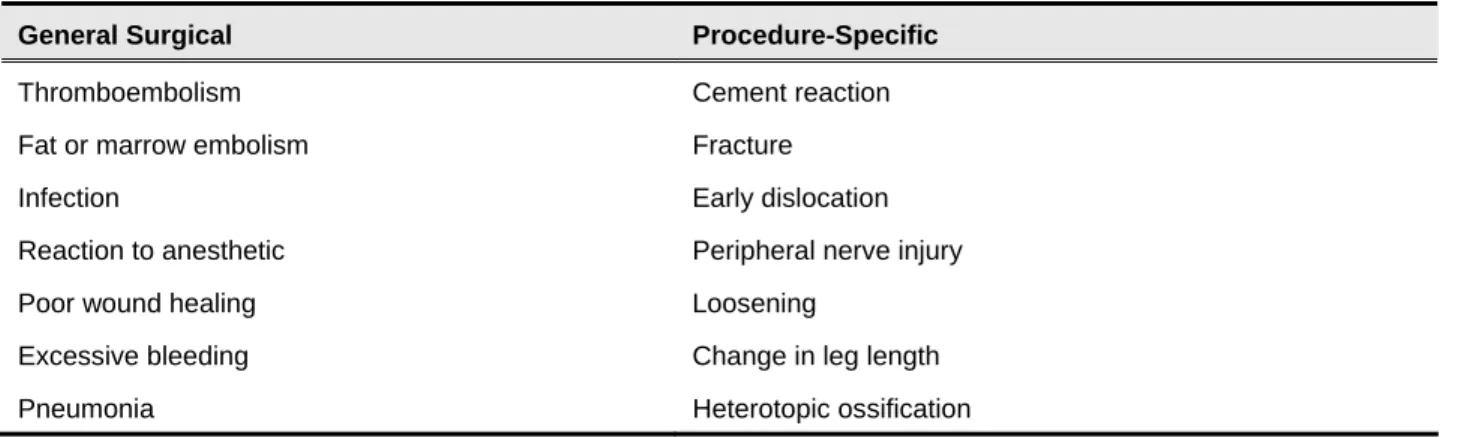
Short-term adverse events include those events that may occur, albeit rarely, following any surgery. Allergic reactions, anesthesia reactions, migration of blood clots, excessive bleeding, infection, heart attack, and pneumonia are all possible. The occurrence rates of these events are not well reported in the literature.
Adverse events specific to hip replacement include dislocation, loosening or breakage of the implant, reaction to the implant materials, breakage of the bone surrounding the implant, locking of the joint and change in the length of the affected leg.(53) Pain, stiffness and nerve damage may occur. In rare cases, leg amputation may be necessary. Adverse event rates are commonly believed to vary according to the surgical approach, the surgical experience of the operating team, implant model used and implant design. Some adverse events may result in a need for additional surgery, including revision of the implant.
Table 3. Short-Term Complications
General Surgical Procedure-Specific
Thromboembolism Cement reaction
Fat or marrow embolism Fracture
Infection Early dislocation
Reaction to anesthetic Peripheral nerve injury
Poor wound healing Loosening
Excessive bleeding Change in leg length
Thrombosis
Deep vein thrombosis (DVT) is a common complication following THR. Many incidents are minor and not clinically evident. Without prophylaxis, asymptomatic DVT will develop in 40% to 60% of patients having THR.(54) Risk factors for thrombosis after THR include increasing age (modest increase with age), sex (women may be at more risk), previous thromboembolism (two to three fold increase in risk), and obesity (twofold with BMI greater than 25). Symptomatic thrombosis often occurs in the calf, leading to local pain and swelling. Most surgeons advocate some form of anticoagulant therapy to help prevent thrombosis or embolism. The ideal agent for prophylaxis has not been identified yet, but randomized controlled trials have shown that low-molecular-weight heparin (LMWH), warfarin, and fondaparinux are safe and effective in
reducing the risk of thrombotic events after THR.(54) The American College of Chest Physicians (ACCP) recommends that patients undergoing THR receive a thromboprophylaxis agent for at least ten days, preferably for 35 days, after surgery.(55)
Pulmonary embolism is the most common surgical adverse event that can lead to death. It occurs most often during the second week after THR and risk declines sharply by the fourth week.(53)
Infection
The most significant early complication of THR is sepsis. It can lead to catastrophic failure requiring explantation.(56) In some cases, permanent resection arthroplasty (Girdlestone arthroplasty) may be necessary to control infection. Improvements in sterile technique and prophylactic antibiotics have dramatically reduced the incidence of sepsis following hip replacement compared to when the technique was first introduced.
Fracture
Bone fracture can occur in the area around the prosthesis, either during the implantation procedure or shortly thereafter. This occurs with approximately 0.1% to 1.0% of cemented implants and 3% to 17.6% of uncemented implants.(53) Risk of fracture increases when bone integrity is compromised by arthritis or other diseases or by previous implantation. Periprosthetic fracture may require revision surgery.(57)
Heterotopic Ossification
Formation of bone in inappropriate places can occur as a result of stress on the bone. Thickening, spur formation and ankylosis are all forms of heterotopic bone formation referred to as
heterotopic ossification (HO). A few weeks after THR, HO begins, and formation is usually complete within three months.(53) While normally harmless, HO can cause pain and may impede joint motion. A recent systematic review of studies of THR found that HO occurred in approximately 42% to 44% of patients.(58) This is significantly more common than was reported in earlier narrative reviews, which reported that one fourth to one third of patients experience HO. The recent review did not indicate how often heterotopic bone led to motion difficulties. However, severe HO or bony ankylosis occurred in 9% of patients. Low dose radiation is frequently used to prevent HO.(53) HO can also be prevented or inhibited by administration of nonsteroidal anti-inflammatory drugs.(58) Radiation and NSAIDs can be used in a combined approach to reduce the incidence of HO after THR.(59)
Dislocation
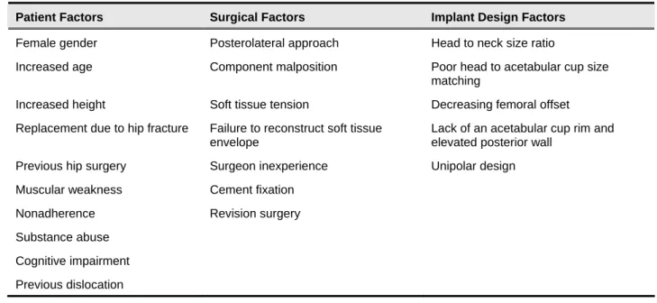
Dislocation of the prosthetic hip has been reported at rates ranging from less than 1% to more than 15%.(28,60) This wide range is probably attributable to the different patients, different devices, and different surgical procedures used in different studies. Rates are similar for THR and hemiarthroplasty, but after hip replacement revision surgery, dislocation rates may be higher than 25%.(28) Most dislocations occur shortly after hip replacement surgery, with 60% to 70% occurring within the first 4 to 6 weeks after surgery.(28)
The most important risk factor for dislocation is previous dislocation. Recurrent dislocation occurs in approximately 33% of cases.(61) Other factors include patient characteristics,
variations in surgical technique, and implant design. THRs performed following hip fracture are at higher risk for dislocation than elective THRs. Instability is also associated with THRs
performed by less experienced surgeons.(60) A list of commonly-reported factors contributing to hip instability is given in Table 4. Some factors may be interrelated. For example, increased age may be associated with decreased cognitive function, which may be associated with inability to adhere to recommended precautions. Other factors may be part of design trade-offs. For
example, a rim around the acetabular cup may decrease hip instability, but also decrease range of motion.
Table 4. Risk Factors for Hip Instability
Patient Factors Surgical Factors Implant Design Factors Female gender Posterolateral approach Head to neck size ratio
Increased age Component malposition Poor head to acetabular cup size matching
Increased height Soft tissue tension Decreasing femoral offset Replacement due to hip fracture Failure to reconstruct soft tissue Lack of an acetabular cup rim and
envelope elevated posterior wall Previous hip surgery Surgeon inexperience Unipolar design Muscular weakness Cement fixation
Nonadherence Revision surgery
Substance abuse Cognitive impairment Previous dislocation
Most dislocated hips can be relocated without surgery, a process known as closed reduction.(61) After closed reduction, the patient may wear a brace or cast for a period so that lax tissue has a chance to tighten as it heals.
Revision surgery is generally reserved for those patients experiencing three or more dislocations. Total revision THR is reportedly successful among these patients in 60% to 75% of cases.(60) Fortunately, the use of modular hip components has made total revision unnecessary in many cases. Instead, hip components can be exchanged. Use of a larger femoral head, or a lipped cup liner can often end an instability problem.(61) Tightening the abductor tissue through
longer femoral neck or lateralizing the acetabular cup can also be effective. During the surgery to exchange components, the surgeon can also alleviate any soft tissue or bony impingement that may contribute to the instability. Such surgery is reportedly successful in 69% to 96% of cases.(60) Use of modular systems in primary THR may also reduce the risk of dislocation.(62) Recurrent dislocation can also be addressed by replacing the acetabular liner with a constrained polyethylene liner.(29,60,63,64) Constrained liners are designed with an extended polyethylene lip that restrains the femoral head within the liner. The capture mechanism reduces the incidence of dislocation but also reduces the range of motion. The greater surface area in contact with the head increases the amount of polyethylene wear. The liner is placed within the acetabular shell component using cement or screws. Constrained liners are still subject to failure due to
loosening, dissociation, breakage, or recurrent dislocation. Reported failure rates range from 4% to 29%.(64)
Long-Term Outcomes
Revision
The most important long-term outcome of hip replacement is surgical revision. The need for surgical revision is the primary definition of failure of a THR.(65) Undergoing additional surgery to repair or replace an implant is extremely inconvenient for the patient and can be dangerous. For this reason, most of the technical innovations in THR have been intended to reduce the need for revision. A more complete discussion of revision surgery can be found below.
Dislocation
Dislocation is usually a short-term complication. However, some patients experience hip instability many years after implantation, despite having never experienced it before. Late dislocation may be associated with increased range of motion and wear of the acetabular cup.(28) Stretching of the soft tissue surrounding the hip by repeated extremes of motion may decrease joint support. Weight loss, decreased muscle mass, or chronic disease
(cancer, rheumatoid arthritis) may also contribute to instability.(64) Dislocation rates have also been reported to increase after surgical replacement of the acetabular cup liner.(30)
Sepsis
Infection associated with implanted prostheses can develop years after surgery and is considered a major complication.(66) The Swedish National Hip Arthroplasty Register annual report for 2004 listed deep infection as the third most common reason for revision surgery (7.9% of all revisions).(67) Deep infection was responsible for 19% of revisions during the first three years after surgery and 1.2% of revisions at more than 10 years after surgery. Management of
periprosthetic infection after THR can involve several different approaches but the standard procedure in North America has been the two-stage exchange revision.(66) The two-stage procedure starts with removal of the infected prosthesis followed by a minimum course of six weeks of parenteral antibiotics. Resolution of the infection is confirmed by repeated
aspiration of the hip. A temporary spacer of antibiotic-loaded cement can be inserted during the first operation and then removed during the second operation when a new prosthesis is put in place. The two-stage revision approach is well accepted but does have some controversial aspects. These include the timing of the procedure, the use of the antibiotic loaded cement at the
second stage, the role of allograft bone grafting, and the use of uncemented components. Other treatment options include antibiotic suppression in patients unable to undergo revision arthroplasty, operative debridement and retention of the infected prosthesis for acute infections, and single-stage exchange revision with post-operative parenteral antibiotics.
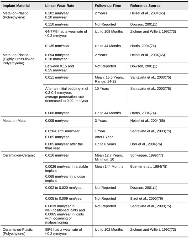
Wear
Wear is the loss of prosthesis material at the interface between the ball of the femur and the acetabular cup. Component wear eventually leads to revision, either because of the component actually wearing out (e.g., the ball breaking through the cup) or because of implant loosening due to osteolysis brought about by wear particles.(68,69) The particles are engulfed by macrophages, which respond by releasing cytokines that encourage resorption of bone. Most often, implants are revised because of osteolysis and implant loosening.(65) Implant wear correlates with osteolysis, loosening and revision.(65) Osteolysis associated with wear debris is the most common cause of revision of prostheses using polyethylene.(70)
Wear can be caused by adhesion of the two components, abrasion caused by the components rubbing against each other or against particles that may find their way between them, or through fatigue. Cracking, pitting or delamination is due to fatigue caused by cyclic stresses placed on the bearing surface.(68)
Wear is often assessed by measuring the linear penetration of the femoral head into the
acetabular cup, reported as mm/year.(71) However, some penetration of the femoral head is the result of “bedding-in,” rather than true wear. “Bedding-in” refers to the settling of the
polyethylene liner within the acetabular shell and the permanent deformation of the plastic due to compression.(72) The deformation due to compression is also known as “creep.” Bedding-in is not considered wear because no material is lost. It slows down within one or two years after implantation.(65) Most of the linear penetration measured after this period is the result of true wear and therefore the wear rate can only be determined after the bedding-in period.(72)
Measurements of linear penetration (as reported in Table 5) are difficult to interpret because the extent of bedding-in may not be accounted for.
In laboratory tests, wear can be measured accurately. However, the relevance of laboratory measurements to wear as it occurs in vivo is unclear. In a clinical situation, wear must be deduced radiologically.(1) Radiologic measurements are inaccurate and insensitive to small changes. Under the best of conditions, meaningful measurements of the penetration of the ball into the socket can only be made when they reach depths of 0.5 mm or more.(1) This level of wear is normally observed years after implantation. Wear of metal-on-metal bearings cannot be measured radiographically at all.(65) Accurate measures of wear can only be made when components are explanted during revision surgery.
Table 5 presents some linear wear rates as reported in various recent reviews and other published reports. At the current time, no national registries or other national data sources are available that provide information on wear rates for different prothesis and their impact on revision rates in clinical practice across the United States. The tabled data only illustrate the range of rates reported and the difficulty of comparing rates reported from different sources. Wear rates reported at different follow-up times may not be comparable because the contribution of the bedding-in process to the overall penetration rate will be different. At shorter follow-up times, linear penetration will be primarily the result of bedding-in rather than true wear. At longer follow-up times, a greater proportion of the penetration will be the result of wear. In addition,
attempts to combine data from studies using the same implant materials may not be valid
because of other differences between studies, particularly patient characteristics. Younger, more active patients have higher rates of wear than older, sedentary patients.(65)
Wear can be reduced by using well-fitted components and wear-resistant materials.
Retrieval studies (studies of implants retrieved after revision surgery) suggest that metal-on-metal prostheses appear to wear more slowly than metal-on-metal-on-plastic.(65) Simulator studies have shown as much as 200 times less wear of metal-on-metal than metal-on-plastic devices.
Ceramic-on-ceramic prostheses may wear even more slowly than metal-on-metal.(65)
Ceramic-on-plastic may wear more slowly than metal-on-plastic.(1) Because surface wear and the subsequent local and systemic effects remain the major cause of THR failure and the need for revision surgery, research into alternative bearing surfaces that minimize wear is still
ongoing.(22) Controversies remain about which type of bearing surface is the most durable. Each surface has its advantages and disadvantages. New developments in bearing surfaces will be discussed in the section on types of replacement hip designs.
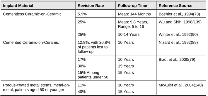
Table 5. Reported Linear Wear Rates of Different Implant Materials
Implant Material Linear Wear Rate Follow-up Time Reference Source Metal-on-Plastic
(Polyethylene)
0.202 mm/year 0.20 mm/year
2 Years Heisel et al., 2004(65)
0.110 mm/year Not Reported Dowson, 2001(1) 64-77% had a wear rate of
<0.2 mm/year
Up to 108 Months Zichner and Willert, 1992(73)
0.135 mm/Year Up to 44 Months Harris, 2004(74)
Metal-on-Plastic (Highly Cross-linked Polyethylene)
0.094 mm/year 0.18 mm/year
2 Years Heisel et al., 2004(65)
Between 0.15 and 0.25 mm/year
Not Reported Dowson, 2001(1)
0.011 mm/year Mean: 15.5 Years, Range: 14-22
Santavirta et al., 2003(75)
After an initial bedding-in of 0.2-0.4 mm/year,
average penetration rate decreased to 0.02 mm/year
10 Years Santavirta et al., 2003(75)
0.008 mm/year Up to 44 Months Harris, 2004(74) Metal-on-Metal 0.005 mm/year 3 Years Heisel et al., 2004(65)
0.020-0.025 mm/Year 0.005 mm/year
1 Year After1 Year
Santavirta et al., 2003(75)
0.005 mm/year after the third year
Up to 8 years Dorr et al., 2004(76)
Ceramic-on-Ceramic 0.016 mm/year Mean 12.7 Years, Minimum 10 Schweppe, 1999(77) 0.0026 mm/year in a stable implant 0.068 mm/year in a loose implant
Mean 144 Months Boehler et al., 1994(78)
0.002 to 0.020 mm/year Not Reported Dowson, 2001(1)
0.005 to 0.009 mm/year Not Reported Bizot et al., 2000(79) 0.0039 mm/year in
well-positioned joints and 0.0065 mm/year in joints with loosening or malpositioning
Not Reported Santavirta et al., 2003(75)
Ceramic-on-Plastic (Polyethylene)
95% had a wear rate of <0.2 mm/year
Loosening
Over time, fixation of the implant to the bone can decline. Micromovements can lead to
fragmentation of acrylic cement, and phagocytosis of acrylic particles can activate macrophages, which respond by releasing cytokines leading to osteolysis. Additional debris derived from friction between the femoral head and the acetabular cup contributes to this process as well.(1,80)
Patients undergoing hip or knee replacement in their 40s or 50s report more rapid onset of loosening, which may be related to increased polyethylene wear.(27) Techniques for prolonging cement life have been developed over the years and incorporated into standard surgical
technique. These include warming the implant prior to cementing,(30) and reducing the porosity of the cement surface. Good surgical technique and avoiding mixing wet cement with blood can help prevent loosening.
Femoral loosening has been associated with small femoral stems implanted into large
intramedullary canals.(56) Other patient characteristics associated with loosening include patient weight, unilateral disease, youth and high activity.
While radiographic monitoring is considered an essential part of THR aftercare, radiographic evaluation frequently underestimates the extent of osteolysis, particularly in the pelvis.(81) Considerable bone loss has to occur before it can be detectable either as radiolucent lines or other cystic changes.(82)
Breakage
The Swedish National Hip Arthroplasty Register annual report for 2004 listed implant fracture as the sixth most common reason for revision surgery. From 1979 to 2004, 1.6% of revisions were needed because of implant breakage.(67) Lindahl et al. analyzed the data in this registry, and concluded that the majority of patients who suffered implant fractures had loose stems at the time of the fracture.(83) The authors of this report suggest that routine radiographic followup to detect loose implants may help prevent implant breakage.
Early ceramic implants were known for their relatively high fracture rates. Later improvements in quality and reductions in grain size appear to have overcome this problem, and more recent surveys have found only one or two breaks in alumina ceramic devices in 10,000 patients.(68) Zirconia ceramic balls are even harder. One survey found only two fractures out of 300,000 implanted devices.(68) However, the quality of zirconia ceramics is highly dependent on the precise manufacturing process used. A change in manufacturing process in 1988 led to an unacceptably high breakage rate (as high as one in three devices from one lot). Nine lots of zirconia balls were eventually recalled.(84) This catastrophic experience was unique to a single type of ball manufactured in a specific manner, but it illustrates the importance of precise control over manufacturing processes and rigid quality control.
Femoral stems can also break. Stem fractures in the early Charnley type prosthesis appeared in the late 1960s.(85) This lead to changes in the design and geometry of the stem to improve corrosion resistance and fatigue properties.
Influence of Patient Factors
Weight
Other factors being equal, heavier patients place more stress on their prostheses than lighter patients. This may lead to greater wear and a higher propensity of the implant to break. At the same time, heavier patients may be less active, which could reduce wear.
Activity
Wear is the result of activity. More active patients will wear out their implants faster.(65) Wear debris contributes to aseptic loosening. Activity also increases micromovement of the implant, leading to release of cement particles and further aseptic loosening. All of these lead to a more rapid need for surgical revision.
Bone Quality
Patients with severe degeneration of the bone due to osteoarthritis, Paget’s disease or other conditions may be more prone to implant loosening, and may lack sufficient structural support for the prosthesis. Patients with weak bone may not be candidates for rigid ceramic prostheses, which do not absorb shocks and may lead to bone damage. Bone thickness is classified according to the system of Dorr et al.(86) from thickest, healthiest cortices (Class A) to thinnest, class C. Dorr class C bone is considered a predictor of less successful THR.(25)
The outcome of THR is also affected by the patients’ Charnley categories.(47) This simple classification scheme describes the extent of patient disability before THR. Category A includes patients with unilateral hip disease, category B includes those with bilateral hip disease, and category C includes patients with multiple joint disease or other disabilities impairing their walking capacity. The results of different clinical trials of THR cannot be compared unless the patients were in comparable Charnley categories. Moreover, patients may move into new Charnley categories as they age. Therefore, clinical trials should report Charnley categories not just at the time of surgery, but at each time of follow-up.(47)
Age
Age by itself is not a factor in deciding whether to perform THR or in deciding which type of implant to use.(87) However, age correlates with other factors that appear to influence THR outcomes. Older patients may not be as active as younger patients, making their prostheses less prone to wear and breakage.(65) Lower wear, in turn, may mean lower incidence of aseptic loosening.(27)
Older patients tend to have lower quality bone, due to ongoing arthritis and osteoporosis. This may influence the type of prosthesis chosen and the longevity of the device. Older patients are also more likely to undergo THR following hip fracture, which is associated with worse outcomes than the typically elective THR performed for other indications in younger individuals. Finally, older patients have shorter life expectancy than younger patients, reducing the number of loading cycles the implants must endure.(65) Younger patients are therefore more likely to outlive their implants, necessitating one or more revisions.
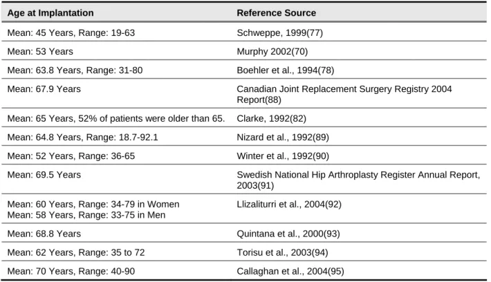
Table 6, below, lists the ages of patients at initial THR reported in recent reviews and other published reports. Because the information was not assembled in a systematic manner, ECRI cannot determine the extent to which the information presented is representative of typical medical practice in the United States. Rather, the table is presented to illustrate two key points. First, patients as young as 19 and as old as 90 have been treated with THR. Younger patients will want protheses with a long functional life-time in order to delay the need for revision surgery. Mean ages in Table 6 range from 45 to 70. Second, the published literature rarely reports data in a way that enables a separate examination of information grouped according to age (for example, patients over 65 years old). While the mean age of patients in a given study may be close to 65, the study will almost certainly include younger and older patients. The effect that age outliers may have on the reported outcomes of a study often cannot be determined.
Table 6. Reported Patient Ages at initial THA
Age at Implantation Reference Source
Mean: 45 Years, Range: 19-63 Schweppe, 1999(77)
Mean: 53 Years Murphy 2002(70)
Mean: 63.8 Years, Range: 31-80 Boehler et al., 1994(78)
Mean: 67.9 Years Canadian Joint Replacement Surgery Registry 2004 Report(88)
Mean: 65 Years, 52% of patients were older than 65. Clarke, 1992(82) Mean: 64.8 Years, Range: 18.7-92.1 Nizard et al., 1992(89) Mean: 52 Years, Range: 36-65 Winter et al., 1992(90)
Mean: 69.5 Years Swedish National Hip Arthroplasty Register Annual Report, 2003(91)
Mean: 60 Years, Range: 34-79 in Women Llizaliturri et al., 2004(92) Mean: 58 Years, Range: 33-75 in Men
Mean: 68.8 Years Quintana et al., 2000(93) Mean: 62 Years, Range: 35 to 72 Torisu et al., 2003(94) Mean: 70 Years, Range: 40-90 Callaghan et al., 2004(95)
Effects of the Clinical Environment
Clinics in which a large number of THR procedures are performed tend to have lower rates of complications or mortality compared to centers with a lower surgical volume.(96,97) Moreover, individual surgeons who perform many procedures a year tend to have superior outcomes compared to surgeons who perform fewer procedures. This phenomenon is particularly noticeable when dislocation rates are examined.(60,98) However, the relationship between surgical volume and complication rates is not strictly linear. In at least one study, the complication rate reached a floor, with no further decrease at higher volumes.(96)
Other factors that may influence between-center differences in complication and mortality rates include the analgesics used, favored surgical methods, favored types of replacement hips, the types of patients typically seen in that facility, and whether or not the facility is a teaching or a research hospital. An analysis of data from the Swedish National Hip Arthroplasty Register indicates that differences in types of patients seen in a facility may account for differences in revision surgery rates between facilities. The Swedish National Hip Arthroplasty Register records both the number of procedures performed and revision rates at a hospital-by-hospital level. The data from the registry are analyzed by the Department of Orthopaedics at Sahlgrenska University Hospital and presented in an annual report. A Cox regression analysis presented in the 2004 Annual Report indicates that revision surgery is approximately 27% higher in patients younger than 60 years of age, older than 75 years of age, or with diagnoses other than primary osteoarthritis. Thus differences in hospital revision rates may be due to differences in the proportion of patients of these types seen by each hospital.
Patients 60 to 75 years of age with primary osteoarthritis represent the most common patient category seen in the Swedish Hip Register.(67) They accounted for 41% of all hip arthroplasties and 3.2% of these patients underwent revisions. The authors of the 2004 Annual Report used this index group of patients as a first step in looking for patient characteristics that could account for differences in THR outcomes between facilities. They compared this group to all other patients and found a significant difference in revision rates between the groups as described above. They also found that this index group varied considerably depending on the type of hospital. In rural and private hospitals, there were more patients in this group and these hospitals tended to have somewhat better implant survival rates. Future analyses will focus on whether having a higher proportion of patients in this index group correlates with reduced medical costs and better outcomes.
That high volume centers may provide superior outcomes may lead more patients or other stakeholders to choose such centers for their procedures. The effect on surgical outcomes of restricting patients to high-volume centers has not been determined. Further increasing the volume at centers that are already working to capacity may lead to increased complication rates due to increased workload. Further research is needed to address this issue.
Types of Replacement Hips
Prosthetic hips come in a wide array of designs. Many models of prosthetic are available in modular designs, so that various combinations of features can be selected to best fit patient needs and clinician preferences. A detailed list of available models and features can be found in the Appendix. A systematic review conducted by the U.K. National Health Service in 1998 noted the “striking paucity of clear and relevant evidence on which to make well-informed choices about prostheses for primary THR.”(99)
Design
The basic design of the hip prosthesis has not changed drastically since hip replacement surgery was introduced decades ago. The most common design features a stem topped by a ball
component that is implanted into the top of the femur to replace the degenerated femoral head that surgeons have removed. The ball fits into a socket (acetabular cup component) placed in the hip bone. Different models vary in details of design, materials and cost. Each component may come in a variety of sizes to accommodate differently-sized patients.
Materials
Over the years, the metal-on-plastic (ultra-high molecular weight polyethylene) design has emerged as the gold standard of hip prostheses.(100,101) In this design, the ball component is metal, while the cup component is lined with plastic. In past years, alternative materials suitable for orthopedic bearings, such as new metal alloys and ceramics, were studied in an attempt to develop more durable joints that provided the same level of functionality. Early metal and
ceramic bearings were abandoned for technical and design deficiencies.(102) The first generation of metal-metal joints were abandoned because of early loosening of the acetabular cup resulting from imprecise fit of the metal ball in the metal socket, while ceramic joints may have had higher fracture rates. Subsequent developments may have reduced or eliminated such problems.(103)
Metal-on-plastic
The first metal-on-plastic hips failed rapidly as the femoral head penetrated the cup.(1) These cups were made of Teflon. Later, polyethylene cups proved to be more durable, but long-term wear remains a problem. Conventional ultra-high molecular weight polyethylene (UHMWPE) cups typically last for 10 to 15 years or longer, depending on the age and activity level of the patient, but the wear and osteolysis remain a problem in individuals who are expected to live longer than this time span.(104,105) This device lifespan is not favorable for younger, more active patients who may require at least one revision procedure in their lifetime to replace a failed prosthetic joint.
The most common cause of metal-on-polyethylene hip joint failures is aseptic inflammatory reaction to the microscopic polyethylene particles released due to joint wear.(70)
This inflammation can cause bone resorption and loosen the bond between bone and prosthesis, causing pain and impairing proper joint function.(1) Loss of bone tissue during the initial device implantation and lower bone quality due to inflammation near the implant means that revision surgery to replace the damaged prosthetic joint is typically more challenging than the initial implantation.
Highly cross-linked polyethylene
More recently, researchers have developed a more durable material called “highly cross-linked polyethylene” (UHMWPE modified in an attempt to change its mechanical properties to decrease wear) to produce next-generation metal-on-plastic hip joints. The use of harder metals and higher levels of cross-linking in the polyethylene may have improved longevity, but have not eliminated the problem of wear. Highly crossed-linked UHMWPE is made by exposure to
gamma or electron beam irradiation.(106) In addition to the cross-linking, the radiation exposure also forms free radicals, which would normally lead to oxidative damage and greater wear of the polyethylene. The free radicals are removed by re-melting the material. The resulting substance has greatly increased wear resistance. This product was first introduced in 1998.(107) Different types of UHMWPE have different biomechanical characteristics. These differences may account for differences in clinical performance. Other factors that may influence wear rates include cross-linking method and sterilization method.(30)
Available data are mixed as to whether highly cross-linked polymers last longer than older materials. A radiographic study comparing conventional and highly cross-linked polyethylene at two-year followup found a 65% reduction in two-dimensional linear wear rate associated with the highly cross-linked polymer, as well as a 54% reduction in three-dimensional wear rate and a 38% reduction in volumetric wear.(30) However, another study that compared polyethylene liners that had been surgically retrieved for reasons other than wear found no difference in damage scores between conventional polyethylene and highly cross-linked polyethylene.(30,68) Although lower wear rates would lead to less wear debris given off by the implant, highly cross-linked polyethylene wear particles tend to be smaller than those given off by standard
polyethylene.(65) These submicrometer particles may induce a greater inflammatory response than larger particles. The question of whether fewer but more active particles lead to more or less osteolysis and implant loosening remains to be answered. The number, size and shape of the particles released by the polyethylene liner depends on the material used, the mode of cross-linking, and patient-related wear factors.(65) Only clinical studies of each specific polyethylene component, controlled for differences between patients, can determine whether cross-linked polyethylene leads to more favorable clinical outcomes.
Highly crossed-linked UHMWPE has many proponents.(108) The lack of metal ion production in particular has been cited as advantage over metal-on-metal systems. However, long-term studies are needed to determine if the purported advantages lead to less wear and longer device life span.(22,106,107)
Metal-on-metal
Early efforts at implanting metal-on-metal prostheses were plagued by rapid dislocations and cup deformation.(109,110) By 1975, metal-on-metal designs had been phased out in favor of the metal-on-polyethylene designs. However, concerns over osteolysis attributed to polyethylene wear debris led to a reintroduction of newer on-metal designs. More recently, new metal-on-metal joints seem to have corrected the previous issues.(111,112) The later generation of metal-on-metal joints has a more precise fit that allows the proper space for lubrication. This has apparently solved the early cup-loosening problem. In particular, a number of manufacturers are marketing metal femoral heads that have a larger diameter than the traditional metal models; these larger heads are designed to decrease the probability of dislocation.(22) Prostheses are available that are made from stainless steel, titanium, or cobalt chrome.(99)
Titanium is no longer a popular choice as a bearing surface due to debris accumulating in tissues as surface oxides detach from the bearing surface.(113) These particles may be associated with a high rate of aseptic loosening. Titanium remains popular as a femoral stem in modular
prostheses.(113)
Some experts have expressed concern that over the long term, the metal ions that are gradually released by these new metal alloys in metal-on-metal couplings may promote some types of systemic or blood-borne cancers or damage internal organs, such as the kidneys.(114) Although cobalt and chrome particles have been shown to induce carcinoma in animal models,
epidemiologic studies have not found any increased risk of cancer among patients with metal-on-metal hip or knee prostheses.(115,116) Toxicity still remains a concern and long-term clinical observations are necessary to determine if the wear resistance of metal-on-metal systems outweighs any associated risks.(117)
Metal-on-metal implants tend to wear rapidly during the period immediately after
implantation.(68) However, this phenomenon is transitory, and wear tends to be slow when considered over the life of the implant. Metal-on-metal joints reportedly have wear rates that are substantially better than metal-on-polyethylene joints, but not quite as good as ceramic-on-ceramic.(65) Metal-on-metal implants may also have the ability to self-heal.(68) Friction between the two components may polish out any imperfections introduced by subluxation or third-body particles.
Proponents of metal-on-metal implants cite this design’s long history, better wear characteristics, and lack of observed biological complications from metal particles or ions.(109,118) Other authors cite concerns over acquired hypersensitivity to metal particles, mutagenicity, and carcinogenicity as reasons not to recommend metal-on-metal designs.(107,110) Long-term comparison studies are needed to determine the extent to which the purported advantages of metal-on-metal lead to better clinical outcomes and if a biological response to metal ions becomes a clinically relevant concern.(22,103,107,117)
Ceramic-on-ceramic
The need for more wear-resistant materials has led to the introduction of ceramic hips. Several types of ceramic have been used for the femur-cup interface, the most popular of which is alumina (aluminum oxide). Although alumina hips have been used, primarily in Europe, for more than 30 years, data derived from these earlier designs may not be relevant because of improvements in manufacturing and materials since that time.
Alumina hips implanted in the 1970s were less dense and more porous than more recent models, with larger grain sizes.(119) Grain size has been reduced from about 40 microns in the earliest models to below 3 microns.(113) This means that the size of flaws in the ceramic structure is likewise reduced, and the surface of the bearing is smoother.(113) In addition, designs,
manufacturing techniques and quality controls have improved.(119) These properties may cause more recent hip joint models to be less prone to breakage and wear than older models.(90) The earliest ceramic models used a ceramic ball mounted on a metal stem with an epoxy resin. Disconnection of ball from stem was a frequent problem, and this design was abandoned in favor of a locking mechanism between ball and stem.(82)
Ceramic-on-ceramic implant joints have the lowest wear rates of any combination investigated thus far.(65,119) Ceramics are hydrophilic, so that the surface of a ceramic joint is more wettable
than other joints, ensuring smooth spread of lubricating synovial fluid throughout the joint. Moreover, ceramics are harder than metal, and can be polished to a smoother finish. Finally, experiments have suggested that ceramic friction debris does not activate macrophages to the same extent as observed for plastic debris.(120) This may lead to greater hip longevity because fewer cytokines and other factors responsible for osteolysis and implant loosening will be released by macrophages. Decreased osteolysis also makes revision surgery easier, should it become necessary.
These properties of greater wear resistance and lower bioactivity may not translate into lower revision rates for ceramic implants. Ceramic components may migrate after a period of firm integration,(90) leading to tilting and malpositioning. In cases of cup malpositioning, loosening, or manufacturing defects leading to increased friction, ceramic hips can produce considerable wear debris.(65,119) Surgical placement must therefore be performed with great precision.(68) Patient complaints of an irritating squeak developing around two years after implantation of ceramic-on-ceramic joints was reported in a presentation at the 2006 meeting of the American Academy of Orthopaedic Surgeons. No formal study has been conducted of this adverse event, but it appears to affect less than 1% of implants.(121)
Ceramic-on-ceramic systems also have their proponents, especially for young active
patients.(122,123) Reduced osteolysis and simpler revision surgery along with reduced cost and reduced risk of ceramic fracture have been cited as ceramic-on-ceramic advantages. Again long-term studies are needed to determine if the wear characteristics lead to extended device use.(101,103,107)
Ceramic-on-Plastic
When zirconia ceramics are used for femoral heads, an ultra-high molecular weight polyethylene cup is used. Zirconia ceramics tend to wear rapidly when they slide against other ceramics.(113) Some researchers believe that polyethylene acetabular cups wear more slowly when coupled with ceramic heads than with metal heads.(1,68) However, not all research supports this contention.
Ceramic-on-Metal
A ceramic ball in a metal cup is said to produce “ten times less metal wear” compared to metal-on-metal.(124) The ceramic ball may act as a polishing stone, further smoothing the cup and decreasing friction with use.(1) Because this design is unusual, data describing wear rates and other factors influencing implant longevity may be difficult to acquire.
Cushion Bearings
Attempts to duplicate the smooth, yielding properties of the natural cartilage joint are
ongoing.(1) As far as we have been able to determine, no THR procedures have been performed using such joints. However, a clinical trial is underway comparing a cushion bearing femoral head hemiarthroplasty with bipolar hemiarthroplasty.(125)
Surface Coatings
A prosthesis that combines the wear properties of a ceramic surface with the bulk properties of metal would be highly desirable. Early attempts to cover a metal surface with a ceramic coating failed when the coating failed to adhere to the metal.(113)