The Role of Sleep in Cognition and Emotion
Matthew P. WalkerSleep and Neuroimaging Laboratory, Department of Psychology & Helen Wills Neuroscience Institute, University of California, Berkeley, California
As critical as waking brain function is to cognition, an extensive literature now indi-cates that sleep supports equally important, different yet complementary operations. This review will consider recent and emerging findings implicating sleep and specific sleep-stage physiologies in the modulation, regulation, and even preparation of cog-nitive and emotional brain processes. First, evidence for the role of sleep in memory processing will be discussed, principally focusing on declarative memory. Second, at a neural level several mechanistic models of sleep-dependent plasticity underlying these effects will be reviewed, with a synthesis of these features offered that may explain the ordered structure of sleep, and the orderly evolution of memory stages. Third, accumu-lating evidence for the role of sleep in associative memory processing will be discussed, suggesting that the long-term goal of sleep may not be the strengthening of individual memory items, but, instead, their abstracted assimilation into a schema of generalized knowledge. Fourth, the newly emerging benefit of sleep in regulating emotional brain reactivity will be considered. Finally, and building on this latter topic, a novel hypoth-esis and framework of sleep-dependent affective brain processing will be proposed, culminating in testable predictions and translational implications for mood disorders. Key words: sleep; learning; memory; encoding; consolidation; association; integration; plasticity; emotion; affect; non-rapid eye movement (NREM) sleep; rapid eye movement (NREM) sleep; offline; slow wave sleep (SWS), slow-wave activity (SWA), sleep spindles
“If sleep does not serve an absolutely vital function, then it is the biggest mistake the evolutionary process has ever made.”
Allan Rechtschaffen University of Chicago Sleep Laboratory Smithsonian, November 1978
Introduction
A perplexing question continues to elude scientific judgment: “Why do we sleep?” In accepting the utility of evolution, as candidly stated by pioneering sleep researcher Allan Rechtschaffen, sleep is likely to support a fun-damental need of the organism. Yet, despite
Address for correspondence: Matthew P. Walker, Sleep and Neuroimaging Laboratory, Department of Psychology & Helen Wills Neuroscience Institute, University of California, Berkeley, California 94720-1650, USA. Voice: 510-642-5292; fax: 510-642-5293. mpwalker@berkeley.edu
the vast amount of time this state takes from our lives, we still lack any consensus function for sleep. In part, this is perhaps because sleep, like its counterpart wakefulness, may serve not one but many functions, for brain and body alike.
Centrally, sleep is a brain phenomenon, and over the past 20 years, an exciting revival has taken place within the neurosciences, one that focuses on the question of why we sleep, and specifically targeting the role of sleep in a num-ber of cognitive and emotional processes. This review aims to provide a synthesis of these re-cent findings in humans, with the goal of ex-tracting consistent themes across domains of brain function that appear to be regulated by sleep. Providing a mechanistic foundation on which to consider these findings, the first sec-tion of this chapter briefly summarizes the brain substrates of sleep: its neurochemistry, neuro-physiology, and functional anatomy. The next
The Year in Cognitive Neuroscience 2009: Ann. N.Y. Acad. Sci. 1156: 168–197 (2009). doi: 10.1111/j.1749-6632.2009.04416.x C 2009 New York Academy of Sciences.
Sleep Stage REM AWAKE NREM-1 NREM-2 NREM-3 NREM-4 Time
12am 1am 2am 3am 4am 5am 6am 7am
R E M S W S
stage-2 N R E M
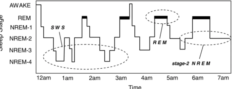
Figure 1. The human sleep cycle. Across the night, NREM and REM sleep cycle every 90 minutes in an ultradian manner, while the ratio of NREM to REM sleep shifts. During the first half of the night, NREM stages 3 and 4 NREM (SWS) dominate, while stage 2 NREM and REM sleep prevail in the latter half of the night. EEG patterns also differ significantly between sleep stages, with electrical oscillations such as slow delta waves developing in SWS, K-complexes and sleep spindles occurring during stage 2 NREM, and theta waves seen during REM.
section explores the role of sleep in memory and brain plasticity and also examines compet-ing models of sleep-dependent learncompet-ing. The third section addresses the role of sleep beyond memory consolidation, in processes of associa-tion, integraassocia-tion, and creativity. The final sec-tion discusses the more recent and emerging role for sleep in emotional and affective brain regulation.
Sleep Neurobiology
The sleep of mammalian species has been broadly classified into two distinct types: non-rapid eye movement (NREM) sleep and non-rapid eye movement (REM) sleep, with NREM sleep being further divided in primates and cats into four substages (1–4) corresponding, in that or-der, to increasing depth of sleep (Rechtschaffen & Kales 1968). In humans, NREM and REM sleep alternate or “cycle” across the night in an ultradian pattern every 90 min (Fig. 1). Although this NREM−REM cycle length re-mains largely stable across the night, the ratio of NREM to REM within each 90-min cycle changes, so that early in the night stages 3 and 4 of NREM dominate, while stage 2 NREM and REM sleep prevail in the latter half of the night. Interestingly, the functional reasons for
this organizing principal (deep NREM early in the night, stage 2 NREM and REM late in the night) remains unknown–another perplex-ing mystery of sleep.
As NREM sleep progresses, electroen-cephalographic (EEG) activity begins to slow in frequency. Throughout stage-2 NREM there is the presence of phasic electrical events includ-ing K-complexes (large electrical sharp waves in the EEG) and sleep spindles (short syn-chronized 10–16 Hz) EEG oscillations (Steri-ade & Amzica 1998). The deepest stages of NREM, stages 3 and 4, are often grouped together under the term “slow wave sleep” (SWS), reflecting the occurrence of low fre-quency waves (1–4 Hz and<1 Hz), which have themselves been termed “slow-wave activity” (SWA), representing an expression of underly-ing mass cortical synchrony (Amzica & Steri-ade 1995). During REM sleep, however, EEG waveforms once again change in their com-position, associated with oscillatory activity in the theta band range (4–7 Hz), together with higher frequency synchronous activity in the 30–80 Hz (“gamma”) range (Llinas & Ribary 1993; Steriade et al. 1996). Periodic bursts of REM also take place, a defining characteristic of REM sleep, associated with the occurrence of phasic endogenous waveforms expressed in, among other regions, the pons (P), lateral
geniculate nuclei of the thalamus (G), and the occipital cortex (O), and as such, have been termed “PGO waves” (Callaway et al. 1987).
As the brain passes through these sleep stages, it also undergoes dramatic alterations in neurochemistry. In NREM sleep subcorti-cal cholinergic systems in the brain stem and forebrain become markedly less active (Hobson et al. 1975; Lydic & Baghdoyan 1988), while firing rates of serotonergic raph´e neurons and noradrenergic locus coeruleus neurons are also reduced relative to waking levels (Aston-Jones & Bloom 1981; Shima et al. 1986). During REM sleep both these aminergic populations are strongly inhibited, while cholinergic sys-tems become as/more active compared to what they are during wake (Kametani & Kawamura 1990; Marrosu et al. 1995), resulting in a brain state largely devoid of aminergic modulation and dominated by acetylcholine.
At a whole-brain systems level, neuroimag-ing techniques have revealed complex and dramatically different patterns of functional anatomy associated with NREM and REM sleep (for review, see Nofzinger 2005). During NREM SWS, rostral brain-stem regions, tha-lamic nuclei, basal ganglia, hypothalamus, pre-frontal cortex, cingulate corticies, and medial regions of the temporal lobe all appear to un-dergo reduced activity. However, during REM sleep significant elevations in activity have been reported in the pontine tegmentum, thalamic nuclei, occipital cortex, mediobasal prefrontal lobes, and associated limbic groups, including the amygdala, hippocampus, and anterior cin-gulate cortex. In contrast, the dorso−lateral prefrontal cortex, posterior cingulate, and pari-etal cortex appear least active in REM sleep.
Although this summary only begins to de-scribe the range of neural processes that are af-fected by the brain’s daily transit through sleep states, it clearly demonstrates that sleep itself cannot be treated as a homogeneous entity, one which may or may not alter cognitive and emo-tional processes. Instead, this constellation of sleep stages offers a range of distinct neurobio-logical mechanisms that can potentially support
the modulation, regulation, and preparation of numerous brain functions.
Memory Processing and Brain Plasticity
In considering the role of sleep in memory processing, it is pertinent one appreciate that memories evolve (Walker & Stickgold 2006). Specifically, memories pass through discrete stages in their “life span.” The conception of a memory begins with the process of encoding, resulting in a stored representation of an expe-rience within the brain (Paller & Wagner 2002). However, it is now understood that a vast num-ber of postencoding memory processes can take place (Stickgold & Walker 2005a). For mem-ories to persist over the longer time course of minutes to years, an offline, nonconscious oper-ation of event consolidoper-ation appears to be nec-essary, affording memories greater resistance to decay (a process of stabilization), or even im-proved recollection (a process of enhancement) (Robertson et al. 2004; Walker 2005). Sleep has been implicated in both the encoding and con-solidation of memory.
Sleep and Memory Encoding One of the earliest human studies to report the effects of sleep and sleep deprivation on declarative memory encoding was by Morris et al. (1960), indicating that “temporal mem-ory” (memory involving when events occur) was significantly disrupted by a night of pre-training sleep loss. These findings have been revisited in a more rigorous study by Harri-son and Horne (2000), again using the tem-poral memory paradigm. Significant impair-ments in retention were evident in a group of subjects deprived of sleep for 36 h, the sub-jects scoring significantly lower than controls, even in a subgroup that received caffeine to overcome nonspecific effects of lower alert-ness. Furthermore, the sleep-deprived subjects displayed significantly worse insight into their
0.0 0.2 0.4 0.6 0.8 1.0 1.2 1.4 1.6
POSITIVE NEGATIVE NEUTRAL
**
ALL STIMULUS TYPES n.s. Sleep Sleep Deprived 0.0 0.2 0.4 0.6 0.8 1.0 1.2*
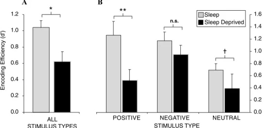
STIMULUS TYPE A B Encoding Ef ficiency (d')Figure 2. Sleep deprivation and encoding of emotional and nonemotional declarative memory. Effects
of 38 h of total sleep deprivation on encoding of human declarative memory (A) when combined across all
emotional and nonemotional categories; (B) When separated by emotional (positive and negative valence)
and nonemotional (neutral valance) categories. †P < 0.08,∗P < 0.05, ∗∗P < 0.01, error bars represent
SEM.
memory-encoding performance, resulting in lower predictive ability of performance.
Pioneering work by Drummond and col-leagues examined the neural basis of sim-ilar memory impairments using functional MRI (fMRI), investigating the effects of 35 h of total sleep deprivation on verbal learning (Drummond et al. 2000). In those who were sleep deprived, regions of the medial temporal lobe were significantly less active during learn-ing, relative to a control group that had slept, while the prefrontal cortex actually expressed greater activation. Most interesting, the pari-etal lobes, which were not activated in the con-trol group during learning, were significantly active in the deprivation group. Such findings suggest that inadequate sleep prior to learn-ing (at least followlearn-ing one night) produces bidi-rectional changes in episodic encoding activity, involving the inability of the medial temporal lobe to engage normally during learning, com-bined with potential compensation attempts by prefrontal regions, which in turn may facilitate recruitment of parietal lobe function (Drum-mond & Brown 2001).
The impact of sleep deprivation on mem-ory formation may be especially pronounced
for emotional material. We have investigated the impact of sleep deprivation on the encod-ing of emotionally negative, positive, and neu-tral words (Walker, unpublished results). When combined across all stimulus types, subjects in the sleep-deprived condition exhibited a strik-ing 40% reduction in the ability to form new human memories under conditions of sleep deprivation (Fig. 2A). However, when these data were separated into the three affective categories (negative, positive, or neutral), the magnitude of encoding impairment differed (Fig. 2B). In those that had slept, both posi-tive and negaposi-tive stimuli were associated with superior retention levels relative to the neutral condition, consistent with the notion that emo-tion facilitates memory encoding (Phelps 2004). However, there was severe disruption of en-coding and hence later retention for neutral and especially positive emotional memory in the sleep-deprived group. In contrast, a relative resistance of negative emotional memory was observed in the deprivation group. These data suggest that, while the effects of sleep depriva-tion are direcdepriva-tionally consistent across memory subcategories, the most profound impact is on the encoding of positive emotional stimuli, and
to a lesser degree, emotionally neutral stimuli. In contrast, the encoding of negative memory appears to be more resistant to the effects of prior sleep loss, at least following one night.
Intriguingly, these data may offer novel in-sights into affective mood disorders that express co-occurring sleep abnormalities (Benca et al. 1992; Buysse 2004). Indeed, if one compares the profiles of memory encoding in Fig. 2B, it is clear that those who slept encoded and retained a balanced mix of both positive and negative memories. In contrast, those who did not sleep displayed a skewed relative distribu-tion of encoding, resulting in an overriding dominance of negative memories, combined with a retention deficit of positive and neutral memories. This selective alteration in mem-ory encoding may provide an experimental explanation for the higher incidence of depres-sion in populations that suffer sleep disruption (Shaffery et al. 2003; Buysse 2004), which, due to these specific deficits, may impose a neg-ative remembering bias, despite the fact that these subjects experienced equally positive- and negative-reinforcing event histories.
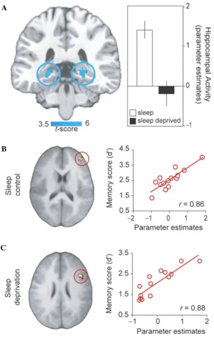
The impact of sleep deprivation on the neu-ral dynamics associated with declarative mem-ory encoding has recently been examined using event-related fMRI (Yoo et al. 2007a). In addi-tion to performance impairments under con-dition of sleep deprivation, and relative to a control group that slept, a highly significant and selective deficit was identified in bilateral re-gions of the hippocampus—a structure known to be critical for learning new episodic infor-mation (Eichenbaum 2004) (Fig. 3A). While these findings indicated that, at a group level, sleep deprivation markedly impairs hippocam-pal memory function, when examined within each group separately, the success of encod-ing, from low to high, was further associated with activity in different regions of the pre-frontal lobe. In those that slept prior to learn-ing, the right dorsal/middle lateral prefrontal cortex showed a strong positive relationship with the proficiency of memory encoding. In contrast, a region in the right inferior frontal
gyrus (IFG) displayed a significant positive, po-tentially compensatory, relationship with mem-ory performance in those who were sleep de-prived (Figs. 3B & C).
Taken together, this collection of findings indicate the critical need for sleep-before-learning in the preparation of key neural structures for efficient next-day learning. With-out adequate sleep, hippocampal function becomes markedly disrupted, resulting in a de-creased ability for recording new experiences, the extent of which appears to be further gov-erned by alterations in prefrontal encoding dynamics.
Sleep and Memory Consolidation Using a variety of behavioral paradigms, ev-idence for the role of sleep in memory consoli-dation has now been reported across a diverse range of phylogeny. Perhaps the earliest refer-ence to the beneficial impact of sleep on mem-ory is by the Roman rhetorician Quintilian, who stated:
[it] is a curious fact, of which the reason is not obvious, that the interval of a single night will greatly increase the strength of the memory. . .. Whatever the cause, things which could not be recalled on the spot are easily coordinated the next day, and time itself, which is generally accounted one of the causes of forgetfulness, actually serves to strengthen the memory.
(Hammond 2004).
In the early eighteenth and twentieth cen-turies respectively, David Hartley (Hartley 1801) and Jenkins and Dallenback (Jenkins & Dallenbach 1924) indicated that the strength of a memory may be better preserved by periods of sleep than it is by equivalent periods of time awake. Following the discovery of discrete sleep stages (Aserinsky & Kleitman 1953), research investigating the influence of sleep on memory has become gradually more complex at both a behavioral and mechanistic level. A robust and consistent literature has demonstrated the need for sleep after learning in the subsequent consolidation and enhancement of procedural memories; the evidence for which has recently been reviewed elsewhere (Walker & Stickgold
Figure 3. Neural basis of sleep-deprivation-induced encoding deficits. (A) Regions of de-creased encoding activation in the sleep deprivation group relative to the sleep control group in bilateral posterior hippocampus, together with a histogram of parameter estimates (effect
size) of averaged hippocampal activity in each group. Effects are significant atP<0.001;
>5 contiguous voxels. (B) correlation analysis with memory performance showing regions
of significant association between encoding-related activation and memory performance (d’) across subjects in the sleep control group (peak – right Middle/dorso-lateral prefrontal cortex),
and (C) in the sleep deprivation group (peak – right Inferior frontal gyrus). Modified from Yoo
et al. 2007a.
2006). Early work focusing on the role for sleep in declarative memory processing was some-what less consistent, but more recent findings have now begun to reveal a robust beneficial
effect of sleep on the consolidation of declar-ative memory—our focus here (Smith 2001; Ellenbogen et al. 2006b; Walker & Stickgold 2006; Marshall & Born 2007).
Several reports by Born and his colleagues showed offline improvement on a word-pair association task following sleep, an improve-ment attributed to early night sleep, rich in SWS (Plihal & Born 1997, 1999; Gais & Born 2004). More recently, the same group demon-strated that, in addition to classically defined slow delta waves (0.5–4 Hz), the very slow corti-cal oscillation (<1 Hz) appears to be important for the consolidation of declarative memories. Following subject learning of a word-pair list, a technique called “direct current stimulation” was used to induce slow oscillation-like field potentials in the prefrontal cortex (in this case, at 0.75 Hz) during early night SWS (Marshall et al. 2006). Direct current stimulation not only increased the amount of slow oscillations dur-ing the simulation period (and for some time after), but also enhanced next-day word-pair retention, suggesting a critical role for SWS neurophysiology in the offline consolidation of episodic facts.
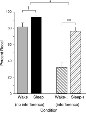
Rather than simply testing memory recall, Ellenbogen and colleagues have since revealed the extent of sleep’s ability to protect declar-ative memories using experimentally induced learning disruption (Ellenbogen et al. 2006a). Taking advantage of a classic interference tech-nique called the A-B–A-C paradigm, subjects first learned unrelated word-paired associates, designated as list A-B (e.g., leaf-wheel, etc.). Af-ter sleep at night, or wakefulness during the day, half of the subjects in each group learned a new, interfering list containing a new associate paired with the first word, designated as list A-C (e.g., leaf-nail, etc.), before being tested on the original A-B list (e.g., leaf-wheel, etc.). In the groups that did not experience the interfering challenge—that is, those who were simply be-ing trained and then tested on list A-B—sleep provided a modest benefit to memory recollec-tion (Fig. 4A). However, when testing the groups that were exposed to interfering list learning (list C) prior to recalling the original list (list A-B), a large and significant protective benefit was seen in those that slept (Fig. 4B). Thus, mem-ories tested after a night of sleep were
signifi-100 80 60 40 20 0 ** *
Wake Sleep Wake-I Sleep-I
(no interference) (interference) Condition
P
ercent Recall
Figure 4. Impact of sleep on the consolidation and stabilization of declarative memory. Percent cor-rect recall for B words from the original A-B pair after a 12-h retention interval of either wake or sleep following no interference or interference
learn-ing (list A-C).†P< 0.10,∗P < 0.05,∗∗P< 0.001;
error bars indicate SEM. Modified from Ellenbogen 2006a.
cantly more resistant to interference, whereas, across a waking day, memories were far more susceptible to this antagonistic learning chal-lenge. Yet it was only by using an interfering challenge, that of the A-C list, that the true benefit of sleep’s protection of memory was re-vealed, a benefit that would not necessarily have been evident in a standard study−test memory paradigm.
One mechanism proposed as underlying these effects on hippocampal-dependent learn-ing tasks (see next section, also) is the reactiva-tion of memory representareactiva-tions at night. A con-siderable number of reports have investigated the firing patterns of large networks of indi-vidual neurons across the wake−sleep cycle in animals. The signature firing patterns of these hippocampal and cortical networks, expressed during waking performance of spatial tasks and
novel experiences, appear to be “replayed” dur-ing subsequent SWS (and in some studies, also REM) (Wilson & McNaughton 1994; Skaggs & McNaughton 1996; Dave et al. 1998; Dave & Margoliash 2000; Poe et al. 2000; Louie & Wilson 2001; Ribeiro et al. 2004; Jones & Wil-son 2005; Ji & WilWil-son 2007). Homologous ev-idence has been reported in the human brain using a virtual maze task in combination with positron emission tomography (PET) scanning (Peigneux et al. 2004). Daytime learning was initially associated with hippocampal activity. Then, during posttraining sleep, there was a reemergence of hippocampal activation, specif-ically during SWS. Most compelling, however, was that the amount of SWS reactivation in the hippocampus was proportional to the amount of next-day task improvement, suggesting that this reactivation is associated with offline mem-ory improvement.
Building on the framework that memories, particularly those involving the hippocampus, are reactivated at night during sleep, Rasch et al. have taken advantage of the classical psychology effect of cue-dependent recall, and translated it into a sleep-dependent consolida-tion paradigm (Rasch et al. 2007). It is well known that memory can be strongly modu-lated by smell (Cann & Ross 1989); most of us have associated the smell of a certain perfume or cologne with a particular person, and when we encounter that same perfume again, it often results in the powerful cued recall of memories of that particular person. In this study, however, following learning of a spatial memory task that was paired with the smell of rose, the odor was not re-presented at retrieval, but instead dur-ing subsequent SWS that night—a time when consolidation was presumed to be occurring. Relative to a control condition where the odor was not presented again during SWS, the re-perfusion of the rose scent at night resulted in significantly improved recall the following day. Moreover, the presentation of the odor re-sulted in greater (re)activation of the hippocam-pus during SWS. These findings support the role of SWS in the consolidation of
individ-ual declarative memories, and may indicate an active reprocessing of hippocampal-bound in-formation during SWS.
Models of Sleep-Dependent Memory Processing
Elucidating the neural mechanisms that control and promote sleep-dependent human memory consolidation remains an active topic of research, and debate (Miller 2007). It is per-haps unlikely that multiple different memory systems, involving diverse cortical and/or sub-cortical networks, require the same underlying neural mechanisms for their modulation. Even if they do, it is not clear that this process would rely on just one type of sleep-stage physiology (Giuditta et al. 1995). At present, two intriguing models of sleep-dependent plasticity, relevant to declarative memory, have been offered to account for the overnight facilitation of recall, which build on different aspects of neural activ-ity during sleep: (1) hippocampal−neocortical dialogue, (2) synaptic homeostasis hypothesis. Hippocampal−Neocortical Dialogue
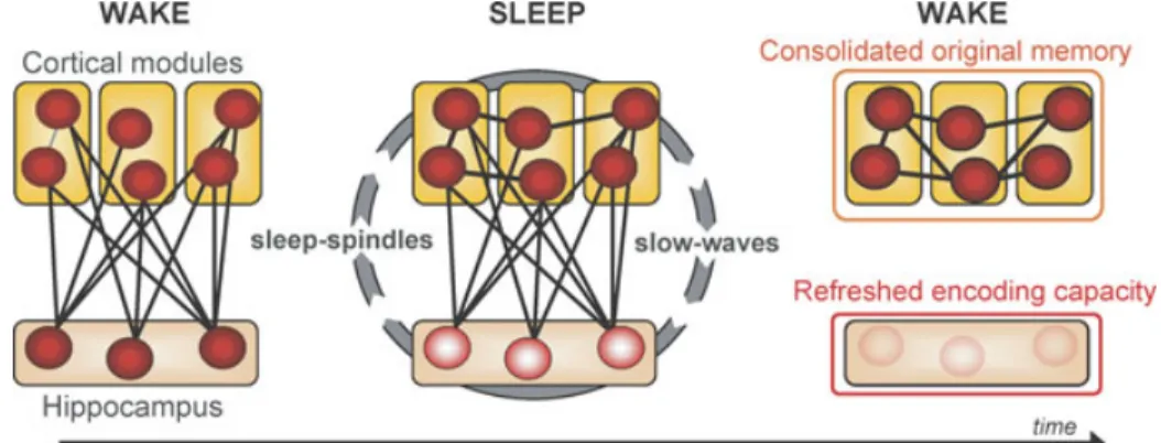
There is considerable agreement that struc-tures within the medial temporal lobe (MTL), most notably the hippocampal complex, are crucial for the formation and retrieval of new declarative memories. These structures are be-lieved to guide the reinstatement of recently formed memories by binding together pat-terns of cortical activation that were present at the time of initial learning. A classical model of declarative memory consolidation suggests that information initially requires MTL bind-ing, but over time, and by way of slow of-fline processes, it is eventually integrated into neocortical circuits (Fig. 5). Neocortical struc-tures thus become the eventual storage site for consolidated episodic memories through cross-cortical connections, and, as a consequence, the MTL is not necessary for these memo-ries’ retrieval. Therefore, the classical model of memory consolidation holds that neocorti-cal structures become increasing important for
Figure 5. Model of sleep-dependent hippocampal-neocortical memory consolidation. At encoding the hippocampus rapidly integrates information within distributed cortical modules. Successive sleep-dependent reactivation of this hippocampal–cortical network leads to pro-gressive strengthening of cortico-cortical connections, which over time, allow these memories to become independent of the hippocampus and gradually integrated with preexisting cortical memories. Modified from Frankland & Bontempi 2005.
the retention and retrieval of successfully con-solidated episodic memories, while the corre-sponding contribution of the hippocampus pro-gressively decreases (Squire 1992; McClelland et al. 1995; Squire & Zola 1996; Squire 2004). It should be noted, however, that controversy remains about the role of these MTL structures in the retrieval of declarative memories after the passage of time. This has led to the emergence of alternate consolidation models, most notably Nadel and Moscovitch’s “multiple trace the-ory” (Nadel & Moscovitch 1997; Moscovitch & Nadel 1998), which posits that hippocampal involvement is always critical for the retrieval of episodic (but not semantic) memories, and that these memories remain permanently de-pendent on hippocampal−neocortical connec-tions (for discussion beyond the scope of this review see Frankl & Bontempi 2005).
In addition to its role in binding distributed cortical memory components, Marr and, also, McClelland et al. suggested that the hippocam-pus plays a critical role in reactivating these networks, specifically during sleep (Marr 1970; McClelland et al. 1995). This process of re-activation, over multiple sleep cycles across a night and/or multiple occurrences of sleep over many nights, would gradually strengthen the initially weak connections between neocortical
sites, thereby reinforcing them (Fig. 5). Eventu-ally, this strengthening would allow the original information to be activated in the cortex, in-dependent of the hippocampus. Buzsaki (1996) has since advanced on these ideas, proposing a model of consolidation that involves two stages or states of hippocampal activity, the first in-volving a mode of “recording” during wake, which shifts to a second stage, involving “play-back” mode during NREM SWS, specifically during bursts of neural activity called “sharp-waves.”
Interestingly, these models make two pre-dictions about the impact of sleep on declar-ative memory. The first is that declardeclar-ative memories from the day prior should be more resistant to interference the next day, due to the increased cortico−cortical connections formed during overnight consolidation (Fig. 5). It is precisely this behavioral effect that was reported in the study by Ellenbogen et al. (2006a) showing greater post-sleep resistance to interference, using the A-B–A-C paradigm. A second and far less considered benefit of this sleep-dependent dialogue is the encod-ing capacity of the hippocampus (Fig. 5). If the strengthening of cortico−cortical connec-tions takes place during sleep, albeit itera-tively, then blocking sleep after hippocampal
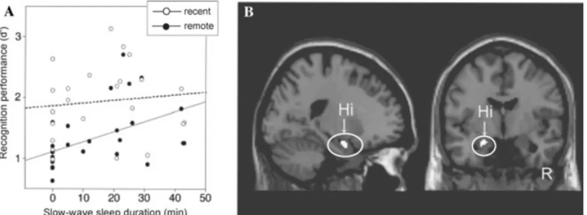
Figure 6. Enhancement of hippocampal declarative memory by daytime naps. (A) Correlation of
recog-nition memory for recent and remote items related to individual slow-wave sleep durations. (B) Correlation
between recognition memory activity and longer slow-wave sleep duration in the left hippocampus (Hi). Modified from Takashima et al. 2006.
learning should negate this offline transfer, pre-venting the development of independence from (or “refreshing” of) the hippocampus, and by doing so, decrease the capacity for new hip-pocampal learning the next day. This sec-ond premise appears to accurately explain the findings discussed in the section above on mem-ory encoding (Yoo et al. 2007b), which de-scribe a significant impairment of hippocam-pal encoding activity when sleep has not taken place (through deprivation) being associated with a decreased ability to form new episodic memories.
Two recent reports have provided further ev-idence in support of this sleep-dependent dia-logue and neural transformation of declarative memory. In the first such report, Takashima and colleagues examined the benefit of day-time naps on episodic declarative memory con-solidation (Takashima et al. 2006). In addi-tion to a long-term evaluaaddi-tion of memory over 3 months, there was also a short-term eval-uation of memory across the first day, which included an intervening nap period (90 min) between training and testing of the original studied (“remote”) stimuli. Interestingly, the duration of NREM SWS during the inter-vening nap correlated positively with later-recognition memory performance (Fig. 6A), yet negatively with retrieval-related activity in the hippocampus (Fig. 6B). Furthermore, with
increasing time following learning, there was progressively greater recall activity in medial prefrontal regions, and a continued dissipa-tion of retrieval-related activity within the hip-pocampus. Advancing on these findings, Ma-quet and colleagues have since demonstrated that one night of posttraining sleep depriva-tion, even following recovery sleep, significantly impairs the normal modulation of hippocam-pal activity associated with episodic memory recollection (Gais et al. 2007). Furthermore, first-night sleep deprivation also prevented an increase in hippocampal connectivity with the medial prefrontal cortex, a development that was only observed in those that slept after learning.
While no one study has yet demonstrated that the neural signature of learning during the day is subsequently reactivated and driven by characteristics of SWS at night, and that the extent of these properties are consequently pro-portional to the degree of next-day recall and memory reorganization, collectively, they offer an empirical foundation on which to entertain this possibility.
Synaptic Homeostasis Hypothesis
In recent years an orthogonal theory of SWS and learning has emerged, one which postu-lates a role for sleep in regulating the synap-tic connectivity of the brain—principally the
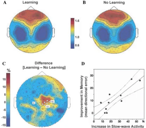
Figure 7. Slow-wave activity and motor-skill memory. Topographical high-density EEG
maps of slow-wave activity (SWA) during NREM sleep following either (A) motor-skill learning
or (B) a nonlearning control condition, and (C) the subtracted difference between SWA in
the learning versus nonlearning condition, demonstrating a local homeostatic increase above
the learning-related central-parietal brain region. (D) the correlation between the amount of
over-night improvement on the task (measured the next day) and the extent of increase in SWA across subjects. Modified from Huber et al. 2004.
neocortex (Tononi & Cirelli 2003, 2006). Their model considers NREM SWS, and specifically the magnitude of slow-wave activity (SWA) of SWS, as a brain-state that promotes the de-crease of synaptic connections, not an inde-crease. Accordingly, plastic processes, such as learning and memory occurring during wakefulness, re-sult in a net increase in synaptic strength in diffuse brain circuits. The role of SWS, there-fore, and the slow oscillation in particular, is to selectively downscale or “depotentiate” synap-tic strength back to baseline levels, preventing synaptic overpotentiation, which would result in saturated brain plasticity. In doing so, this rescaling would leave behind more efficient and refined memory representations the next day, affording improved recall.
A number of human studies by Huber, Tononi, and colleagues have provided evi-dence supporting their model. For example, it has been shown that learning of a motor-skill adaptation task during the day subsequently triggers locally specific increases in cortical SWA at night, the extent of which is propor-tional to both the amount of initial daytime learning and the degree of next-day improve-ment (Fig. 7) (Huber et al. 2004). Further-more, experimentally impairing the amount of experience-dependent activity during the day (arm immobilization) produced the opposite effect—reduced amounts of SWA activity in associated cortical regions (Huber et al. 2006). These findings substantiate the concept of lo-cal sleep-dependent neural pruning by SWS,
the goal of which may be to regulate neural ar-chitecture at a highly specific anatomical level, mapping onto corresponding locations of the memory representation.
How can the two concepts of neural reacti-vation (such as increased fMRI activity dur-ing SWS; e.g., Rasch et al. 2007) and neu-ral homeostasis (such as increased slow wave EEG activity; e.g., Huber et al. 2004) be inter-preted at a neural synaptic level? It could be argued that both these reported changes reflect either an increased neural reactivation, or an increase in SWA associated with homeostasis and synaptic downscaling. Furthermore, both hypotheses, while distinctly different in a mech-anistic sense, could offer complementary ben-efits at the network level in terms of signal-to-noise ratio (SNR) and may account for overnight memory improvements (Robertson, unpublished). Specifically, homeostatic synap-tic downscaling could result in the removal of superfluous neural connections, resulting in improved SNR. However, neural reactivation and strengthening of experience-dependent circuits, done without removing redundant synaptic connects, may equally improve SNR. Therefore, both mechanisms, while different, could produce a similar outcome: enhanced fi-delity of the memory representation. Presum-ably the combination of both would produce perhaps the most optimal and efficient mem-ory trace, yet a careful delineation of these pos-sibilities remains an important goal for future studies.
Interestingly, the homeostasis model would also predict that sleep deprivation, specifically the prevention of SWS, would also negate effec-tive next-day new learning, due to overpotenti-ation of synaptic connections. Thus, any region that exhibits SWA and is involved in represent-ing memory (e.g., hippocampus), would display a corresponding inability to code further in-formation beyond a normal waking duration (∼16 hr in humans). Such a premise may of-fer an alternative explanation to the marked hippocampal encoding deficits reported under conditions of sleep loss (Yoo et al. 2007b).
A Role for Sleep Spindles?
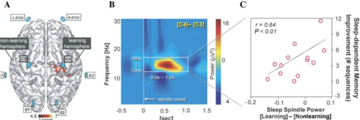
Independent of both these models, which consider the role of SWS in memory pro-cessing, a number of reports have also de-scribed an association between learning and the hallmark feature of stage-2 NREM: sleep spindles; short (∼1 s) synchronous bursts of activity expressed in the EEG in the 10– 16 Hz frequency range (Sejnowski & Destexhe 2000; Steriade 2001; Smith et al. 2004). For example, following learning of a motor-skill memory task, Nishida and Walker (2007) ex-amined posttraining sleep spindles over the motor cortex, evaluating the difference in spin-dle activity in the learning hemisphere (since subjects perform the task with their left, non-dominant, hand), relative to the nonlearning hemisphere (Fig. 8A). Remarkably, when sleep spindle power at electrode sites above the pri-mary motor cortex of the nonlearning hemi-sphere (left) were subtracted from those in the learning hemisphere (right), representing the within-subject, between-hemisphere difference in spindle activity following learning, a strong predictive relationship to the amount of mem-ory improvement emerged (Figs. 8B & C). Simi-larly, Fogel et al. (2001) reported increased spin-dle density after intensive training on a pursuit motor skill task, and Fogel and Smith (2006) reported increased spindle density after com-bined training on several simple procedural motor tasks.
Such findings indicate that the enhancement of specific memory representations is associ-ated with electrophysiological events expressed at local, anatomically discrete locations of the brain. Contrasting with the proposed impact of SWS, the mechanistic benefit of sleep spin-dles may be related to their faster stimulating frequency; a range suggested to facilitate long-term potentiation (LTP; a foundational prin-cipal of synaptic strengthening in the brain) (Sejnowski & Destexhe 2000; Steriade 2001; Smith et al. 2004), and not synaptic depres-sion. This increase in spindle activity may rep-resent a local, endogenous trigger of intrinsic
Figure 8. Sleep spindles and motor-skill memory plasticity. (A) Sleep-EEG array (blue discs) superimposed
on the known overnight plastic reorganization of motor memory, including the right motor cortex (red). (B)
Difference in sleep spindle activity (power) following task training in the Learning (relative to Non-learning)
hemisphere, which (C) accurately predicts the amount of postsleep memory improvement across subjects.
Modified from Nishida & Walker 2007.
synaptic plasticity, again corresponding topo-graphically to the underlying memory repre-sentation (Nishida & Walker 2007).
Increases in posttraining spindle activity are not limited to procedural memory tasks. For example, Gais et al. have shown that there is significantly higher sleep spindle density in subjects that underwent a daytime episodic learning session (encoding of word-pair as-sociates) compared to a control group that did not perform the learning session. More-over, the spindle density was associated with the proficiency of memory recalled the next day in the learning group (Gais et al. 2002). These findings mirror previous observations by Meier-Koll et al. (1999), who reported a sim-ilar increase in spindles following learning of a hippocampally dependent maze task, and by Clemens et al. (2005), who have since identi-fied a correlation between spindle density and overnight verbal memory retention (although not memory for faces). Intriguingly, the study by Huber et al. (2004), which implicates slow oscillatory activity associated with offline mem-ory improvement, also describes similar, albeit near-significant, associations with activity in the sleep spindle frequency range. There may be a combinatory role for spindles in regulating plasticity, together with SWS.
Continued evidence suggests that sleep spin-dles can be separated into two subtypes: “slow”
(11–13 Hz), which have a more anterior dis-tribution, and “fast” (13–15 Hz), which have a more posterior localization (Werth et al. 1997; Zeitlhofer et al. 1997). With this di-vision has also come an interest in under-standing functional differences between each of these spindles subtypes. The relevance of this separation from a memory consolidation perspective is highlighted by a recent neu-roimaging study demonstrating that fast spin-dles are associated with, among other re-gions, significantly greater activation within the hippocampal complex (Schabus et al. 2007). Investigating the role of hippocampal- and extra−hippocampal-dependent memory con-solidation in relation to spindle subtypes will be an important challenge for future research. Reconciling Models
None of these models necessarily is wrong. Instead, aspects of each may afford com-plementary and synergistically beneficial out-comes for memory. Clues to this possibility lie within the ordered structure of human sleep (Fig. 1), with NREM SWS dominating early in the night and stage-2 NREM and REM prevailing later in the night. When placed in this temporal framework a progression of events emerges that may be optimal for the neuroplastic modulation of memory represen-tations. From a reactivation perspective, the
predominance of hippocampal−neocortical interaction would take place in the early SWS-rich phase of the night, leaving cortico−cortical connections on offer for later processing dur-ing stage-2 NREM and REM. Similarly, and even in coincidence, SWS may downscale cor-tical (and possibly subcorcor-tical) plasticity, and it may do so in a learning-dependent man-ner, again leaving only those representations (or aspects of these representations) which are strongest—including those strengthened by hippocampal−neocortical interplay—for pro-cessing during these latter periods of sleep, dominated by faster frequency oscillations.
This concept is analogous to the art of sculpture. During the day, through experi-ence, substantial informational “clay” is ac-quired on the cortical pedestal; some of it relevant, some not. Once accumulated, the brain’s next step is carving out and select-ing the strongest and most salient memory representations (“statues”)—a mechanism that SWS, occurring first and predominately early in the night, may be ideally suited for. Follow-ing such downscalFollow-ing and/or dynamic selection of memory through translocation, the remain-ing cortical representations—the rough outline of the sculpted form—may finally be strength-ened by faster frequency oscillations, including those of sleep spindles (and potentially PGO-waveburst during REM; (Datta 2000; Datta et al. 2008), more associated with the potentia-tion of synaptic connecpotentia-tions, not their depoten-tiation. This final step is akin to polishing and improving the detailed features of the mem-ory statue, which, in terms of computational modeling, would offer improved SNR quality within the system. Such a cooperative mech-anism, which appreciates the temporal order of the wake−sleep cycle (acquisition, followed by postprocessing), and, within sleep, the ultra-dian pattern of sleep-stage progression across a night (selection and removal, followed by strengthening), would produce a network of stored information that is not only more effi-cient, but for those representations remaining, more enhanced. Both these processes would
predict improved recall of remodeled individ-ual memories from the prior day and further afford the synaptic capacity for efficient acqui-sition of new “information clay” the next day.
Association, Integration, and Creativity
As critical as consolidation may be—an op-eration classically concerned with individual memory items—the association and integra-tion of new experience into preexisting net-works of knowledge is equally as important, if not more so. The resulting creation of associa-tive webs of information offers numerous and powerful advantages. Indeed, the final goal of sleep-dependent memory processing may not be the enhancement of individual memories in isolation, but, instead, their integration into a common schema, and by this enhancement, facilitation of the development of universal con-cepts, a process that forms the basis of general-ized knowledge and even creativity.
Association and Integration Perhaps the earliest demonstration that sleep may be involved in a form of memory general-ization was by Fenn et al. (2003). Utilizing an ar-tificial grammar task, subjects were trained and later tested on their ability to transfer phono-logical categories across different acoustic patterns. The task required forming new map-pings from complex acoustical sounds to pre-existing linguistic categories, which then gener-alized to new stimuli. As such, it involved both a declarative process of forming specific mem-ories associated with the learned stimuli, to-gether with a procedural component involving mapping across the set of learned sounds that supports generalization to novel stimuli. During the initial training session there was a signifi-cant improvement in recognition performance on the task. However, when retested after a 12-h waking interval, performance had de-cayed. Yet, if subjects were retested following
a night of sleep, this ability for memory gener-alization was restored. Supporting this concept, Dumay and Gaskell (2007) also demonstrated that sleep and not equivalent time awake can integrate related but novel phonemes into pre-existing, long-term lexical memory stores overnight.
In a related study by Gomez and colleagues (Gomez et al. 2006), infants were exposed to “phrases” from an artificial language during a learning session—for example, phrases like “pel–wadim–jic”—until the infants became fa-miliar (as indexed by look responses). However, these three syllable units had an embedded rule, which was that the first and last unit formed a relationship of nonadjacency; in this case, pel predicts jic. The infants were then retested sev-eral hours later, yet some infants took normally scheduled naps, while others were scheduled at a time when they would not sleep after learn-ing. At later testing, infants again heard the recordings, along with novel phrases in which the predictive relationship between the first and last word was new. Infants who did not sleep recognized the phrases they had learned ear-lier, yet those who had slept demonstrated a generalization of the predictive relationship to new phrases, suggesting that the intervening process of sleep allowed the reinterpretation of prior experience, and supported the abstrac-tion of commonalities—that is, the ability to detect a general pattern in new information.
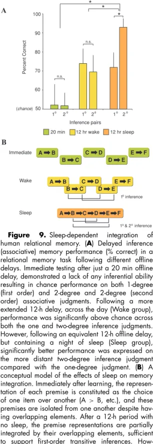
Ellenbogen et al. (2007) have since tested this sleep-dependent hypothesis of integration by examining human relational memory—the ability to generalize previously acquired associ-ations to novel situassoci-ations. Participants initially learned five “premise pairs” (A> B, B > C, C > D, D > E, E > F). Unknown to sub-jects, the pairs contained an embedded hier-archy (A >B>C >D >E >F). Following an offline delay of 20 min, 12 h across the day, or 12 h containing a night of sleep, knowledge of this hierarchy was tested by examining re-lational judgments for novel “inference” pairs, either separated by one degree of associative distance (B>D, C>E pairs) or by two degrees
of associative distance (B>E pair). Despite all groups achieving near identical premise-pair retention after the offline delay (i.e., the build-ing block pair of the hierarchy), a strikbuild-ing disso-ciation was evident in the ability to make rela-tional inference judgments. Subjects that were tested soon after learning in the 20 min group showed no evidence of inferential ability, per-forming at chance levels (Fig. 9A). In contrast, the two 12 h groups displayed highly significant relational memory development. Most remark-able, however, was the observation that if the 12-h period contained a night of sleep, a near 25% advantage in relational memory over the 12 h across the day group was seen for the most distantly connected inferential judgment (the B>E pair; Fig. 9A). Together, these find-ings demonstrate that human memory integra-tion takes time to develop, requiring slow, of-fline associative processes. Furthermore, sleep appears to preferentially facilitate this integra-tion by enhancing hierarchical memory bind-ing, biasing the development of the most dis-tant/weak associative links amongst related yet separate memory items (Fig. 9B). It is also inter-esting to note a further advantage of this sleep-dependent assimilation process. When it stores individual premise-pairs (top row, Fig. 9B) the size/number of items (“bits”) of information the brain has to code is ten (A-B, B-C, C-D, D-E, E-F). However, when it the items are formed into a hierarchy, the informational load is compressed, reduced by nearly 50% to just six bits (A-B-C-D-E-F). Therefore, a supple-mentary benefit of sleep-dependent memory association may be the improved efficiency of memory storage, in addition to a more gener-alized representation.
Thus, the overnight strengthening and con-solidation of individual item memories (re-viewed above), may not be the ultimate ob-jective of sleep-dependent memory processing, especially when one considers that declarative (nonemotional) memories decay over the long-term (Wixted & Carpenter 2007). It is then interesting to speculate whether sleep serves to facilitate two complementary objectives for
A B C D E F A B C D E F A B A B B C B C C D C D D E D E E F E F B C B C D ED E A B A B C DC D E FE F Immediate Wake Sleep
20 min 12 hr wake 12 hr sleep
Percent Correct 60 70 80 90 100 (chance) 50 Inference pairs * n.s. * * n.s. A B 1 inference 1 & 2 inference o o o 1 2 1 2 1 2 o o o o o o
Figure 9. Sleep-dependent integration of
human relational memory. (A) Delayed inference
(associative) memory performance (% correct) in a relational memory task following different offline delays. Immediate testing after just a 20 min offline delay, demonstrated a lack of any inferential ability resulting in chance performance on both 1-degree (first order) and 2-degree and 2-degree (second order) associative judgments. Following a more extended 12-h delay, across the day (Wake group), performance was significantly above chance across both the one and two-degree inference judgments. However, following an equivalent 12-h offline delay, but containing a night of sleep (Sleep group), significantly better performance was expressed on the more distant two-degree inference judgment
compared with the one-degree judgment. (B) A
conceptual model of the effects of sleep on memory integration. Immediately after learning, the represen-tation of each premise is constituted as the choice
of one item over another (A > B, etc.), and these
premises are isolated from one another despite hav-ing overlapphav-ing elements. After a 12-h period with no sleep, the premise representations are partially integrated by their overlapping elements, sufficient to support first-order transitive inferences. How-ever, following a 12-h offline period with sleep, the
declarative memory, which span different time courses. The first may be an initial process of consolidating individual item (episodic) mem-ories that are novel, which may occur in the relative short term. Over a longer time course, however, and utilizing these recently consol-idated item memories prior to their fading, sleep may begin the process of the brain’s ex-traction (of meaning) and absex-traction (building associational links with existing information), thereby creating more adaptive semantic net-works (McClelland et al. 1995; Squire 2007; Tse et al. 2007). Ultimately, individual item memories would no longer be necessary for the goal that sleep is trying to achieve, and only the conceptual meaning of such experiences would remain. Whether the subsequent loss of item memories is passive or whether sleep plays an active role in this process (Crick & Mitchi-son 1983) remains to be examined, but this is a testable hypothesis, i.e., forgetting (individ-ual items) is the price we pay for remembering (general rules).
Creativity
One potential advantage of testing asso-ciative connections and building cross-linked systems of knowledge is creativity—the ability to take existing pieces of information and com-bine them in novel ways that lead to greater understanding and offer new, advantageous be-havioral repertoires. The link between creativ-ity and sleep, especially dreaming, has long been a topic of intense speculation. Even sci-entific examples of creativity occurring dur-ing sleep are common: from the dreams of both August Kekul´e that led to the concep-tion of a simple structure for benzene (Hubert 1985) to those of Dmitry Mendeleyev that ini-tiated the creation of the periodic table of ←−−−−−−−−−−−−−−−−−−−−−−−−−−−−−−−
premise representations are fully interleaved, support-ing both first- and second-order transitive inferences.
∗P <0.05; error bars indicate SEM. Modified from
elements (Strathern 2000) to the late night dreaming of Otto Loewi that inspired the experimental demonstration of neurochemi-cal transmission (Mazzarello 2000).Quantita-tive data have further demonstrated that solu-tion performance on tests of cognitive flexibility using anagram word puzzles is more than 30% better following awakenings from REM sleep compared with NREM awakenings (Walker et al. 2002). Similarly, a study of semantic prim-ing has demonstrated that, in contrast to the sit-uation in waking, performance following REM sleep awakenings shows a greater priming effect by weakly related words than by strong primes, while strong priming exceeds weak priming in NREM sleep (Stickgold et al. 1999), again in-dicating the highly associative properties of the REM sleep brain. Even the study of mental ac-tivity (dreams) from REM sleep indicates that there is not a concrete episodic replay of day-time experiences, but instead, a much more as-sociative process of semantic integration during sleep (Fosse et al. 2003).
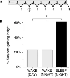
Yet the most striking experimental evidence of sleep-inspired insight is arguably that re-ported by Wagner and colleagues (Wagner et al. 2004). Using a mathematical “number reduc-tion task” (Thurstone & Thurstone 1941), a process of sleep-dependent creative insight was elegantly demonstrated. Subjects analyzed and worked through a series of 8-digit string prob-lems, using specific addition rules (Fig. 10A). Following initial training, after various peri-ods of wake or sleep, subjects returned for an additional series of trials. When retested after a night of sleep, subjects solved the task, us-ing this “standard” procedure, 16.5% faster. In contrast, subjects who did not sleep prior to retesting averaged less than a 6% improve-ment. However, hidden in the construction of the task was a much simpler way to solve the problem. On every trial, the last three response digits (e.g., “4–1–9” in Fig. 10A) were the mirror image of the preceding three (i.e., “9–1–4”). As a result, the second response digit always pro-vided the answer to the problem, and using such “insight,” subjects could stop after producing
WAKE (NIGHT) WAKE (DAY) 0% 20% 40% 60%
% Subjects gaining insight
9 1 1 4 4 9 4 9 4 1 4 4 1 9 1 SLEEP (NIGHT) A B
*
Figure 10. Sleep-dependent production of
cre-ative insight. (A) Example of the Number Reduction
Task. Subjects analyze digits in a 8-digit string of 1s, 4s, and 9s, from left to right, using two rules: (1) If two digits are the same, respond with that digit. Thus, starting from the left, the first two digits are both “1,” and hence the response (listed below and to the right of the second digit) is also “1.” (2) If two digits are different, respond with the remaining digit. Thus, hav-ing produced the response “1,” this response and the next digits are analyzed. Since they differ (“1” and “4”), the next response is “the remaining digit, or “9.” This response and the next digit, “4” also differ (“9” and “4”) and so the next response is the remain-ing digit, “1.” The analysis is continued to the end, and the final response, “9” in this case, is the solu-tion to the problem. This final response is then entered as the answer the problem. However, on every trial, the last three response digits (e.g., “4–1–9” in figure above) are the mirror image of the preceding three (i.e., “9–1–4”). As a result, the second response digit (circled “9”) always provides the answer to the prob-lem, resulting in a “shortcut” to solving the probprob-lem,
if the subject gains this hidden insight. (B)
Percent-age of subjects that gained insight into this hidden rule following an offline delay while awake across the day, awake across the night of following sleep
across the night.∗P<0.05. Modified from Wagner
et al. 2004.
the second response digit. Most dramatically, nearly 60% of the subjects who slept for a night between training and retesting discovered this shortcut the following morning (Fig. 10B). In
contrast, no more than 25% of subjects in any of four different control groups that did not sleep had this insight. Sleeping after exposure to the problem therefore more than doubled the likelihood of solving it (although it is inter-esting to note that this insight was not present immediately following sleep, but took over 100 trials on average to emerge the next day).
In summary, substantial evidence now sug-gests that sleep serves a metalevel role in memory processing that moves far beyond the consolidation and strengthening of individual memories and, instead, aims to intelligently assimilate and generalize these details offline. In doing so, sleep may offer the ability to test and build common informational schemas of knowledge, providing for increasingly accurate statistic predictions about the world and allow-ing for the discovery of novel, even creative, next-day solution insights.
Emotional Regulation
Despite substantial research focusing on the interaction between sleep and cognition, espe-cially memory, the impact of sleep and sleep loss on affective and emotional regulation has received more limited research attention. This absence of investigation is perhaps surprising considering that nearly all psychiatric and neu-rological mood disorders express co-occurring abnormalities of sleep, suggesting an intimate relationship between sleep and emotion. Nev-ertheless, a number of recent studies evaluating subjective as well as objective measures of mood and affect, combined with insights from clinical domains, offer an emerging understanding for the critical role of sleep in regulating emotional brain function.
Affective Reactivity
Together with impairments of attention and alertness, sleep deprivation is commonly asso-ciated with increased subjective reports of ir-ritability and affective volatility (Horne 1985).
Using a sleep restriction paradigm (5 h/night), Dinges et al. (1997) reported a progressive in-crease in emotional disturbance across a one-week period on the basis of questionnaire mood scales. In addition, subjective descrip-tions in participants’ daily journals also indi-cated increasing complaints of emotional dif-ficulties. Zohar et al. (2005) investigated the effects of sleep disruption on emotional re-activity to daytime work events in medical residents. Sleep loss was shown to amplify nega-tive emotional consequences of disrupnega-tive day-time events while blunting the positive benefit associated with rewarding or goal-enhancing activities.
Although these findings help to characterize the behavioral irregularities imposed by sleep loss, evidence for the role of sleep in regulat-ing our emotional brain is surprisregulat-ingly scarce. To date, only one such study has investigated whether a lack of sleep inappropriately mod-ulates human emotional brain reactivity (Yoo et al. 2007a). Healthy young participants were allowed to sleep normally prior to an fMRI scanning session or were sleep deprived for one night (accumulating approximately 35 h of total sleep loss). During scanning, subjects performed an affective stimulus viewing task in-volving the presentation of picture slides rang-ing in a gradient from emotionally neutral to increasingly negative and aversive.
While both groups expressed significant amygdala activation in response to increas-ingly negative picture stimuli, those in the sleep-deprivation condition exhibited a re-markable +60% greater magnitude of amyg-dala reactivity, relative to the control group (Figs. 11A & B). In addition to this increased intensity of activation, there was also a three-fold increase in the extent of amygdala vol-ume recruited in response to the aversive stim-uli in the sleep-deprivation group (Fig. 11B). Perhaps most interestingly, relative to the sleep control group, there was a significant loss of functional connectivity identified between the amygdala and the medial prefrontal cortex (mPFC) in those who were sleep deprived—a
Figure 11. The impact of sleep deprivation on emotional brain reactivity and functional connectivity.
(A) Amygdala response to increasingly negative emotional stimuli in the sleep deprivation and sleep control
groups, and (B) corresponding differences in intensity and volumetric extent of amygdala activation between
the two groups (average±SEM. of left and right amygdala). (C) Depiction of associated changes in functional
connectivity between the medial prefrontal cortex (mPFC) and the amygdala. With sleep, the prefrontal lobe
was strongly connected to the amygdala, regulating and exerting and inhibitory top-down control, yet (D)
Without sleep, however, mPFC connection was decreased, potentially negating top-down control and resulting
in an overactive amygdala.∗P<0.01; error bars indicate SEM. Modified from Yoo et al. 2007b.
region known to have strong inhibitory pro-jections and hence modulatory impact on the amygdala (Sotres-Bayon et al. 2004). In con-trast, significantly greater connectivity in the deprivation group was observed between the amygdala and the autonomic-activating cen-ters of the locus coeruleus.
Thus, without sleep, an amplified hyper-limbic reaction by the human amygdala was observed in response to negative emotional stimuli. Furthermore, this altered magnitude of limbic activity is associated with a loss of functional connectivity with the mPFC in the sleep deprivation condition implying a failure of top-down inhibition by the prefrontal lobe
(Figs. 11C & D). It would therefore appear that a night of sleep may “reset” the correct affec-tive brain reactivity to next-day emotional chal-lenges by maintaining functional integrity of this mPFC−amygdala circuit and thus govern appropriate behavioral repertoires (e.g., opti-mal social judgments and rational decisions). Intriguingly, a similar pattern of anatomical dysfunction has been implicated in a number of psychiatric mood disorders that express co-occurring sleep abnormalities (Davidson 2002; Davidson et al. 2002; New et al. 2007), directly raising the issues of whether such factors (sleep loss and clinical mood disorders) are causally related.
Emotional Information Processing Sleep’s role in declarative memory consoli-dation, rather than being absolute, may depend on more intricate aspects of the information be-ing learned, such as novelty, meanbe-ing to extract, and also the affective salience of the material. Independent of the field of sleep and memory, there is a wealth of evidence demonstrating that memory processing is modulated by emotion (Cahill 2000; McGaugh 2004; Phelps 2004). Experiences which evoke emotions not only en-code more strongly, but appear to persist and even improve over time as the delay between learning and testing increases (from hours to days) (Kleinsmith & Kaplan 1963; Walker & Tarte 1963; Levonian 1972; LaBar & Phelps 1998; Sharot & Phelps 2004).
Although these findings indicate a strong in-fluence of emotion on slow, time-dependent consolidation processes, based on the coinci-dent neurophysiology that REM sleep pro-vides and the neurobiological requirements of emotional memory processing (Cahill 2000; McGaugh 2004), work has now begun to test a selective REM-dependent hypothesis of affec-tive human memory consolidation. For exam-ple, Hu et al. (2006) compared the consolida-tion of emoconsolida-tionally arousing and nonarousing picture-stimuli following a 12-h period across a day or following a night of sleep. A specific emotional memory benefit was observed only following sleep and not cross an equivalent time awake. Wagner and colleagues (Wagner et al. 2001) have also shown that sleep selectively fa-vors the retention of previously learned emo-tional texts relative to neutral texts, and that this affective memory benefit is only present following late-night sleep (a time period rich in stage-2 NREM and REM sleep). Furthermore, this emotional memory enhancement has been shown to persist for several years (Wagner et al. 2006).
Using a nap paradigm, researchers have most recently demonstrated that sleep, and specifically REM neurophysiology, may under-lie this consolidation benefit (Nishida et al. in
press). Subjects performed two study sessions in which they learned emotionally negative and neutral picture stimuli: one 4 h prior to a recognition memory test, and one 15 min prior to it. In one group, participants slept (90 min nap) after the first study session, while in the other group, participants remained awake. Thus, items from the study sessions tested after 4 h transitioned through different brain-states in each group prior to testing—sleep in the Nap group and no sleep in the No-Nap group— yet experienced identical brain-state conditions when tested after 15 min.
No change in memory for emotional (or neutral stimuli), occurred across the offline delay in the no-nap group. However, a sig-nificant and selective offline enhancement of emotional memory was observed in the nap group (Fig. 12A), the extent of which was corre-lated with the amount of REM sleep (Fig. 12B), and the speed of entry into REM (latency; not shown in figure). Furthermore, spectral analysis of the EEG demonstrated that the magnitude of right-dominant prefrontal theta power dur-ing REM (activity in the frequency range of 4.0–7.0 Hz) exhibited a significant and posi-tive relationship with the amount of emotional memory improvement (Figs. 12C & D).
These findings go beyond demonstrating that affective memories are preferentially en-hanced across periods of sleep, and indicate that the extent of emotional memory im-provement is associated with specific REM sleep characteristics—both quantity and qual-ity. Corroborating these correlations, it has pre-viously been hypothesized that REM sleep rep-resents a brain state particularly amenable to emotional memory consolidation, based on its unique biology (Pare et al. 2002; Hu et al. 2006). Neurochemically, levels of limbic and forebrain ACh are markedly elevated during REM (Vazquez & Baghdoyan 2001), report-edly quadruple those seen during NREM and double those measured in quite waking (Mar-rosu et al. 1995). Considering the known im-portance of ACh in the long-term consolidation of emotional learning (McGaugh 2004), this
Figure 12. REM sleep enhancement of negative emotional memory consolidation. (A) Offline benefit (change in memory recall for 4 h versus 15 min old memories) across the day (wake, grey bar) or following a
90 min nap (sleep, filled bar) (B) Correlation between the amount of offline emotional memory improvement
in the nap group (i.e. the offline benefit expressed in filled bar of figure A), and the amount of REM sleep
obtained within the nap, (C) Correlation strength (Pearson’sr-value) between offline benefit for emotional
memory in the sleep group (the benefit expressed in filled bar of figure A) and the relative right versus left prefrontal spectral-band power ([F4 – F3]) within the delta, alpha, theta, and beta spectral bands, expressed in average 0.5 Hz bins increments. Correlation strength is represented by the color range, demonstrating
significant correlations within the theta frequency band (hot colors), and (D) exhibiting a maximum significance
at the 5.75 Hz bin.∗P<0.05; error bars indicate SEM. Modified from Nishida et al. unpublished.
pro-cholinergic REM state may result in a se-lective facilitation of affective memories, similar to that reported using experimental manipula-tions of ACh (Power 2004). Neurophysiologi-cally, theta oscillations have been proposed as a
carrier frequency allowing disparate brain re-gions that initially encode information to se-lectively interact offline, in a coupled relation-ship. By doing so, REM theta may afford the ability to promote the strengthening of specific