101
THE RESULTS OF VOLAR LOCKING PLATE FIXATION
FOR THE FRAGILITY FRACTURE POPULATION WITH
DISTAL RADIUS FRACTURE IN JAPANESE WOMEN
SHUICHI KATO1, MASAHIRO TATEBE2, MICHIRO YAMAMOTO1, KATSUYUKI IWATSUKI1, TAKANOBU NISHIZUKA1, and HITOSHI HIRATA1 1Department of Hand Surgery, Nagoya University Graduate School of Medicine, Nagoya, Japan
2Anjo Kosei Hospital, Anjo, Japan
ABSTRACT
The purpose of this study was to determine whether volar locking plate fixation for distal radius fracture benefits the fragility fracture population as much as it benefits the non-fragility fracture population. This matched case-control study was conducted based on a multi-center clinical prospective cohort. A comparison of treatment outcomes after volar locking plate fixation was made between females 55 years of age and older (fragility fracture population) and males less than 75 years of age (non-fragility fracture population) by evaluating clinical, radiological, and subjective outcomes using Hand20, a validated patient-rated disability instrument. A total of 170 patients were enrolled in this study. The two cohorts were matched in terms of AO fracture type. The fragility fracture population group and the non-fragility fracture population group each consisted of 50 patients. All objective measurements including wrist range of motion and radiological evaluations, but excluding grip strength, were not significantly different between the two groups. However, the Hand20 at 18 months after surgery was worse in the fragility fracture population group than in the non-fragility fracture population group. Carpal tunnel syndrome was the most frequently encountered complication in the fragility fracture population group, with one case (2%) in the non-fragility fracture population group and six cases (12%) in the fragility fracture population group, but the difference was not significant. In conclusion, there was a significant deficit in the improvement in disability despite favorable radiological and functional outcomes in fragility fracture population patients. Therefore, the fragility fracture population, especially middle-aged or older women, needs to be informed about prolonged disability and the higher risk of upper extremity disorders prior to surgery.
Key Words: distal radius fracture, elderly, Japanese
INTRODUCTION
Distal radius fracture (DRF) is one of the most common fractures in the elderly with osteoporosis. Previous studies suggested that almost 10% of women aged 65 years and older are expected to sustain a DRF in their lifetime.1) The current upsurge of interest in using more aggressive fracture fixation in the elderly in the hope of quicker and better functional recovery appears rational. Chung investigated trends in the treatment of DRF for the elderly in the Received: December 27, 2013; accepted: January 16, 2014
Corresponding author: Shuichi KATO, MD
Department of Hand Surgery, Nagoya University Graduate School of Medicine, 65 Tsurumai-cho, Showa-ku, Nagoya 466-8550, Japan
United States between 1996 and 2005.2, 3) Although closed reduction/cast fixation was still the predominant method of treatment even as late as 2005, there has actually been a significant increase in the use of the volar locking plate, associated with a concurrent decrease in the rate of conservative treatment.
Despite a plethora of clinical and biomechanical evidence proving that the volar locking plate system can provide stable internal fixation to allow anatomical healing even in elderly osteoporotic patients,4, 5) the optimal treatment for DRF in this age group remains controversial. Some authors insist that radiological parameters of adequate reduction might not translate into good functional or clinical outcomes in elderly patients who have low functional demands.6–11)
Although DRF can occur at any age, there exists a remarkable sex difference in age distribu-tion in adulthood. After the age of 45 years, the incidence rate in women increases rapidly, whereas in men, the incidence remains fairly constant up to 75 years.12) Low bone mineral density (BMD) is one fracture risk, and BMD is known to decrease markedly in middle age or later in women. In fact, the Dutch osteoporosis guideline strongly recommends implementation of Vitamin D supplementation regardless of the BMD value for women over 50 years of age with risks of osteoporosis, such as a history of fracture, and use of osteoporosis drugs including bisphosphonates for women over 50 years with low BMD.13) In addition, the decrease in BMD is compatible with the age group in which DRF increases in women.14–16) Consistent with these reports, in the present cohort, the number of surgical treatments for DRFs in women increased rapidly after 55 years of age, while it was consistently low in men. Since the trend was sym-metric with a graph of the BMD, it was decided to define the fragility fracture population as women aged 55 years and older.
The purpose of this study was to evaluate whether anatomical repair of the DRF with the volar locking plate benefits Japanese fragility fracture population patients (women aged 55 years and older) in terms of complications, as well as function and disability.i
PATIENTS AND METHODS
A multi-center, prospective, clinical follow-up study was conducted to clarify the clinical outcome after internal fixation for DRF using a validated, patient-rated outcome instrument. The study was approved by the institutional review boards of all participating centers. During the 18-month prospective study period (April 2006 to March 2007), 241 patients with a DRF were treated with a volar locking plate, and all patients gave their informed consent prior to their inclusion in the study. Enrolled patients were included in the present series if they were at least 20 years old. They were then assigned to one of following two cohorts: male patients <75 years old (cohort A) and female patients ≥55 years old (cohort B). The exclusion criteria were as follows: 1) <20 years old (8 cases); 2) open fractures (0 cases); 3) bilateral fractures (3 cases); 4) concomitant upper extremity injuries other than ulnar styloid fractures (3 cases); 5) systemic, multiple organ, or head injuries (4 cases); 6) dropout within 3 months (30 cases); and 7) missing data (3 cases). Overall, the original group consisted of 199 patients, of whom 170 were adult males aged 75 years and younger or adult females aged 55 years and older. There was a significant difference in fracture type between the two groups. The two cohorts were matched in terms of AO fracture type for the matched case-control study (Table 1). No significant differences in demographic variables, except patients’ age and the bone graft rate, were found between the two groups; the bone graft rate was significantly higher in group B than in group A.
Three different volar locking plates, the Matrix Smart Lock system (Stryker Osteosynthesis, Freiburg, Germany), the Acu-Lock (Acumed, Hillsboro, OR, USA), and the Locking DRP system (SYNTHES, Paoli, PA, USA) were used in this study at the surgeons’ discretion. Plate removal after bone union was also performed at the discretion of the surgeon.
Outcome Evaluations
All fractures were classified with preoperative X-rays according to the AO classification system. Enrolled patients were followed up clinically and radiographically. At each scheduled
Table 1 Description of cohorts (matched groups)
Parameter Group A
(20–74 yrs male)
Group B
(≥55 yrs female) P Value
Number of patients 50 50
Male : emale 50 : 0 0 : 50 .000†
Age, years 44.6 (range 20–73) 68.8 (range 55–84) .000*
Number of weeks at final examination 26 ( ±10) 24 ( ± 9) .181‡
Injured side (right : left) 21 : 29 22 : 28 .840†
AO classification 1.000† A2 4 4 A3 4 4 C1 7 7 C2 19 19 C3 16 16 Ulnar fracture 27 25 .849† Plate type .910† Acu-Loc 7 (14%) 7 (14%) Matrix 18 (36%) 20 (40%) LCP DRP 25 (50%) 23 (46%) Cast type .929† Long arm 21 21 Short arm 28 20 Unknown 1 2
Casting period, weeks (±SD) 2.6 ( ±1.0) 2.7 ( ±1.2) .672‡
Position of the plate, mm (±SD) 3.3 ( ±2.1) 2.6 ( ±1.5) .066‡
Bone graft 6 (12%) 17 (34%) .009†
Plate removal 43% 31% .349†
Patients with complication, n 9 (18%) 15 (30%) .160†
† Based on chi-square test from comparing demographics between Group A and B.
* Based on Student’s t-test from comparing outcomes at final examination between Group A and B. ‡ Based on Mann-Whitney U test from comparing demographics between Group A and B.
follow-up visit, the patient was evaluated by a hand surgeon for the presence of postoperative complications. Independently, certified hand therapists performed commonly used hand function tests, including grip strength and range of motion. Grip strength was assessed by comparing with that of the uninjured hand. Standard plain radiographs (posteroanterior and lateral) were used to measure volar tilt (VT), radial inclination (RI), ulnar variance (UV), articular congruity, and plate position. The Hand20 questionnaire was mailed at 6, 12, and 18 months after surgery as a patient-rated outcome assessment. The Hand20, a validated outcome measure, consists of 20 self-reported questions designed to measure upper extremity disability and symptoms. The Hand20 score ranges from 0 to 100, with higher scores indicating greater levels of disability.17–20) Intensity of pain was indicated on a 0 to 10-point numeric rating scale (NRS) in this questionnaire.ii
Statistical Analysis
Differences in clinical characteristics were assessed using the Chi-square test for categorical variables. Objective functional outcomes, the Hand20, and radiological parameters were analyzed using Student’s t-test or the Mann-Whitney U test, as appropriate. Student’s t-test was used if the samples were assumed to have a normal distribution, and the Mann-Whitney U test was used otherwise. Changes in continuous outcomes between follow-up investigations were evalu-ated with ANOVA if the data distribution was normal or with the Friedman test if the data were not normally distributed, followed by a series of Wilcoxon signed-rank tests for discrete variables. Missing values (total score) were circumvented using a classical method known as the last-observation-carried-forward method. In order to determine the most important confounding factors from among age, duration of cast immobilization, plate selection, implant removal, bone grafting, intra-articular fracture, ulnar fracture, articular step-off (>1 mm), volar tilt angle, radial inclination angle, ulnar variance, ROM, grip strength, surgical approach (additional dorsal ap-proach), plate position, injury compensation status, and complications associated with the response variables of disability score and pain while accounting for any confounding (inter-relationship) among explanatory variables, bivariate analysis looking at differences between two variables was conducted first. Data were compared by the Chi-square test, Fisher’s exact test, Student’s t-test, the Mann-Whitney U test, Pearson correlation, or Spearman correlation as appropriate. Any explanatory variables with a significant association (p<0.05) or a tendency toward association (p<0.10) in the bivariate analysis were then included in a multiple linear regression analysis to determine the explanatory variables that accounted best for the variation in response variables, independent of any confounding among the variables. All variables are reported as the mean values ± SD. All tests of significance were two-tailed. The level of significance was set at P < 0.05. All data analyses were performed using SPSS ver.21 (SPSS Inc, Chicago, IL).
RESULTS Objective Measures
The mean grip strength at the final examination was 82.5% ± 30.0% in group A and 65.5% ± 20.0% in group B, which was significantly different (Table 2). The mean flexion-extension and pronation-supination arc ranges of motion at the final examination were 79.1% ± 16.6% and 90.6% ± 12.1%, respectively, in group A, and 74.5% ± 16.1% and 92.3% ± 8.5%, respectively, in group B; they were not significantly different.
Similarly, radiographic assessment, i.e., VT, RI, and UV, showed no significant differences between the two groups.
Patient-Rated Measures
The Hand20 scores were consistently lower in group A than in group B at all follow-up time points, and they were significantly different at 18 months (P = 0.014) (Fig. 1). Furthermore, it should be noted that a significant difference was found in the improvement pattern of patient-rated disability between the two groups. The Freidman test showed a significant improvement between 6 and 18 months in group A (P = 0.000), while no appreciable improvement was detected in group B (P = 0.066). Post hoc analysis in group A showed a significant reduction in the Hand20 score between 6 and 12 months (P = 0.007).
Meanwhile, no significant differences were found by direct comparison between the two groups at any of the follow-up time points in the NRS for pain (P=0.173) (Fig. 1). Similarly, there was no significant difference in the improvement pattern (P=0.149).
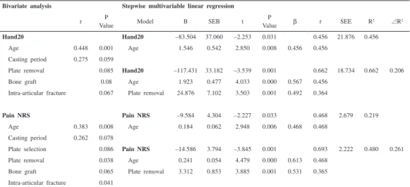
On multiple linear regression analysis, age and plate removal were identified as variables affecting the final Hand20 score and the NRS score for pain in group B, while no variables were found to affect the disability score and pain in group A (Table 3). These results show that increasing age was associated with the Hand20 and pain scores, with contributions of ap-proximately 46% and 22%, respectively.iii
iii Abbreviations: carpal tunnel syndrome (CTS)
Table 2 Postoperative objective outcomes
Measurement Group A Group B P Value
Grip strength § 82.5 ± 30.0 65.5 ± 20.0 .002‡
Flexion and extension § 79.1 ± 16.6 74.5 ± 16.1 .182*
Pronation and supination § 90.6 ± 12.1 92.3 ± 8.5 .064‡
VT (degrees) postoperative 4.4 ± 5.1 5.8 ± 6.0 .193* final 4.3 ± 5.7 4.1 ± 6.9 .729‡ VT –0.1 ± 3.1 –1.8 ± 4.0 .009‡ RI (degrees) postoperative 21.5 ± 5.4 21.5 ± 5.3 .970* final 22.5 ± 5.5 21.8 ± 5.7 .594‡ RI 1.0 ± 2.8 0.3 ± 2.8 .135‡ UV (mm) postoperative –0.1 ± 1.9 0.1 ± 1.9 .407‡ final 0.7 ± 2.0 1.0 ± 2.4 .456‡ UV 0.8 ± 1.2 0.9 ± 1.9 .557‡
§ Data given as the mean percentage of the value for the uninjured side ± standard deviation. ‡ Based on Mann-Whitney U test from comparing outcomes between Group A and B. * Based on Student’s t-test from comparing outcomes between Group A and B. expresses the difference between final and postoperative values.
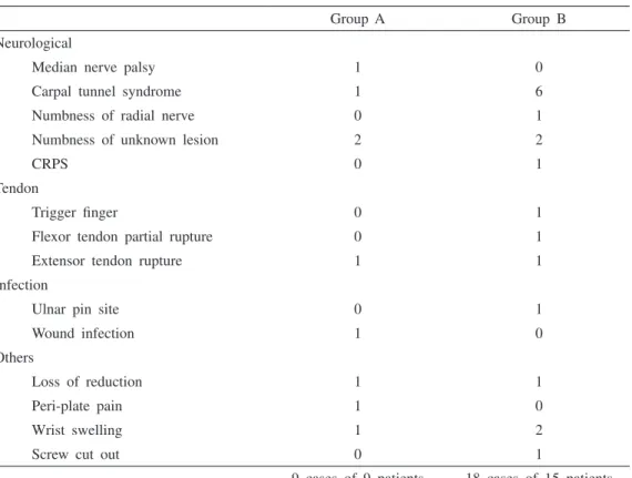
Complications
A total of 9 complications was diagnosed in group A (18%), with 18 in group B (30%), which was not significantly different (P = 0.160) (Table 4). Carpal tunnel syndrome (CTS) was the most frequently encountered complication in group B (one case in group A and six cases in group B), but the difference was not significant (P=0.056).
Fig. 1
‡ Significant difference between Group A and B based on the Mann-Whitney U test (P<0.05).
* Significant differences between follow-up investigations based on the Wilcoxon signed-rank test (P<0.017) following the Friedman test (P<0.05).
0
20
40
60
80
100
6mo
12mo
18mo
Hand20
‡
*
*
0
2
4
6
8
10
6mo
12mo
18mo
Numeric Ra
ting Scal
e for Pain
Group A
Group B
Table 3 Multiple regression analysis assessing the effect of confounding factors on disability score or pain score at 18 months in Group B
Bivariate analysis Stepwise multivariable linear regression
r P
Value Model B SEB t
P
Value b r SEE R2 R2
Hand20 Hand20 –83.504 37.060 –2.253 0.031 0.456 21.876 0.456
Age 0.448 0.001 Age 1.546 0.542 2.850 0.008 0.456 0.456
Casting period 0.275 0.059
Plate removal 0.085 Hand20 –117.431 33.182 –3.539 0.001 0.662 18.734 0.662 0.206
Bone graft 0.08 Age 1.923 0.477 4.033 0.000 0.567 0.456
Intra-articular fracture 0.067 Plate removal 24.876 7.102 3.503 0.001 0.492 0.364
Pain NRS Pain NRS –9.584 4.304 –2.227 0.033 0.468 2.679 0.219
Age 0.383 0.008 Age 0.184 0.062 2.948 0.006 0.468 0.468
Casting period 0.262 0.078
Plate selection 0.086 Pain NRS –14.586 3.794 –3.845 0.001 0.693 2.222 0.480 0.261
Plate removal 0.038 Age 0.241 0.054 4.479 0.000 0.613 0.468
Bone graft 0.065 Plate removal 3.312 0.853 3.885 0.001 0.531 0.365
Intra-articular fracture 0.041
All variables with significant (p<0.05) or tendency toward association (p<0.10) in the bivariate analysis were entered in the stepwise multivariable linear regression analysis.
DISCUSSION
DRF is one of the most common fractures and accounts for 1/6 of all fractures.21) The fracture can occur at any age; however, the epidemiology of DRF changes drastically with age. In older adults, especially women, the fracture more often results from low-energy or moderate trauma, such as falling from a standing height. There is a general notion that the number of osteoporosis-related fractures has been increasing drastically with the progressive aging of the world population.22) In the case of DRF in Japan, however, the age-adjusted incidence rate has only increased in women.23) These data indicate that the recent abrupt upsurge in the number of surgeries for DRF may be due to an increase in the number of osteoporotic Japanese women with a high risk of sustaining fractures. In fact, the age distribution of surgically treated patients with DRF shows a clear difference between sexes.12) Furthermore, the fracture type was different between the fragility population and the non-fragility population. Taking this background into consideration, a subset of controls was selected from the cohort that is comparable to patients in group B in terms of patient demographics and fracture type to conduct a matched case-control study.
The fact that DRF in the elderly is generally caused by relatively low-energy trauma does not necessarily mean that these fractures are less severe than those in young adults caused by high-energy trauma. According to Clayton, a definite correlation between BMD and the severity of the DRFs exists in the osteoporotic population.24) In addition, Dias demonstrated that deterioration of the bony deformity in the healing phase is significantly greater in patients with osteoporosis
Table 4 Complications
Group A Group B
Neurological
Median nerve palsy 1 0
Carpal tunnel syndrome 1 6
Numbness of radial nerve 0 1
Numbness of unknown lesion 2 2
CRPS 0 1
Tendon
Trigger finger 0 1
Flexor tendon partial rupture 0 1
Extensor tendon rupture 1 1
Infection
Ulnar pin site 0 1
Wound infection 1 0
Others
Loss of reduction 1 1
Peri-plate pain 1 0
Wrist swelling 1 2
Screw cut out 0 1
than in those with good bone quality.25) Therefore, it seems rational to consider the use of open reduction internal fixation (ORIF) for this particular population. Orbay and Fernandez, who popularized the use of the volar locking plate for DRFs, reported that the volar locking plate can minimize morbidity in the elderly population by successfully handling osteopenic bone, allowing early return of function, providing good final results, and resulting in a low complication rate.4) iv
ROM of the wrist and the forearm and grip strength were evaluated in this study as indicators of functional outcome. Still, no significant differences were seen in ROM at the final evaluation. Despite these favorable radiological and functional outcomes, the present data demonstrated a significant difference in serial changes of the disability scores between the two groups. Arora conducted a randomized trial to compare treatment outcomes of volar locking plate fixation for unstable DRFs with that of non-operative treatment in the elderly. They found that, despite significantly better radiological outcomes and grip strength in the volar locking plate group, there were no significant differences between the two treatment groups in terms of active ROM, patient-rated wrist evaluation (PRWE) scores, and disability of the arm, shoulder, and hand (DASH) scores at both 6 and 12 months after surgery.26) Recently, Diaz-Garcia conducted a systematic review to compare outcomes of the following five common techniques for DRF: the volar locking plate system; non-bridging external fixation; bridging external fixation; percutaneous Kirschner wire fixation; and cast immobilization. Significant differences were detected for wrist arc of motion, grip strength, and DASH score, but the observed differences could not be regarded as “clinically significant differences”.27) These data, as well as the data from the present study, seem to indicate that, in contrast to young adults for whom there is a high correlation between the anatomical result and the functional outcome,28–30) achieving anatomical reduction does not necessarily translate into better functional and health status in the elderly with a high risk of osteoporosis-related wrist fractures. In the present study, there was a significant deficit in grip strength and disability improvement in the fragility fracture group. Similar results were reported by Chung et al.2, 3) Moreover, multiple linear regression analysis showed that increasing age was associate with the Hand20 and pain scores in the fragility fracture group in the present study. Considering the fact that both the Hand20 and the DASH used by Chung are well validated, patient-rated, disability assessment tools, we strongly believe that these results have important implications for further advancement of surgical treatment of fragility fractures of the forearm.
Multiple linear regression analysis showed that aging was identified as a variable affecting the Hand20 score in the fragility fracture group in the present study. In the previous study, the Hand20 scores of the nonclinical population tended to be higher in women and older people.20) It is well known that basic motor control generally deteriorates with aging.31) In addition, previous studies confirmed that the prevalence of musculoskeletal disorders increases with age.32) Taking these issues into consideration, the fragility fracture population, especially middle-aged or older women, needs to be informed about prolonged disability with upper extremity disorders when obtaining informed consent.
There were 9 complications in group A and 18 complications in group B, but this difference was not significant. Diaz-Garcia specifically focused on complications and found no significant differences in the rates of all types of complications among the different treatments.27) Analysis of their compiled data showed overall rates of minor complications, major complications not requiring surgery, and major complications requiring surgery of 1%, 6%, and 11%, respectively, for volar locking plate fixation of DRF in the elderly. Though a different classification system was used in the present study, if the complications in the present study were re-classified using their system, the present data appear consistent with their study. However, in the present study,
iv Abbreviations: open reduction internal fixation (ORIF), patient-rated wrist evaluation (PRWE), disability of
12% of the fragility fracture population developed CTS during the follow-up period, while the rate of CTS in Diaz-Garcia’s compiled data was less than 3%. The cause of the difference is not clear, but a recent meta-analysis identified the volar approach as a contributing factor to increased incidence of carpal tunnel syndrome. The small hands of Asian elderly women may help explain the difference.33) Nakamichi found an inverse relationship between hand size and risk of carpal tunnel syndrome.34) Although the present study could not produce any conclusive evidence on this issue, Itsubo closely analyzed 105 patients who developed CTS after DRFs and reported that elderly women with low-energy DRF have a higher risk of subacute or delayed onset CTS.35) They suggested that pre-existing median nerve dysfunction in the form of prolonged distal motor latency of the median nerve on the uninjured side was a factor in CTS.
The present study had the following limitations. First, it was not possible to specifically clarify what percentages of fragility fractures were represented in each group, because it was not possible to obtain BMD values, especially in the non-fragility population. However, it was decided to define the fragility fracture population as women aged 55 years and older, because the decrease in BMD is compatible with the age group in which DRF increases in women.14–16) In fact, with respect to the cause of DRFs, the rate of falling down from standing was 24% in Group A and 82% in Group B in the present study, even though the fracture type was matched (P=0.000). Therefore, this definition appears justified. Second, although plate removal was identified as a variable affecting the final Hand20 score and the NRS score for pain in the fragility fracture group, it was not possible to determine whether the score was potentially worse in the patients with plate removal, or whether second surgical invasion for plate removal itself worsened the score. Despite these limitations, we believe that these results, including the complications, represent useful information for patients, especially elderly women, when determining the treatment options.
In conclusion, this matched case-control study demonstrated that volar locking plate fixation for DRF can provide favorable radiological and functional outcomes for the fragility fracture population to the same extent as it does for non-fragility patients. However, these favorable objective findings do not appear to translate into better subjective outcomes. Multiple linear regression analysis showed that age was the variable affecting the final Hand20 score and the pain NRS score in the fragility fracture population. Therefore, we believe that the fragility fracture population, especially middle-aged or older women, needs to be informed about these issues prior to surgery.
ACKNOWLEDGMENTS
The authors would like to thank all participants and all members of the Hand Frontier for their year-long dedication to this cohort study.
DECLARATION OF CONFLICTING INTERESTS
The authors declare that there are no conflicts of interest.
REFERENCES
1) Cummings SR, Black DM, Rubin SM. Lifetime risks of hip, Colles’, or vertebral fracture and coronary heart disease among white postmenopausal women. Arch Intern Med 1989; 149: 2445–8.
2) Chung KC, Shauver MJ, Birkmeyer JD. Trends in the United States in the Treatment of Distal Radial Fractures in the Elderly. J Bone Joint Surg Am 2009; 91A: 1868–73.
3) Chung KC, Squitieri L, Kim HM. Comparative outcomes study using the volar locking plating system for distal radius fractures in both young adults and adults older than 60 years. J Hand Surg Am 2008; 33A: 809–19.
4) Orbay JL, Fernandez DL. Volar fixed-angle plate fixation for unstable distal radius fractures in the elderly patient. J Hand Surg Am 2004; 29A: 96–102.
5) Sobky K, Baldini T, Thomas K, Bach J, Williams A, Wolf JM. Biomechanical comparison of different volar fracture fixation plates for distal radius fractures. Hand (N Y) 2008; 3: 96–101.
6) Anzarut A, Johnson JA, Rowe BH, Lambert RGW, Blitz S, Majumdar SR. Radiologic and patient-reported functional outcomes in an elderly cohort with conservatively treated distal radius fractures. J Hand Surg Am 2004; 29A: 1121–7.
7) Beumer A, McQueen MM. Fractures of the distal radius in low-demand elderly patients - Closed reduction of no value in 53 of 60 wrists. Acta Orthop Scand 2003; 74: 98–100.
8) Dayican A, Unal VS, Ozkurt B, Portakal S, Nuhoglu E, Tumoz MA. Conservative treatment in intra-articular fractures of the distal radius: A study on the functional and anatomic outcome in elderly patients. Yonsei Med J 2003; 44: 836–40.
9) Földhazy Z, Tornkvist H, Elmstedt E, Andersson G, Hagsten B, Ahrengart L. Long-term outcome of nonsurgically treated distal radius fractures. J Hand Surg Am 2007; 32A: 1374–84.
10) Grewal R, MacDermid JC. The risk of adverse outcomes in extra-articular distal radius fractures is increased with malalignment in patients of all ages but mitigated in older patients. J Hand Surg Am 2007; 32A: 962–70.
11) Young BT, Rayan GM. Outcome following nonoperative treatment of displaced distal radius fractures in low-demand patients older than 60 years. J Hand Surg Am 2000; 25A: 19–28.
12) Wilcke MK, Hammarberg H, Adolphson PY. Epidemiology and changed surgical treatment methods for fractures of the distal radius. Acta Orthop. 2013; 84(3): 292–6.
13) Geusens PP, Lems WF, Verhaar HJ, Leusink G, Goemaere S, Zmierczack H, Compston J. Review and evaluation of the Dutch guidelines for osteoporosis. J Eval Clin Pract 2006; 12(5): 539–48.
14) Riggs BL, Melton LJ 3rd. Involutional osteoporosis. N Engl J Med. 1986; 26; 314(26): 1676–86. 15) Lofthus CM, Frihagen F, Meyer HE, Nordsletten MK, Falch JA. Epidemiology of distal forearm fractures
in Oslo, Norway. Osteoporos Int 2008; 19: 781–6.
16) Nellans KW, Kowalski E, Chung KC. The epidemiology of distal radius fractures. Hand Clin. 2012; 28: 113–25.
17) Kurimoto S, Suzuki M, Yamamoto M, Okui N, Imaeda T, Hirata H. Development and validation of a ten-item questionnaire with explanatory illustrations to assess upper extremity disorders: favorable effect of illustrations in the item reduction process. J Orthop Sci 2011; 16: 737–44.
18) Kurimoto S, Yamamoto M, Shinohara T, Tatebe M, Katsuyuki I, Hirata H. Favorable effects of explanatory illustrations attached to a self-administered questionnaire for upper extremity disorders. Qual Life Res 2013; 22: 1145–9.
19) Suzuki M, Kurimoto S, Shinohara T, Tatebe M, Imaeda T, Hirata H. Development and validation of an illustrated questionnaire to evaluate disabilities of the upper limb. J Bone Joint Surg Br 2010; 92B: 963–9. 20) Onishi T, Kurimoto S, Suzuki M, Imaeda T, Hirata H. Work-related musculoskeletal disorders in the upper
extremity among the staff of a Japanese university hospital. Int Arch Occup Environ Health 2013 Jul. 21) Jupiter JB. Fractures of the distal end of the radius. J Bone Joint Surg Am 1991; 73A: 461–9.
22) Dontas IA, Yiannakopoulos CK. Risk factors and prevention of osteoporosis-related fractures. J Musculoskelet Neuronal Interact 2007; 7: 268–72.
23) Hagino H, Yamamoto K, Ohshiro H, Nakamura T, Kishimoto H, Nose T. Changing incidence of hip, distal radius, and proximal humerus fractures in Tottori prefecture, Japan. Bone 1999; 24: 265–70.
24) Clayton RAE, Gaston MS, Ralston SH, Court-Brown CM, McQueen MM. Association Between Decreased Bone Mineral Density and Severity of Distal Radial Fractures. J Bone Joint Surg Am 2009; 91A: 613–9. 25) Dias JJ, Wray CC, Jones JM. The radiological deformity of Colles’ fractures. Injury 1987; 18: 304–8. 26) Arora R, Gabl M, Gschwentner M, Denil C, Krappinger D, Lutz M. A Comparative Study of Clinical and
Radiologic Outcomes of Unstable Colles Type Distal Radius Fractures in Patients Older Than 70 Years: Nonoperative Treatment Versus Volar Locking Plating. J Orthop Trauma 2009; 23: 237–42.
27) Diaz-Garcia RJ, Oda T, Shauver MJ, Chung KC. A Systematic Review of Outcomes and Complications of Treating Unstable Distal Radius Fractures in the Elderly. J Hand Surg Am 2011; 36A: 824–35.
Surg Br 1988; 70: 649–51.
29) Porter M, Stockley I. Fractures of the distal radius. Intermediate and end results in relation to radiologic parameters. Clin Orthop Relat Res 1987: 241–52.
30) Villar RN, Marsh D, Rushton N, Greatorex RA. Three years after Colles’ fracture. A prospective review.
J Bone Joint Surg Br 1987; 69: 635–8.
31) Laidlaw DH, Bilodeau M, Enoka RM. Steadiness is reduced and motor unit discharge is more variable in old adults. Muscle Nerve 2000; 23: 600–612
32) Roquelaure Y, Ha C, Leclerc A, Touranchet A, Sauteron M, Melchior M, Imbernon E, Goldberg M. Epidemiologic surveillance of upper-extremity musculoskeletal disorders in the working population. Arthritis Rheum 2006; 55: 765–778
33) Wei J, Yang TB, Luo W, Qin JB, Kong FJ. Complications following dorsal versus volar plate fixation of distal radius fracture: a meta-analysis. J Int Med Res. 2013; 41: 265–75.
34) Nakamichi KI, Tachibana S. Small hand as a risk factor for idiopathic carpal tunnel syndrome. Muscle & Nerve 1995; 18: 664–6.
35) Itsubo T, Hayashi M, Uchiyama S, Hirachi K, Minami A, Kato H. Differential onset patterns and causes of carpal tunnel syndrome after distal radius fracture: a retrospective study of 105 wrists. J Orthop Sci. 2010; 15: 518–23.