One-stage full-mouth disinfection
and systemic antimicrobial therapy in
generalized aggressive periodontitis
One-stage full-mouth disinfection ed antibiotico terapia
sistemica nel trattamento della parodontite aggressiva
generalizzata
Romano F.
1, Guzzi N.
1, Poma M.
1, Aimetti M.
11University of Torino, Department of Biomedical Sciences and Human Oncology,
Division of Periodontoloy, Torino, Italy
PROCEEDINGS BOOK RESEARCH SESSION . “HENRY M. GOLDMAN PRIZE” 2011 – ATTI DELLA SESSIONE DI RICERCA “PREMIO H.M. GOLDMAN” 2011
Italian Society of Periodontology
Summary
The amoxicillin-metronidazole combination as an adjunct to the one-stage full-mouth disinfection represents a viable approach to deal with generalized aggressive periodontitis. In highly compliant patients it resulted in clinical as well as microbiological improvement over 6 months.
Riassunto
L’associazione one-stage full-mouth disinfection ed amoxicillina-metronidazolo si è dimostrata efficace nel trattamento della parodontite aggressiva generalizzata. Nei pazienti con elevata compliance è possibile mantenere un controllo dei parametri clinici e microbiologici nei 6 mesi successivi al trattamento.
Introduction
The treatment of the generalized aggressive periodontitis (GAgP) has always represented a challenge for clinicians, because there are no established protocols and guidelines for efficiently controlling the disease. Treatment plans have traditionally centred on quadrant/sextant scaling and root planing combined with effective supragingival plaque control to reduce and/or eradicate periodontal pathogens. However despite this therapy some patients may experience ongoing periodontal attachment loss. It has been demonstrated that Aggregatibacter actinomycetemcomitans and Porphyromonas
gingivalis, which are frequently isolated in subgingival plaque samples from GAgP patients, are able to invade gingival
epithelium as well as connective tissue where mechanical debridement is not effective. In addition, they were found to colonize intra-oral reservoirs such as the tongue dorsum, the mucosa, and the tonsils and to translocate to treated peri-odontal pockets. In this context, two approaches have been introduced to reduce the risk of bacterial cross-contamination and to improve clinical and microbiological outcomes of non-surgical periodontal therapy: the one-stage full-mouth dis-infection (OSFMD) and the use of adjunctive antibiotics. The OSFMD, proposed by Quirynen et al. (1995)1, consisted in the instrumentation of all pockets within a 24-hour period in combination with a disinfection of intra-oral niches by means of chlorhexidine applications. Despite recent studies, performed in chronic periodontitis patients, questioned the advantage of the full-mouth disinfection over quadrant scaling aggressive periodontitis patients may benefit of this non-surgical approach Reports of the European Federation of Periodontology and the American Academy of Periodontology suggest that patients with G-AgP appear to benefit from the adjunctive use of systemic antimicrobials.2,3 Among the possible regimens the combination of amoxicillin and metronidazole may be a more effective therapy because of the syn-ergistic effect on A. actinomycetemcomitans and its wide spectrum of activity against anaerobic periodontal pathogens.4 To the best of our knowledge no information is available regarding the effects of the OSFMD associated with the systemic administration of amoxicillin and metronidazole. Thus, the aim of the present investigation was to evaluate the adjunctive clinical and microbiological effects of the use of amoxicillin and metronidazole in the full-mouth disinfection protocol of G-AgP patients.
Materials and Methods
The present study was designed as a randomized, double-masked and controlled clinical trial. Consecutive patients from those referred for treatment to the Department of Periodontology, University of Turin, were enrolled from January 2009 to February 2010. Subjects who were invited to participate met the following inclusion criteria: 1) diagnosis of G-AgP ac-cording to criteria described by the AAP5; 2) good general health; 3) absence of smoking habits. Subjects were randomly assigned by a balance random permuted block approach in one of the two treatment groups: a test group consisting of 21 patients received OSMFD and systemic antimicrobials and a control group comprising 21 patients received OSFMD and placebo. During the first appointment the following clinical baseline parameters were recorded: plaque index (FMPS), bleeding index (FMBS), probing depth (PD) and clinical attachment level (CAL). One week later, patients were subjected to non-surgical therapy. Immediately before the first session of OSFMD bacterial samples were collected at 4 sites, 2 with moderate PD (4-5 mm) and the other 2 with severe PD (≥ 6 mm). Periodontal sites for bacterial sampling were ran-domly selected by means a computer generated list. The OSFMD was performed in two sessions within 24 h according to the protocol by Quirynen et al. (1995) 1 modified by Bollen et al. (1998) 6. Upon the completion of the first session of OSFMD each subject received a neutral package containing the test or the placebo medication (metronidazole, 500 mg, and amoxicillin, 500 mg, three times a day for 7 days). The clinician responsible for the research (A.M.) opened the subject-coded envelope containing the treatment allocation.
The periodontal charting, the microbial samples and the non-surgical therapy were performed by two different experienced and calibrated clinicians (R.F. and G.N.) for the entire study period. All subjects were recalled every two weeks during the first month and at 2-month interval up to the 6-month evaluation. In every maintenance session professional supragingival plaque control and reinforcement of oral hygiene motivation and instructions were performed. Full-mouth clinical exami-nation was performed at 3 and 6 months after the completion of the second session of OSFMD and bacterial sampling at 15 days, 3 and 6 months. The microbiologist who analyzed subgingival samples was not aware of the treatment that the patient had received. First the amplification of bacterial 16S rRNA was performed, followed by a nested PCR with primers for Aggregatibacter actinomycetemcomitans (A.a), Tannerella forsythia (T.f), Prevotella intermedia (P.i), Treponema
denti-cola (T.d), e Porphyromonas gingivalis (P.g) according to a procedure described in a previous report.7 The PCR detection
limit was 10 genome equivalents.
Statistical analysis: The primary outcome measure of the study was reduction in mean full-mouth PD values after 6 months. According to a systematic review of Herrera et al.3 a difference of 0.5 mm between test and control groups for mean full-mouth PD reduction was considered to be clinically relevant. The sample size calculation determined that 17 subjects per treatment group would provide 80% power to detect a true difference of 0.5 mm between test and placebo, assuming 0.5 mm as the common standard deviation. To compensate for possible drop-out 21 patients were recruited per treatment group.
To test the effect of time and treatment on response variables the repeated measures of analysis of variance (PD, CAL) and the Friedman’s test (FMPS, FMBS) were used. Post hoc multiple comparisons were performed to explore intra- and inter-treatment differences. The proportions of sites with PD ≥ 5 mm were analysed by the chi-square test. The prevalence of the five pathogens was analyzed separately in sites with moderate and severe PD. A Fisher’s exact test was used to compare the detection frequency of specific microorganism at all time points. Differences were considered statistically significant when p < 0.05.
Results
Demographic and clinical parameters are displayed in Table 1. Thirty-nine patients completed the study: 19 in the test group, and 20 in the control one. The mean age group of the participants was 36.3 ± 3.2 years for the test group and 35.7 ± 2.8 years for the placebo group. No serious adverse effects were observed or reported from antibiotic intake other than a mild gastrointestinal discomfort in three subjects.
At baseline no statistically significant difference was detected between test and control groups (p<0.05, Table 1). The FMBS was high in both groups (61.5 ± 17.7% in the test group and 56.2 ± 18.2% in the control one) and correlated to plaque scores (59.7 ± 19.0 % in the test group and to 60.4 ± 27.1% in the control one). Participants presented with severe G-AgP as indicated by full-mouth mean PD values (4.3 ± 1.1 mm and 4.5 ± 1.1 mm, respectively, for test and control group) and mean CAL values (4.7 ± 1.1 mm in the test group and 5.0 ± 1.2 mm in the control group).
As reported in Table 2, both treatments led to a statistically significant decrease in all clinical parameters compared to baseline (p<0.001). Plaque scores decreased in both treatments below 15% during the experimental phase, and FMBS
decreased to 9.4 ± 0.6% in the test group and to 15.5 ± 3.8% in the control one at 6-month evaluation. At this time PD reduction and CAL gain amounted to 1.6 ± 0.5 mm and 1.4 ± 0.6 mm, respectively, in the test group and to 1.2 ±0.3 and 1.0 ± 0.3 mm, respectively, in the placebo group. The differences between the groups were statistically significant (p<0.0001).
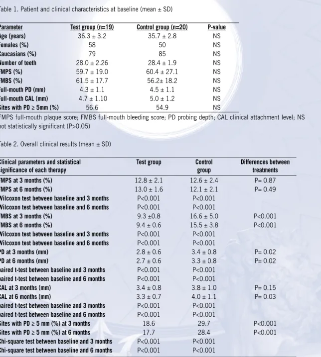
Table 1. Patient and clinical characteristics at baseline (mean ± SD)
Parameter Test group (n=19) Control group (n=20) P-value
Age (years) 36.3 ± 3.2 35.7 ± 2.8 NS Females (%) 58 50 NS Caucasians (%) 79 85 NS Number of teeth 28.0 ± 2.26 28.4 ± 1.9 NS FMPS (%) 59.7 ± 19.0 60.4 ± 27.1 NS FMBS (%) 61.5 ± 17.7 56.2± 18.2 NS Full-mouth PD (mm) 4.3 ± 1.1 4.5 ± 1.1 NS Full-mouth CAL (mm) 4.7 ± 1.10 5.0 ± 1.2 NS Sites with PD ≥ 5mm (%) 56.6 54.9 NS
FMPS full-mouth plaque score; FMBS full-mouth bleeding score; PD probing depth; CAL clinical attachment level; NS not statistically significant (P>0.05)
Table 2. Overall clinical results (mean ± SD)
Clinical parameters and statistical Test group Control Differences between
significance of each therapy group treatments
FMPS at 3 months (%) 12.8 ± 2.1 12.6 ± 2.4 P= 0.87
FMPS at 6 months (%) 13.0 ± 1.6 12.1 ± 2.1 P= 0.49
Wilcoxon test between baseline and 3 months P<0.001 P<0.001
Wilcoxon test between baseline and 6 months P<0.001 P<0.001
FMBS at 3 months (%) 9.3 ±0.8 16.6 ± 5.0 P<0.001
FMBS at 6 months (%) 9.4 ± 0.6 15.5 ± 3.8 P<0.001
Wilcoxon test between baseline and 3 months P<0.001 P<0.001
Wilcoxon test between baseline and 6 months P<0.001 P<0.001
PD at 3 months (mm) 2.8 ± 0.6 3.4 ± 0.8 P= 0.02
PD at 6 months (mm) 2.7 ± 0.6 3.3 ± 0.8 P= 0.02
paired t-test between baseline and 3 months P<0.001 P<0.001
paired t-test between baseline and 6 months P<0.001 P<0.001
CAL at 3 months (mm) 3.4 ± 0.8 3.8 ± 1.0 P= 0.15
CAL at 6 months (mm) 3.3 ± 0.7 4.0 ± 1.1 P= 0.03
paired t-test between baseline and 3 months P<0.001 P<0.001
paired t-test between baseline and 6 months P<0.001 P<0.001
Sites with PD ≥ 5 mm (%) at 3 months 18.6 29.7 P<0.001
Sites with PD ≥ 5 mm (%) at 6 months 17.7 28.4 P<0.001
Chi-square test between baseline and 3 months P<0.001 P<0.001
Chi-square test between baseline and 6 months P<0.001 P<0.001
FMPS full-mouth plaque score; FMBS full-mouth bleeding score; PD probing depth; CAL clinical attachment level When sites were stratified according to initial PD, the clearest advantage of antibiotics over placebo was noted in pockets initially ≥ 6 mm (p=0.003), while no difference between two groups was detected in moderate pockets sites (p> 0.05). Subjects receiving the antibiotics showed a decrease in PD at deep sites from 6.9 ± 0.7 mm at baseline to 3.8 ± 0.8 mm at 6 months, whereas patients receiving placebo showed a decrease from 7.1 ± 0.6 mm at baseline to 4.7 ± 0.8 mm at 6 months.
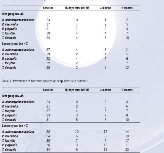
Microbiological data are summarized in Table 3 and 4. In the baseline samples Aa and Td were the most prevalent micro-organisms found in the subgingival biofilm of moderate pockets, whereas. Tf and Td in deep sites.
Table 3. Prevalence of bacterial species at moderate sites (site number)
Baseline 15 days after OSFMF 3 months 6 months Test group (n= 38) A. actinomycetemcomitans 23 0 1 2 P. intermedia 17 1 2 5 P. gingivalis 21 1 2 4 T. forsythia 19 4 4 7 T. denticola 24 4 6 10 Control group (n= 40) A. actinomycetemcomitans 27 4 8 11 P. intermedia 19 2 4 5 P. gingivalis 23 4 6 6 T. forsythia 22 2 3 7 T. denticola 25 2 6 12
Table 4. Prevalence of bacterial species at deep sites (site number)
Baseline 15 days after OSFMF 3 months 6 months
Test group (n= 38) A. actinomycetemcomitans 23 0 3 4 P. intermedia 21 2 5 6 T. forsythia 27 4 6 7 P. gingivalis 23 3 7 8 T. denticola 31 5 8 10 Control group (n= 40) A. actinomycetemcomitans 22 10 12 14 P. intermedia 24 6 8 10 T. forsythia 30 7 7 17 P. gingivalis 26 3 10 11 T. denticola 34 5 18 23
The combination OSFMD and antibiotic intake decreased the number of sites positive for the monitored species more than OSFMD alone. With regard 4-5 mm sampled pockets 65.8% of test sites unlike 37.5% of control sites were free of pathogens at 6 months. Among pockets initially ≥ 6 mm 57.9% of test sites were free of pathogens at 6-month examina-tion unlike 15% of control sites, respectively (p< 0.001).
When analyzing bacterial recolonization at moderate sites, in contrast to SRP administration of antimicrobials resulted in elimination of A. actinomycetemcomitans 15 days after the OSFMD (Table 3). At 6 months A. actinomycetemcomitans were detected in 5.3% of test sites compared to 35% of control ones (p=0.002). No differences were observed between treatment groups in the prevalence of P. gingivalis, P.intermedia, T. forsythia, T. denticola at any time point.
At deep sites the combined therapy was more efficient than SRP alone to achieve a lower prevalence of A.
actinomycetem-comitans and T. denticola at 3 (p= 0.03 and p= 0.04, respectively) and 6 months (p= 0.02 and p= 0.03, respectively),
and of T. forsythia at 6-month examination (p= 0.04). The proportion of P. gingivalis and P. intermedia was not signifi-cantly lower in the test sites than in the scaling group at any interval (p>0.05).
Discussion
Limited information is available on the effectiveness of the OSFMD in the treatment of aggressive periodontitis patients and no data are reported on its association with systemic antimicrobial agents. The present investigation demonstrated that the OSFMD plus systemic amoxicillin/metronidazole resulted in statistically significant improvement in microbio-logical as well clinical parameters. The high dosage of both antibiotics (500 mg of each, 3 times a day, for 7 days) was proposed by recent randomized controlled clinical trials to reach effective antimicrobial concentration in soft tissues and crevicular fluids. 4,8
With regard to microbiological analysis, the systemic administration of amoxicillin-metronidazole decreased the number of sites positive for the monitored species more than OSFMD alone. In the test group all the target bacteria were suppressed below the detection level in 65.8% and 57.9% of moderate and deep sites, respectively, at 6-month evaluation. The per-centage of moderate and deep sites free of pathogens amounted to 37.5% and 15% in the control group at the same time point (p<0.001). When individual species were analyzed the combined therapy was more effective than OSFMD alone in controlling the recolonization of A. actinomycetemcomitans in moderate sites and of A. actinomycetemcomitans, T.
for-sythia and T. denticola in deep pocket sites. At test sites, it could not be detected 15 days following treatment and
recolo-nized only 5.3% and 7.9% of moderate and deep periodontal pockets, respectively, at 6 months. This finding suggests the important role that bacteria of green and red complex may be playing in the stability of periodontal treatment outcome. The clearest clinical advantage of antibiotics over placebo was noted on inflammatory values and in pockets initially ≥ 6 mm. In the test group the FMBS remained below 10%, whereas in the control group the percentage was superior to 15% over the experimental period. In deep pockets the test treatment resulted in an additional 1 mm in PD reduction, while no statistically significant differences were observed in moderate periodontal sites.
It is important to point out the pivotal role of the supportive periodontal care. Plaque levels were maintained at a low level (< 15%) through the study in both treatment groups. The stringent recall protocol might be relevant to the reduction of plaque because full-mouth instrumentation is performed in two visits and thus the therapist has fewer opportunities to check and reinforce the necessary oral hygiene instructions.
Only a few randomized controlled clinical trials focused on the additional effects of adjunctive amoxicillin-metronidazole in the therapy for well-defined G-AgP.4,8-10 In these reports the combination of antimicrobials with quadrant or full-mouth SRP resulted in average values of 1.2-1.5 mm for PD reduction and of 0.5-0.9 mm for CAL gain over a 6-month period. A direct comparison of these measures of clinical outcomes is unfeasible because of discrepancy among study designs, especially regarding the schedule for debridement, dosage and timing of drug administration.
It is important to point out that because of the experimental design we did not perform any subgingival re-instrumentation of periodontal pockets exhibiting bleeding on probing. It is our opinion that in the clinical practice re-instrumentation have to be performed at each maintenance treatment session.
References
1. Quirynen, M., Bollen, C.M.L., Vandekerckhove, B.N., Dekeyser, C., Papaionnou, W. & Eyssen, H. (1995) Full - versus partial-mouth disinfection in the treatment of periodontal infection. Short-term clinical and microbiological observa-tions. Journal of Dental Research 74, 1459-1467.
2. Haffajee, A., Socransky, S. & Gunsolley, J. (2003) Systematic anti-infective periodontal therapy. A systematic review. Annals of Periodontology 8, 115-181.
3. Herrera, D., Sanz, M., Jepsen, S., Needleman, I. & Roldàn, S. (2002) A systematic review on the effect of systemic antimicrobials as an adjunct to scaling and root planning in the periodontal patients. Journal of Clinical Periodontology 29 (Suppl 3), 136-159.
4. Xajigeorgiou, C., Sakellari, D., Slini, T., Baka, A. & Konstantinidis, A. (2006) Clinical and microbiological effects of different antimicrobials on generalized aggressive periodontitis. Journal of Clinical Periodontology 33, 254-264. 5. American Academy of Periodontology (2000) Parameters on aggressive periodontitis. Journal of Periodontology 71,
867-869.
6. Bollen, C.M.L., Mongardini, C., Papaioannou, W., van Steenberghe, D. & Quirynen, M. (1998) The effect of a one stage full mouth disinfection on different intra-oral niches. Clinical and microbiological observations. Journal of Clinical Periodontology 25, 56-66.
plaques in advanced chronic periodontitis patients. Journal of Periodontology 78, 1718-1723.
8. Guerrero, A., Griffiths, G.S., Nibali, L., Suvan, J., MOles, D.R., Laurell, L. & Tonetti, M.S. (2005) Adjunctive benefits of systemic amoxicillin and metronidazole in non-surgical treatment of generalized aggressive periodontitis: a randomized placebo-controlled clinical trial. Journal of Periodontology 32, 1096-1107.
9. Kaner, D., Christan, C., Dietrich, T., Bernimoulin, J.P., Kleber, B.M., & Friedmann, A. (2007) Timing affects the clinical outcomes of adjunctive systemic antibiotic therapy for generalized aggressive periodontitis. Journal of Periodontology 78, 1201-1208.
10. Yek, E.C., Cintan, S., Topcuoglu, N., Kulekci, G., Issever, H. & Kantarci, A. (2010) Efficacy of amoxicillin and met-ronidazole combination for the management of generalized aggressive periodontitits. Journal of Periodontology 81, 964-974.