Very Small Embryonic-Like Stem Cells
with Maximum Regenerative Potential Get Discarded
During Cord Blood Banking and Bone Marrow
Processing for Autologous Stem Cell Therapy
Deepa Bhartiya,1Ambreen Shaikh,1Punam Nagvenkar,1Sandhya Kasiviswanathan,1Prasad Pethe,1 Harsha Pawani,1Sujata Mohanty,2 S.G. Ananda Rao,3 Kusum Zaveri,4and Indira Hinduja4
Very small embryonic-like stem cells (VSELs) are possibly lost during cord blood banking and bone marrow (BM) processing for autologus stem cell therapy mainly because of their small size. The present study was conducted on human umbilical cord blood (UCB,n=6) and discarded red blood cells (RBC) fraction obtained after separation of mononuclear cells from human BM (n=6), to test this hypothesis. The results show that VSELs, which are pluripotent stem cells with maximum regenerative potential, settle along with the RBCs during Ficoll-Hypaque density separation. These cells are very small in size (3–5mm), have high nucleo-cyto-plasmic ratio, and express nuclear Oct-4, cell surface protein SSEA-4, and other pluripotent markers such as
Nanog, Sox-2, Rex-1,and Tert as indicated by immunolocalization and quantitative polymerase chain reaction (Q-PCR) studies. Interestingly, a distinct population of slightly larger, round hematopoietic stem cells (HSCs) with cytoplasmic Oct-4 were detected in the ‘‘buffy’’ coat, which usually gets banked or used during autologus stem cell therapy. Immunohistochemical studies on the umbilical cord tissue (UCT) sections (n=3) showed the presence of nuclear Oct-4–positive VSELs and many fibroblast-like mesenchymal stem cells (MSCs) with cy-toplasmic Oct-4. These VSELs with nuclear Oct-4, detected in UCB, UCT, and discarded RBC fraction obtained after BM processing, may persist throughout life, maintain tissue homeostasis, and undergo asymmetric cell division to self-renew as well as produce larger progenitor stem cells, viz. HSCs or MSCs, which follow dif-ferentiation trajectories depending on the somatic niche. Hence, it can be concluded that the true stem cells in adult body tissues are the VSELs, whereas the HSCs and MSCs are actually progenitor stem cells that arise by asymmetric cell division of VSELs. The results of the present study may help explain low efficacy reported during adult autologous stem cell trials, wherein unknowingly progenitor stem cells are injected rather than the pluripotent stem cells with maximum regenerative potential.
Introduction
B
one marrow transplantation (BMT), nowadays termed as hematopoietic stem cell (HSC) transplantation, is considered as one of the major medical breakthroughs of the 21st century. Besides bone marrow (BM), even peripheral blood (PB) and umbilical cord blood (UCB) are considered as standard sources of ‘‘autologus’’ HSCs and mesenchymal stem cells (MSCs). The stem cells are present many folds higher in cord blood when compared with PB and BM. Thus, there has been a lot of interest to cryopreserve cord blood since mid-1990s for harnessing their regenerative potential in the future. Traditionally, cord blood banking and BMTtherapy use isolation methods targeting progenitor cells present in the buffy coat using Ficoll-Hypaque gradient separation method. The isolated stem cells have been found to be largely committed toward hematopoiesis and hence have been used to treat leukemia, aplastic anemia, and some immune deficiency diseases. However, it has also been claimed that these stem cells may also transdifferentiate and contribute to skeletal muscle, liver, neural, and myocardial regeneration when transplanted following tissue injury. As a result, several cord blood banks have been established, which promote private and public cord blood banking for future autologous or allogeneic stem cell therapy toward regeneration.
1
Stem Cell Biology Department, National Institute for Research in Reproductive Health, Parel, Mumbai, India. 2Stem Cell Facility, All India Institute of Medical Sciences, New Delhi, India.
3International Stem Cell Services Ltd., Dahisar (West), Mumbai, India. 4
INKUS IVF Centre, Mumbai, India. Volume 00, Number 0, 2011
!Mary Ann Liebert, Inc. DOI: 10.1089/scd.2011.0311
Growing evidence suggests that besides the HSCs and MSCs, a small population of pluripotent stem cells, termed very small embryonic-like stem cells (VSELs), are also pres-ent in the UCB, which could potpres-entially contribute to organ and tissue regeneration [1]. Recently, VSELs were isolated from various adult mice organs, blood, and BM as well as from human cord blood and BM by fluorescent activated cell sorting [2]. Our group has reported the presence of VSELs in situ in adult human gonads [3,4]. On processing the VSELs, isolated by scraping the sheep ovary surface epithelium, we observed that these cells with high nucleo-cytoplasmic ratio were invariably lost during processing by centrifugation. We hypothesized that, similarly, during processing of cord blood and BM on Ficoll-Hypaque gradient, VSELs may be lost and get discarded during processing. Thus, the present study was undertaken and our results may have serious implica-tions, because VSELs being pluripotent are expected to have more regenerative potential than the HSCs and MSCs.
Materials and Methods
Sample collection and processing
For the study, UCB (n=6) from full-term deliveries scheduled for caesarean section, umbilical cord tissue (UCT; n=3), and discarded fraction of red blood cell pellet obtained after processing BM for autologus use (BM,n=6) were used. The UCB was collected in heparinized tubes from deliveries performed by Dr. Hinduja and Dr. Zaveri, whereas dis-carded fraction of red blood cell pellet from BM samples were transported from Dr. S.G.A. Rao’s clinic in Mumbai suburbs to Stem Cell Biology Department at NIRRH for further studies. Small piece of UCT was fixed in normal buffered formalin and slides were prepared for immuno-histochemical studies using standard protocols, whereas the UCB and discarded fractions of BM were processed to isolate cells by Ficoll-Hypaque centrifugation method.
Ficoll-Hypaque density centrifugation
Collected UCB was diluted 1:1 in phosphate-buffered sa-line (PBS) and carefully overlaid in 1:2 ratio onto research-grade Ficoll-Hypaque solution (HISTOPAQUE-1077; Sigma; d: 1.077 g/cm3), followed by centrifugation at 1,500 rpm for
30 min at room temperature (RT). After centrifugation, all the 4 layers (1–4) (Fig. 1A) were easily visualized and collected in separate tubes.
Processing of mononuclear cells
The layer 2 (buffy coat) was diluted 3 times its volume with DMEM F12 (Invitrogen) containing 5% FBS (Invitrogen) and centrifuged for 10 min at RT. The pellet was collected and resuspended in PBS to make a single-cell suspension. The platelet (layer 1) and Ficoll (layer 3) were also processed similarly.
Processing of red blood cells (4) to further enrich VSELs
The red blood cells (RBC) pellet (layer 4) collected from both UCB and BM was diluted 3 times its volume with DMEM F12 and centrifuged at 800 g for 15 min at RT to
remove most of RBCs. The supernatant was collected in a fresh centrifuge tube and spun at 1,000gfor 10 min at RT. The collected pellet was resuspended in PBS to make a sin-gle-cell suspension (layer 5).
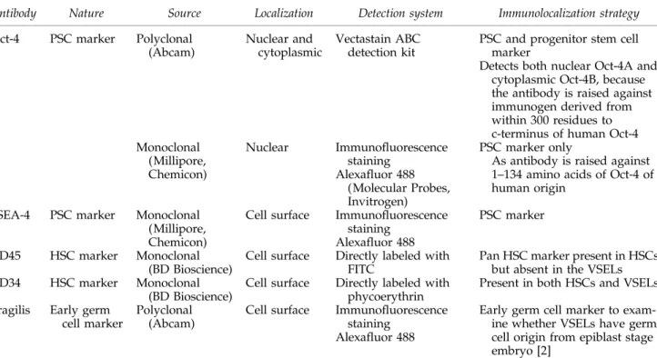
The cell suspensions obtained from layers 1, 2, 3, and 5 were used to make cell smears for immunolocalization studies, whereas part of the cell pellet (2 and 5) was stored in TRIZOL (Invitrogen) for RNA extraction. Table 1 is a com-prehensive representation of various immunolocalization studies undertaken.
The air-dried cell smears were fixed with 4% parafor-maldehyde for 10 min, washed twice with PBS, air dried, and stored at 4"C till further use. Smears were stained with he-matoxylin and eosin (H&E) using a standard method and viewed under a bright-field microscope. Representative fields were photographed under a Nikon 90i microscope (Nikon). The smears prepared from layers 2 and 5 were used for immunolocalization and confocal studies.
Immunolocalization studies
Immunolocalization studies were performed on paraffin-embedded sections of cord tissue and on cell smears from layers 2 and 5.
Immunocytochemistry. The cell smears were processed for immunolocalization of Oct-4 using polyclonal Oct-4 anti-body (Abcam) and Vectastain Elite ABC kit (Vector La-boratories) as detection system.
In brief, the endogenous peroxidase was blocked using 0.3% hydrogen peroxide for 45 min in dark at RT. Antigen retrieval was done by treating the smears with sodium citrate buffer (10 mM sodium citrate, pH 6.0) at high power for 5 min in a microwave oven. This was followed by a per-meabilization step with 0.3% Triton X-100 for 10 min. Blocking was done with 10% normal goat serum for 1 hr. The cells were then incubated with the primary polyclonal Oct-4 antibody (1:100) at 4"C overnight. Primary antibody was omitted and smears were incubated with the blocking solu-tion for negative control. After washing with PBS, the cells were incubated with the biotinylated secondary antibody (anti-rabbit IgG) for 30 min followed by avidin–biotin com-plex formation step for 30 min; the cells were then ex-tensively washed with PBS and detected using 3,3¢ diaminobenzidine (DAB) (Sigma-Aldrich) and later coun-terstained with hematoxylin.
Immunohistochemistry on UCT sections. The paraffin sec-tions were deparaffinzed and rehydrated through a graded methanol series. Endogenous peroxidase was blocked using 0.3% hydrogen peroxide for 90 min in dark at RT and then the sections were processed in a similar manner to the cell smears.
Representative areas of smears and sections were photo-graphed under Nikon 90i microscope and results were recorded.
Immunofluorescent staining. The expression of nuclear antigen Oct-4A, cell surface antigen SSEA-4, CD45-FITC, CD34-PE, and Fragilis were studied on cell smears from layers 2 and 5 only. Immunofluorescent staining was per-formed using specific antibodies listed in Table 1. Briefly, fixed cell smears were washed with wash buffer (PBS con-taining 0.3% bovine serum albumin and 0.1 mM EDTA), permeabilized by 0.3% Triton X-100 for 10 min, washed in
PBS, preblocked with 15% normal goat serum, and subse-quently incubated with antibodies to Oct-4, CD45 FITC, CD34 PE, Fragilis, and SSEA-4. For detection, cell smears were incubated with secondary antibody indicated in Table 1; however, for CD34 and CD45 this step was omitted as the primary antibodies were directly labeled with the flur-ochrome. Negative controls were incubated overnight in the blocking solution with the omission of primary antibody. Counterstaining was done using (1 mg/mL) 4¢ ,6-diamidino-2-phenylindole (DAPI; Molecular Probes). All images were
captured by laser scanning confocal microscope (Carl Ziess) at 63·magnification with 5·optical zoom using argon laser atk=488 nm and blue diode laser atk=405 nm for FITC and DAPI staining, respectively. For staining of cell surface markers, permeabilization with Triton X-100 was omitted. RNA extraction and cDNA synthesis
Total RNA was isolated from cell pellets (2 and 5) obtained after processing UCB and discarded fractions of BM, using
FIG. 1. (A)Procedure to enrich and isolate very small embryonic-like stem cells (VSELs) from umbilical cord blood (UCB). UCB is overlaid on Ficoll-Hypaque and centrifuged and 4 layers (1–4) get separated. Layer 4 is further centrifuged to separate RBCs and then the pellet obtained (layer 5) contains enriched VSELs with few contaminating blood cells (for details, see Materials and Methods section).(B)Hematoxylin and eosin staining of cell smears of the 4 layers 1–3 and 5. As evident, layer 1 is comprised of platelets, which appear pink and is devoid of nuclei; layer 2 comprised a mix of mononuclear cells (MNCs)—‘‘buffy coat’’; layer 3 was mainly the large granulocytes with characteristic multilobed nuclei; layer 5 (obtained after further processing of layer 4) comprised very small sized cells with high nucleo-cytoplasmic ratio, which are the VSELs (arrow) (40·).(C) VSELs under Hoffman optics. They appear round, have a slightly shiny appearance (20·), and tend to aggregate (inset, 40·) in vitro.(D)ICC with polyclonal Oct-4 antibody on MNCs and VSELs. As evident, a distinct big cell, possibly the hematopoietic stem cell (HSC,arrow), had cytoplasmic Oct-4, whereas the VSELs had distinct nuclear Oct-4 (asterisk) (40·).(E)IHC with polyclonal Oct-4 antibody on umbilical cord tissue. Two distinct cell types could be visualized in the Wharton jelly region, viz. spindle-shaped mesenchymal stem cells with cytoplasmic Oct-4 (arrow) and very small-sized VSELs with nuclear Oct-4 (asterisk) (40·).(F)Q-PCR analysis of MNCs and VSELs. Higher expression of pluripotent markers was observed in cells from layer 5 (VSELs) when compared with cells from layer 2 (buffy coat).(G)Confocal microscopy studies for pluripotent, germ cell, and hematopoietic markers. The VSELs exhibited nuclear Oct-4 and cell-surface SSEA-4 and Fragilis. VSELs were negative for SSEA-1 (arrow); however, cell surface staining was observed on granulocytes. The slightly bigger HSCs displayed cytoplasmic Oct-4 and minimal SSEA-4. CD45 antibody stained all the blood cell types; however, the VSELs were negative (asterisk), whereas CD34 stained all the blood cells including VSELs, which were dis-tinguished by their size and negative DAPI staining (asterisk) because of the presence of abundant euchromatin (discussed earlier) [4]. All 63·, except that CD45 is at 40·. RBC, red blood cells; ICC, immunocytochemistry, IHC, im-munohistochemistry; Q-PCR, quantitative polymerase chain reaction.
TRIZOL according to the manufacturer’s instructions. The extracted RNA was treated with ribonuclease-free deoxyri-bonuclease (DNase 1; Qiagen) to remove any contaminating genomic DNA. Two micrograms of total RNA was reverse-transcribed using the iScript RT Kit according to the manu-facturer’s instructions (Biorad) to synthesize the first-strand cDNA in a GSTORM thermocycler (Gene Technologies). Quantitative polymerase chain reaction
Expression of various pluripotent markers, viz. Oct-4A, Oct-4, Nanog, Rex-1, Tert, and Sox-2, was studied by
Q-PCR using a CFX96 real-time PCR system (Bio-Rad Laboratories) and SYBR Green chemistry (Bio-Rad Labora-tories) (Table 2).
The amplification conditions for Oct-4 were as follows: initial denaturation at 95"C for 5 min, followed by 40 cycles comprising denaturation at 95"C for 10 s, primer annealing at 57"C for 30 s, and extension at 72"C for 30 s. For the rest of gene transcripts, viz.Oct-4A, Nanog, Rex-1, Sox-2, Tert,and 18S rRNA, all the amplification conditions were the same except that the annealing temperatures were optimized at 62"C.
The threshold cycle (Ct) was determined subsequently using the CFX Manager software (Bio-Rad Laboratories) and normalized to the housekeeping gene (18S RNA). Relative fold change ofOct-4 andNanog mRNA over the calibrator was expressed as 2-DDCt, where DCt=Ct of target genes (Oct-4, Nanog)-Ct of endogenous control gene (18s), and
DDCt=DCt of samples for target gene-DCt of calibrator. Melt curve analysis was performed at the end of every run to confirm the homogeneity of the PCR products, which was also confirmed by running the same on 2% agarose gel.
Results
Enrichment of VSELs from UCB and BM
Careful processing of different fractions obtained after Ficoll-Hypaque separation of UCB showed that the VSELs separated along with the RBCs in layer 4. Layers 1, 2, and 3 comprised of platelets, mononuclear cells (MNCs), and mostly granulocytes, respectively (Fig. 1A), as evident from the H&E staining (Fig. 1B). Further centrifugation of the RBC pellet (layer 4) at different speeds enabled separation of RBCs from the VSELs, as RBCs settled down at 800gand
Table1. Details of Antibodies Used for Immunolocalization Studies
Antibody Nature Source Localization Detection system Immunolocalization strategy
Oct-4 PSC marker Polyclonal (Abcam)
Nuclear and cytoplasmic
Vectastain ABC detection kit
PSC and progenitor stem cell marker
Detects both nuclear Oct-4A and cytoplasmic Oct-4B, because the antibody is raised against immunogen derived from within 300 residues to c-terminus of human Oct-4 Monoclonal (Millipore, Chemicon) Nuclear Immunofluorescence staining Alexafluor 488 (Molecular Probes, Invitrogen) PSC marker only
As antibody is raised against 1–134 amino acids of Oct-4 of human origin
SSEA-4 PSC marker Monoclonal (Millipore, Chemicon)
Cell surface Immunofluorescence staining
Alexafluor 488
PSC marker CD45 HSC marker Monoclonal
(BD Bioscience)
Cell surface Directly labeled with FITC
Pan HSC marker present in HSCs but absent in the VSELs CD34 HSC marker Monoclonal
(BD Bioscience)
Cell surface Directly labeled with phycoerythrin
Present in both HSCs and VSELs Fragilis Early germ
cell marker
Polyclonal (Abcam)
Cell surface Immunofluorescence staining
Alexafluor 488
Early germ cell marker to exam-ine whether VSELs have germ cell origin from epiblast stage embryo [2]
HSC, hematopoietic stem cell; PSC, pluripotent stem cell.
Table2. Primers Used in Quantitative Polymerase Chain Reaction Studies
Primer set Sequence Product size (bp) Oct-4A F AGCCCTCATTTCACCAGGCC 448 Oct-4A R TGGGACTCCTCCGGGTTTTG Oct-4 F CTTGCTGCAGAAGTGGGTGGAGGAA 168 Oct-4 R CTGCAGTGTGGGTTTCGGGCA Nanog F AGTCCCAAAGGCAAACAACCCACTTC 164 Nanog R TGCTGGAGGCTGAGGTATTTCTGTCTC Tert F AGCTATGCCCGGACCTCCAT 185 Tert R GCCTGCAGCAGGAGGATCTT Sox-2 F ATGCACCGCTACGACGTGA 448 Sox-2 R CTTTTGCACCCCTCCCATTT Rex F GCGTACGCAAATTAAAGTCCACA 306 Rex R CAGCATCCTAAACAGCTCGCAGAAT CXCR4 F GGCAGCAGGTAGCAAAGTGACGC 334 CXCR4 R AGAGGAGGTCGGCCACTGACA 18S F GGAGAGGGAGCCTGAGAAAC 171 18S R CCTCCAATGGATCCTCGTTA
later the VSELs were pelleted at 1,000g. H&E staining of the smears prepared from this layer comprised of very small round cells with high nucleo-cytoplasmic ratio, which ran-ged in size from 3 to 5mm (Fig. 1B) along with few multi-lobed nucleated cells. Similarly, it was possible to separate the BM VSELs from the RBC pellet collected after separation of the MNCs. The VSELs enriched in layer 5 were present in large numbers, were round, and had a characteristic shiny appearance as seen under Hoffman optics (Fig. 1C). They had striking similarity in their appearance to the VSELs isolated by scraping the ovarian surface epithelium, reported earlier by our group [4].
Immunocytochemical localization of Oct-4
Polyclonal Oct-4 antibody was used to study cell smears from layers 2 and 5 only. As evident in layer 2, few large cells with cytoplasmic Oct-4 were observed (possibly the HSCs; Fig. 1D), whereas majority of cells remained unstained. In layer 5, the VSELs showed distinct nuclear staining for Oct-4. Immunohistochemical localization of Oct-4
in UCT sections
The cell population in the mucous connective tissue also termed as the Wharton’s jelly of the umbilical cord com-prised mainly of spindle-shaped fibroblasts after H&E staining. However, 2 distinct cell populations were easily identified after immunolocalization studies using polyclonal Oct-4 antibody, viz. very small cells with nuclear Oct-4 and the fibroblasts (possibly the MSCs) with cytoplasmic Oct-4. Similar results were obtained using VSELs isolated from BM samples.
Immunofluorescence staining and confocal microscopy
Various pluripotent, germ cell, and hematopoietic markers were studied by confocal microscopy.
(i) Pluripotent markers Oct-4 and SSEA-4 revealed char-acteristic nuclear and cell surface staining, respectively, on VSELs in layer 5 only (Fig. 1G). A distinct larger cell with abundant cytoplasm revealed minimal cytoplasmic staining for both Oct-4 and SSEA-4. All other cell types in layer 2 remained unstained for Oct-4 and SSEA-4.
(ii) Primordial germ cell marker Fragilis showed cell sur-face staining only on the VSELs (Fig. 1G).
(iii) Hematopoietic marker CD45 stained all the blood cells in the MNC fraction (layer 2), whereas the VSELs remained distinctly negative.
(iv) CD34, a more primitive marker for hematopoietic progenitor cells, stained all the cells including the small cells, which were easily identified because of their size and be-cause they stained negative for DAPI. Interestingly, SSEA-1 also stained the granulocytes but VSELs were distinctly negative for SSEA-1, confirming their primitive status.
Similar results were obtained using VSELs isolated from BM samples.
Q-PCR results
As evident, gene transcripts for various pluripotent markers including Oct-4, Nanog, and TERT were present and exhibited several fold higher expression in the VSELs (layer 5) when compared with the MNC fraction (layer 2) isolated from the UCB. Similar results were obtained using VSELs isolated from BM samples.
Discussion
The present study describes a simple method based on differential centrifugation to isolate pluripotent stem cells, which are termed VSELs (because of their size), from both UCB and BM. This method is relatively cheaper, is practical, and can be easily carried out in a laboratory setting when compared with available sophisticated approach using spe-cific markers by FACS [2]. It is an Indian Council of Medical Research (ICMR), Government of India, New Delhi, patent-protected technology (1257/DEL/2011). Multiparameteric analysis was employed to characterize and confirm the pluripotent nature of the VSELs by visualizing morphology under Hoffman optics, H&E staining, immunolocalization for pluripotent, germ cell, and hematopoietic markers, and Q-PCR. The VSELs exhibit a characteristic round shape and high nucleo-cytoplasmic ratio and showed expression of pluripotent markers, such as Oct-4A, Nanog, Rex, and TERT, at the mRNA level and immunolocalization for nu-clear Oct-4 and cell surface SSEA-4. The VSELs present in layer 5 were strikingly similar to the VSELs obtained by scraping ovary surface epithelium, recently reported by our group [4].
Results clearly show that UCB and BM VSELs settle down along with RBCs in the lowermost layer (Fig. 1A, layer 4) during Ficoll-Hypaque processing. RBCs settle down, be-cause they possess hemoglobin, whereas the VSELs possibly settle down as per laws of physics. VSELs being smaller in size have lower mass and so require higher centrifugation speeds to pellet when compared with larger HSCs, which get separated in the upper buffy coat (layer 2). This is similar to
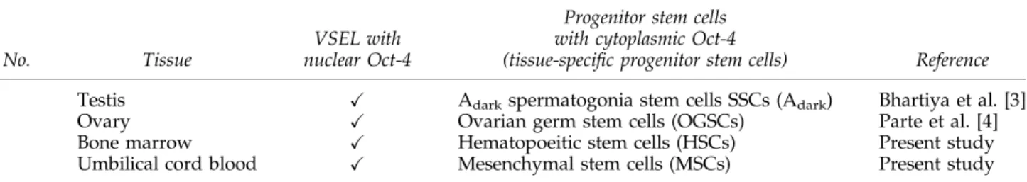
Table3. Details of Very Small Embryonic-Like Stem Cell–Derived Progenitors in Adult Human Tissues
S. No. Tissue
VSEL with nuclear Oct-4
Progenitor stem cells with cytoplasmic Oct-4
(tissue-specific progenitor stem cells) Reference 1 Testis X Adarkspermatogonia stem cells SSCs (Adark) Bhartiya et al. [3]
2 Ovary X Ovarian germ stem cells (OGSCs) Parte et al. [4]
3 Bone marrow X Hematopoeitic stem cells (HSCs) Present study
the situation wherein ship (HSCs) floats despite being bigger in size, whereas small bullets (VSELs) sink in water.
We have observed several folds higher expression of various pluripotent makers in layer 5 compared with layer 2 (Fig. 1F), which further confirms the pluripotent nature of these cells. Oct-4A mRNA was expressed several folds higher in layer 5 compared with layer 2. Interestingly, total Oct-4was also detected in layer 2, suggesting presence of an alternative spliced variant Oct-4B in the HSCs rather than pluripotent Oct-4A. Immunolocalization studies also con-firmed that MNC in layer 2 had HSCs with cytoplasmic Oct-4, whereas the VSELs in layer 5 had predominantly nuclear Oct-4–positive cells. Immunohistochemical studies on UCT sections also yielded intriguing results, because a heteroge-neous population of both VSELs with nuclear Oct-4 and spindle-shaped, fibroblast-like MSCs with cytoplasmic Oct-4 were identified (Fig. 1E).
Current banking protocols cryopreserve only cells of the buffy coat for future autologus stem cell therapy and large sums of money in tune of $2,000 and yearly fees are charged for the same. However, our results imply that instead of true stem cells (VSELs), only the progenitors are being banked for therapeutic use. On a similar note, huge expenses are in-curred for stem cell therapy using autologus BM, because the public is vulnerable and at any cost wish to save lives of their dear ones.
Oct-4 studies have confused stem cell researchers in the past, because it has several transcripts and pseudogenes. However, using specific primers and 3 different antibodies, we have earlier reported the presence of similar VSELs with nuclear Oct-4 and slightly bigger progenitor stem cells with cytoplasmic Oct-4 in the adult mammalian testis (Adark
spermatogonial stem cell) [3] and also ovaries (ovarian germ stem cells [OGSCs]) [4]. Results are depicted in Table 3.
These results are indeed startling. It is apparent that there is nothing like adult stem cells in somatic body tissues and embryonic stem cells isolated from spare human embryos, which has divided the scientific community into 2 groups. Embryonic-like stem cells, that is, VSELs, are present in various adult body tissues. They are relatively quiescent, serve as a backup, and give rise to the progenitor stem cells (which are currently termed the adult stem cells) by asym-metric cell division.
The progenitor stem cells exhibit high proliferation activ-ity and differentiate further to give rise to various differen-tiated progenies. The question that arises is if it is the same VSEL in different body tissues then why the progenitors differ from each other and give rise to different end points? We propose that this indeed highlights the importance of the somatic microenvironment, which differs from tissue to tis-sue and thus directs the progenitors to follow different dif-ferentiation pathways. Thus, in contradiction to earlier reports that Oct-4 is dispensable to somatic tissues, the present study results in contrast show that indeed Oct-4 pluripotency network is crucial for proper homeostasis of body tissues throughout life. Being pluripotent in nature, VSELs have the ability to differentiate into any cell lineage based on what genes get turned ON when the pluripotent stem cell chromatin gets reprogrammed during the transition of the VSELs into the progenitor. The present study results are in agreement with the huge body of literature published by Ratajczak and group and Li and Clevers [5], who have
proposed that 2 distinct populations of stem cells exist in various body tissues. The results are also in agreement with Virchow and Connheim, who had proposed almost 150 years back that adult tissues contain dormant embryonic remnants.
It is concluded that adult stem cells are actually progenitor stem cells derived from VSELs in the adult body. It has al-ways been the pluripotent embryonic-like stem cells, which bring about homeostasis in the adult body. The debates be-tween the groups who work on adult versus embryonic stem cells and the pros and cons of embryonic versus adult stem cells research become irrelevant. Hopefully, VSELs will unify both the groups and new paradigms will emerge wherein more emphasis will be to better understand the somatic niche that controls the fate of VSELs.
Acknowledgments
This study (BT/PR14026/MED/31/100/2010) was finan-cially supported by Department of Biotechnology and Indian Council of Medical Research, New Delhi, India.
Author Disclosure Statement
No competing financial interests exist.
References
1. Ratajczak J, E Zuba-Surma, E Paczkowska, M Kucia, P Nowack and MZ Rataczak. (2011). Stem cells for neural regeneration—a potential application of very small embry-onic-like stem cells. J Physiol Pharmacol 62:3–12.
2. Zuba-Surma EK, M Kucia, J Ratajczak and MZ Ratajczak. (2009). ‘‘Small stem cells’’ in adult tissues: very small embryonic-like stem cells stand up! Cytometry 75:4–13. 3. Bhartiya D, S Kasiviswanathan, SK Unni, P Pethe, JV
Dha-balia, S Patwardhan and HB Tongaonkar. (2010). Newer in-sights into premeiotic development of germ cells in adult human testis using Oct-4 as a stem cell marker. J Histochem Cytochem 58:1093–1106.
4. Parte S, D Bhartiya, J Telang, V Daithankar, V Salvi, K Zaveri and I Hinduja. (2011). Detection, characterization and spon-taneous differentiationin vitroof very small embryonic-like putative stem cells in adult mammalian ovary. Stem Cells Dev 2011 Mar 23 [Epub ahead of print]; DOI: 10.1089/ scd.2010.0461.
5. Li L and H Clevers. (2010). Coexistence of quiescent and ac-tive adult stem cells in mammals. Science 327:542–545.
Address correspondence to: Dr. Deepa Bhartiya Stem Cell Biology Department National Institute for Research in Reproductive Health Parel Mumbai 400 012 India E-mail:[email protected]; [email protected] Received for publication June 17, 2011 Accepted after revision July 22, 2011 Prepublished on Liebert Instant Online XXXX XX, XXXX