Spinal Anesthesia
Kati Ja¨rvela¨,
MD*, Seppo E. Honkonen,
MD,
PhD†, Timo Ja¨rvela¨,
MD†, Tiit Ko¨o¨bi,
MD,
PhD‡, and
Seppo Kaukinen,
MD,
PhD*Departments of *Anaesthesia and Intensive Care, †Orthopaedics, and ‡Clinical Physiology, Tampere University Hospital, Tampere, Finland
Hypertonic saline can be used for initial fluid adminis-tration before spinal anesthesia. It is effective in small-volume fluid resuscitation. This randomized double-blinded study compared the effects of 7.5% hypertonic saline (HS) and 0.9% normal saline (NS) in doses con-taining 2 mmol/kg of sodium in 40 ASA physical status I–II patients undergoing arthroscopy or other lower limb surgery under spinal anesthesia. We infused 1.6 mL/kg of HS or 13 mL/kg of NS for initial fluid administration before spinal anesthesia induced with a 10-mg dose of 0.5% hyperbaric bupivacaine. Etilefrine was administered to maintain mean arterial pressure at ⱖ80% of its control value. Systolic and diastolic blood
pressure, heart rate, and cardiac index did not differ between the groups, and the amount of etilefrine ad-ministered was similar in the treatment groups. In all our patients, the plasma sodium concentrations were within the normal range after surgery and serum osmo-lality was within the normal range after spinal anesthe-sia. The time and the volume of the first micturition were similar in both groups, despite the much smaller amount of infused free water in the HS group. We con-clude that 7.5% HS was as good as NS for the initial fluid administration before spinal anesthesia when the amount of sodium was kept unchanged.
(Anesth Analg 2000;91:1461–5)
S
pinal anesthesia produces sympathetic block-ade; systemic vascular resistance decreases, and the blood pressure may decrease (1). Initial fluid administration with isotonic fluids is often used for the prevention of hypotension, and it is usually well tolerated by healthy young patients. However, excess free water administration is not desirable in patients with cardiovascular restrictions.Infusion of hypertonic saline (HS) increases plasma osmolality and causes fluid shift from the intracellular to the extracellular space (2). This leads to intravascu-lar and interstitial volume expansion, and thereby im-proves hemodynamics (3). HS is inexpensive and in-volves no risks of allergic reactions compared with other artificial plasma expanders (4). In addition, there is no risk of transmission of infectious substances, as with human plasma (4). HS solutions of varying con-centrations (1.8%–7.5%) have been investigated earlier
in hemorrhagic (5), cardiogenic (6), and refractory hy-povolemic shock (7) and in postoperative use (8).
Initial fluid administration with 5% saline solution before extradural anesthesia maintained adequate ar-terial pressure as effectively as isotonic saline or lac-tated Ringer’s solution when an equal amount of so-dium (2 mmol/kg) was given (9). Spinal anesthesia causes more rapid changes in hemodynamics than does extradural anesthesia. The effect of hypertonic saline is rapid and short-lasting (10). Therefore, it could be even more suitable for the initial fluid ad-ministration before spinal anesthesia than before ex-tradural anesthesia. Only a few studies have been performed on the use of HS solution in this situation (11,12). However, in these studies the investigators have used 7 mL/kg of 3% saline and compared it with the same volume of either normal saline or lactated Ringer’s solution. Thus, the sodium load was mark-edly greater in the HS group. Initial fluid administra-tion with 3% saline maintained cardiovascular stabil-ity as effectively as 0.9% saline containing the same amount of sodium (2 mmol/kg) (13). Free water ad-ministration could be reduced even more by using 7.5% saline.
Supported, in part, by the Medical Research Fund of Tampere University Hospital, Tampere, Finland.
Accepted for publication August 1, 2000.
Address correspondence and reprint requests to Kati Ja¨rvela¨, MD, Department of Anaesthesia and Intensive Care, Tampere University Hospital, P.O. Box 2000, FIN-33521 Tampere, Finland.
©2000 by the International Anesthesia Research Society
In this randomized, double-blinded study, our pur-pose was to evaluate the effects of the initial fluid administration before spinal anesthesia when using either 7.5% hypertonic saline or normal saline contain-ing the same amount of sodium (2 mmol/kg).
Methods
Forty ASA physical status I–II patients scheduled for knee arthroscopy or other lower limb orthopedic sur-gery under spinal anesthesia gave their informed con-sent to participate in this randomized, double-blinded study. The study was approved by the ethics commit-tee of the hospital. The exclusion criteria included any contraindications to spinal anesthesia.
If premedication was needed, 1 mL of fentanyl was administered IV after the insertion of venous cannula. A radial arterial catheter was inserted for blood sam-ples and monitoring of real-time arterial pressure dur-ing the surgical procedure. Monitordur-ing of circulation was started in a separate room for baseline mea-surements before spinal anesthesia. CircMon B202 (JR Medical Ltd, Tallinn, Estonia) was used for the mea-surement of whole-body impedance cardiography-derived cardiac output (CO) and heart rate (HR). Dis-posable electrocardiogram electrodes (Blue sensor type R-00S; Medicotest A/S, Ølstykke, Denmark) were used. A pair of electrically connected current electrodes was placed on the distal parts of the ex-tremities just proximal to the wrists and ankles. Volt-age electrodes were placed 5 cm proximally from the current electrodes (14). The patient’s limbs were iso-lated from the trunk to prevent an electrical connec-tion during the bioimpedance measurements. Systolic blood pressure (SBP) and diastolic blood pressure (DBP) were measured noninvasively by using Accu-torr 4 (Datascope Corp., Montvale, NJ).
Then, the patients were transferred to the operating room; a 16-gauge cannula was inserted in a peripheral vein in the cubital fossa. Through this cannula, the patients received either 1.6 mL/kg of HS (NaCl 7.5%) or 13 mL/kg of NS (NaCl 0.9%) according to random-ization, for initial fluid administration over 10 –15 min.
All patients received the same amount of sodium (2 mmol/kg) in this initial fluid administration, which was given by the anesthesia nurse caring for the pa-tient in the operating room. The investigators were blinded to the infusion. After the initial fluid admin-istration, 0.45% saline infusion was started as mainte-nance fluid at the rate of 2 mL 䡠kg⫺1䡠h⫺1.
Spinal anesthesia was induced immediately after the end of the study fluid infusion. It was performed by using a 27-gauge Quinke-type spinal needle (Spi-nocan; B. Braun, Melsungen, Germany) at the L2-3 or L3-4 intravertebral space with the patient in lateral decubitus position with the operative side dependent. All patients received 2.0 mL of 0.5% bupivacaine (hy-perbaric). The patients were kept in lateral decubitus position for 5 min and then repositioned in the supine position. The surgical procedure was started when the level of sensory block was satisfactory for the opera-tion. During the surgical procedure, invasive arterial pressure was monitored continuously by using a Hewlett Packard monitor (Hewlett-Packard GmbH; Bo¨blingen, Germany). A 2-mg bolus of etilefrine was administered IV during the course of spinal anesthesia whenever the mean arterial pressure (MAP) stabilized
⬍80% of its baseline value. The baseline value of MAP was measured before the initial fluid administration in the operating room. The highest cutaneous level of the sensory block was determined by cold sensation, and the patients were asked about possible side effects.
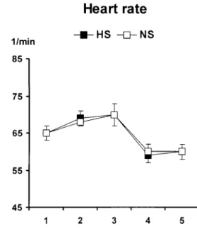
Blood pressure, HR, and CO were measured, and blood samples were taken from the radial arterial cannula before the initial fluid administration, after repositioning the patient in the supine position after Figure 1. Heart rate before (1) and after (2) preload, after spinal puncture (3), after completion of the surgery (4), and after recovery from spinal anesthesia (5) in the hypertonic saline (HS) and normal saline (NS) groups. Values are expressed as mean ⫾ sem. The groups did not differ at any time point.
Table 1. Demographic Data and Effects of Spinal Anesthesia HS (n⫽20) NS (n⫽20) Sex (F/M) 7/13 3/17 Age (yr) 44 (18–63) 42 (23–64) Height (cm) 174 (8) 177 (11) Weight (kg) 81 (16) 79 (12)
Sensory level of anesthesia T6 (T4–10) T7 (T5–10) Duration of anesthesia (min) 154 (36) 156 (43)
Values are mean (sdor range).
the spinal puncture, after the surgical procedure, and after recovery from the spinal anesthesia (plantar and dorsiflexion of both ankles recovered). Plasma concen-trations of sodium, potassium, and chloride and se-rum osmolality were measured. The time of voiding after spinal anesthesia (min after the induction of spi-nal anesthesia) and the volume of urine were recorded.
Before the trial, a power calculation for a 15 mm Hg difference in SBP with a probability level of 0.05 and power of 0.80 (1-) yielded a sample size of 15–16 patients. Accordingly, 20 patients were enrolled in both groups.
Statistical analysis was performed by using the SPSS for Windows (version 9.0) (SPSS Inc., Chicago, IL). The results were analyzed by using an analysis of variance for repeated measures with group as the factor, and time as the repeating factor (before the initial fluid administration, after repositioning the patient in the supine position after the spinal puncture, after the surgical procedure, and after recovery from the spinal anesthesia). Student’s t-test for independent samples was performed at different time points. Dichotomous variables were tested by using the2test. Results are expressed as mean ⫾ sd; P ⬍ 0.05 was considered statistically significant.
Results
The two groups were comparable for patient charac-teristics and the number of blocked segments (Ta-ble 1). The duration of spinal anesthesia did not differ
between the treatment groups (Table 1). The volume of initial fluid administration was 129⫾25 mL in the HS group and 1031 ⫾ 161 mL in the NS group. No notable intraoperative blood loss occurred in either of the study groups.
The groups were similar for the total amount of etilefrine administered (HS: 1.0 ⫾ 2.4 vs NS: 1.0 ⫾ 2.1 mg; not significant). Five patients (25%) in both groups needed etilefrine administration. Baseline val-ues of HR, SBP, DBP, and cardiac index (CI) did not differ between the two groups. No significant differ-ences were observed between the groups in the HR (Fig. 1), SBP and DBP (Fig. 2), or CI (Fig. 3) values during the study.
The plasma sodium and chloride levels as well as the serum osmolality increased after the initial fluid administration. The highest plasma sodium value af-ter the initial fluid administration was 150 mmol/L. All values were within normal range after the surgery. The plasma sodium, chloride, and potassium values, and the serum osmolality are presented in Table 2.
There were no significant differences in the time of voiding after spinal anesthesia (HS 319⫾68 min; NS 290 ⫾ 71 min) or in the volume of urine (HS 449 ⫾ 282 mL; NS 433⫾270 mL). Adverse effects, including sensation of heat and compression around the arm during the study fluid infusion and sensation of thirst, were more common in the HS group (75% of patients) than in the NS group (no adverse effects;P ⬍0.001).
Discussion
We found that 1.6 mL/kg of 7.5% HS was as effective as 13 mL/kg of 0.9% NS in the prophylaxis of hemody-namic changes in ASA physical status I–II patients un-dergoing knee arthroscopy or other lower limb orthope-dic surgery. When the same amount of sodium was Figure 2. Systolic blood pressure (SBP) and diastolic blood
pres-sure (DBP before (1) and after (2) preload, after spinal puncture (3), after completion of the surgery (4), and after recovery from spinal anesthesia (5) in the hypertonic saline (HS) and normal saline (NS) groups. Values are expressed as mean⫾sem.
Figure 3. Cardiac index (CI before (1) and after (2) preload, after spinal puncture (3), after completion of the surgery (4), and after recovery from spinal anesthesia (5) in the hypertonic saline (HS) and normal saline (NS) groups. Values are expressed as mean⫾sem.
infused during the initial fluid administration, the pa-tients required similar amounts of etilefrine to maintain adequate arterial pressure during spinal anesthesia. Both study fluids were able to keep MAP greater than the acceptable limit in 75% of patients. This is in line with the results of Veroli and Benhamou’s group (9). They used 5% saline for the initial fluid administration before ex-tradural anesthesia.
The ideal treatment for hypotension related to spi-nal anesthesia is controversial, as the etiology is still uncertain. Some investigations suggest that hypoten-sion is primarily the result of a reduction in cardiac filling or the initial fluid administration and systemic vascular resistance (15). Initial fluid administration might provide a protection against undesired cardio-vascular side effects (16), but not all studies have shown this (17). The augmentation of blood volume with initial fluid administration, regardless of the fluid used, must be large enough to result in a significant increase in CO for effective prevention of hypotension (18). However, excess free water administration is to be avoided in patients with cardiovascular restric-tions. Fluid administration reduces the incidence of early events, but a vasopressor is needed for preven-tion of the late events (19). We used etilefrine, which is as effective as ephedrine in restoring SBP (20).
The level of sensory nervous block did not differ between the treatment groups. Hyperbaric bupiva-caine 0.5% in a dose of 10 mg was chosen for spinal anesthesia because it provides tolerance to pneumatic tourniquet for 70 min (21), which is usually enough for arthroscopic anterior cruciate ligament-reconstruction and other orthopedic lower limb surgery. This dose may, however, delay the ability to void in some pa-tients (22). In our study, the time of the first micturi-tion in both groups was comparable to previous re-sults (22), despite the much smaller amount of infused free water in the HS group. In addition to the ability to maintain adequate MAP, HS solution improves kid-ney function by reducing renal vascular resistance (23). This may even counteract the adverse effects of
etilefrine on renal vasculature, when vasoconstriction is required.
HS administration causes a rapid increase in serum sodium concentration and osmolality related to the sodium dose. This has been associated with central pontine myelinolysis in chronically debilitated pa-tients with a prolonged period of hyperosmolality or hypernatremia (24). However, 7.5% HS is safe in small-volume resuscitation, and the usual dose is 4 mL/kg (25). In our study, the highest plasma so-dium value after the initial fluid administration was 150 mmol/L. The plasma sodium concentration of all patients was within the normal range after surgery, and the serum osmolality level was within the normal range after spinal anesthesia. Hypokalemia may de-velop after rapid expansion of the plasma volume with HS containing no potassium. This may cause arrhythmia. Hypokalemia was not seen in our pa-tients, however. In the HS group, the patients experi-enced the sensation of heat and compression around the arm during the HS infusion. These symptoms were well tolerated and disappeared immediately af-ter the completion of the HS infusion. Heat and pain are probably caused mainly by the high osmolality of the HS solution and cannot be eliminated totally (26). The patients of the HS group also felt thirsty until they were allowed to drink.
Whole-body impedance cardiography (ICGWB) is a
reliable method compared with other methods of mea-suring CO. Comparison with thermodilution (TD) and direct Fick methods showed that ICGWBmeasures CO
accurately in different conditions (in the supine posi-tion, during head-up tilt, after the induction of anes-thesia and after coronary artery bypass surgery) (14,27,28). The differences in CO values between ICGWB and TD were comparable with those attained
between direct Fick and TD, and the repeatability of ICGWB was nearly twice as good as in TD (27,28).
Therefore, ICGWB is an adequate method to estimate CO and its changes. In our patients, CI remained at the same level in the HS group as in the NS group. HR did
Table 2. Plasma Sodium (Na), Potassium (K) and Chloride (Cl), and Serum Osmolality
Group Baseline After preload After surgery
After recovery from spinal anesthesia Plasma Na (mmol/L) HS 140⫾1 145⫾2* 142⫾2* 141⫾1* NS 140⫾1 140⫾1 140⫾2 139⫾1 Plasma K (mmol/L) HS 3.9⫾0.2 3.8⫾0.2 4.0⫾0.3 4.0⫾0.2 NS 3.9⫾0.2 3.9⫾0.3 3.9⫾0.2 3.9⫾0.2 Plasma Cl (mmol/L) HS 107⫾2 114⫾3* 111⫾3† 111⫾2 NS 107⫾2 110⫾2 109⫾3 109⫾3
Serum osmolality (mosm/kg) HS 291⫾4 302⫾5* 297⫾4* 295⫾3*
NS 292⫾3 292⫾3 290⫾3 290⫾3
HS⫽hypertonic saline, NS⫽normal saline.
not differ between the groups. HS solution increases myocardial contractility (29). This effect of HS proba-bly explains the improved CO after HS infusion. Be-cause osmolality is the driving force for volume dis-tribution, HS causes fluid shift from the intracellular space into the intravascular and interstitial spaces (30). Therefore, it increases the plasma volume more than by its own volume. The increase of myocardial con-tractility and plasma volume together lead to in-creased CO and thus help stabilize the hemodynamics during spinal anesthesia.
We conclude that 7.5% HS was as good as NS for the initial fluid administration before spinal anesthesia when the amount of sodium was kept unchanged. It was effective in small doses of 1.6 mL/kg. Also, the potential disadvantages of HS solution, including hy-pernatremia, hyperosmolality, and hypokalemia, could be avoided. HS can be recommended for the initial fluid administration in situations in which ex-cess free water administration is not desired. We have studied ASA physical status I–II patients, but initial fluid administration with HS may also be beneficial in other patient groups, such as elderly patients with cardiovascular restrictions.
We thank Pirjo Ja¨rventausta, RN, and Satu Ruusuvuori, RN, for their valuable technical assistance.
References
1. Carpenter RL, Caplan RA, Brown DL. Incidence and risk factors for side effects of spinal anesthesia. Anesthesiology 1992;76: 906 –16.
2. Onarheim H. Fluid shifts following 7% hypertonic saline (2400 mosmol/L) infusion. Shock 1995;3:350 – 4.
3. Tollofsrud S, Noddeland H. Hypertonic saline and dextran after coronary artery surgery mobilises fluid excess and improves cardiorespiratory functions. Acta Anaesthesiol Scand 1998;42: 154 – 61.
4. Vassar MJ, Perry CA, Holcroft JW. Analysis of potential risks associated with 7.5% sodium chloride resuscitation of traumatic shock. Arch Surg 1990;125:1309 –15.
5. Bitterman H, Triolo J, Lefer AM. Use of hypertonic saline in the treatment of hemorrhagic shock. Circ Shock 1987;21:271– 83. 6. Ramires JAF, Serrano CV Jr, Cesar LAM, et al. Acute
hemody-namic effects of hypertonic (7.5%) saline infusion in patients with cardiogenic shock due to right ventricular infarction. Circ Shock 1992;37:220 –5.
7. De Filippe J, Timoner J, Velasco IT, et al. Treatment of refractory hypovolemic shock by 7.5% sodium chloride injections. Lancet 1980;2:1002– 4.
8. Cross JS, Gruper DP, Burchard KW, et al. Hypertonic saline fluid therapy following surgery: a prospective study. J Trauma 1989;29:817–25.
9. Veroli P, Benhamou D. Comparison of hypertonic saline (5%), isotonic saline and Ringer’s lactate solutions for fluid preload-ing before lumbar extradural anaesthesia. Br J Anaesth 1992;69: 461– 4.
10. Smith GJ, Kramer GC, Perron P, et al. A comparison of several hypertonic solutions for resuscitation of bled sheep. J Surg Res 1985;39:517–39.
11. Baraka A, Taha S, Ghabach M, et al. Hypertonic saline prehy-dration in patients undergoing transurethral resection of pros-tate under spinal anaesthesia. Br J Anaesth 1994;72:227– 8. 12. Wang BW, Chiou YH, Chen WB, et al. Intravenous pretreatment
of hypertonic saline can prevent systemic hypotension induced by spinal anesthesia. Acta Anaesthesiol Sin 1997;35:85–90. 13. Karabeyog˘lu I, Go¨g˘ u¨s N, Gu¨mu¨s˛ S, et al. Effects of preloading
with NaCl 0.9%, HES (450/0.7) 6% and NaCl 3% on haemody-namic changes and serum osmolality in patients undergoing TURP under spinal anaesthesia [abstract]. Br J Anaesth 1998;80: suppl1:A365.
14. Ko¨o¨bi T, Kaukinen S, Turjanmaa VM, Uusitalo AJ. Whole-body impedance cardiography in the measurement of cardiac output. Crit Care Med 1997;25:779 – 85.
15. Marhofer P, Faryniak B, Oismu¨ller et al. Cardiovascular effects of 6% hetastarch and lactated Ringer’s solution during spinal anesthesia. Reg Anesth Pain Med 1999;24:399 – 404.
16. Casati A, Fanelli G, Berti M, et al. Cardiac performance during unilateral lumbar spinal block after crystalloid preload. Can J Anaesth 1997;44:623– 8.
17. Coe AJ, Revana¨s B. Is crystalloid preload useful in spinal an-aesthesia in the elderly? Anan-aesthesia 1990;45:241–3.
18. Ueyama H, He Yan-ling, Tanigami H, et al. Effects of crystalloid and colloid preload on blood volume in the parturient under-going spinal anesthesia for elective cesarean section. Anesthesi-ology 1999;91:1571– 6.
19. Arndt JO, Bo¨mer W, Krauth J, et al. Incidence and time course of cardiovascular side effects during spinal anesthesia after prophylactic administration of intravenous fluids or vasocon-strictors. Anesth Analg 1998;87:347–54.
20. Taivainen T. Comparison of ephedrine and etilefrine for the treatment of arterial hypotension during spinal anaesthesia in elderly patients. Acta Anaesthesiol Scand 1991;35:164 –9. 21. Liu SS, Ware PD, Allen HW, et al. Dose-response characteristics
of spinal bupivacaine in volunteers: clinical implications for ambulatory anesthesia. Anesthesiology 1996;85:729 –36. 22. Tarkkila P, Huhtala J, Tuominen M. Home-readiness after
spi-nal anaesthesia with small doses of hyperbaric 0.5% bupiva-caine. Anaesthesia 1997;52:1157– 60.
23. Fujita T, Matsuda Y, Shibamoto T, et al. Effect of hypertonic saline infusion on renal vascular resistance in anesthetized dogs. Jpn J Physiol 1991;41:653– 63.
24. Mc Kee AC, Winkelman MD, Banker BQ. Central pontine my-elinolysis in severely burned patients: relationship to serum hyperosmolality. Neurology 1988;38:1211–7.
25. Kreimeier U, Messmer K. Small-volume resuscitation. Baillieres Clin Anaesthesiol 1988;2:545.
26. Himi K, Takemoto A, Himi S, et al. Heat and pain sensation induced by arterial injection of low-osmolality contrast media: a comparison of patients’ discomfort with ionic saline, nonionic glucose and vasodilator nitrate. Acad Radiol 1996;3 Suppl 2:S214 –7.
27. Ko¨o¨bi T, Kaukinen S, Ahola T, Turjanmaa VMH. Non-invasive measurement of cardiac output: whole-body impedance cardi-ography in simultaneous comparison with thermodilution and direct oxygen Fick methods. Intensive Care Med 1997;23:1132–7. 28. Ko¨o¨bi T, Kaukinen S, Turjanmaa VMH. Cardiac output can be reliably measured noninvasively after coronary artery bypass grafting operation. Crit Care Med 1999;27:2206 –11.
29. Mouren S, Delayance S, Mion G, et al. Mechanism of increased myocardial contractility with hypertonic saline solutions in iso-lated blood-perfused rabbit hearts. Anesth Analg 1995;81: 777– 82.
30. Mazzoni MC, Borgstro¨m P, Arfors KE, et al. Dynamic fluid redistribution in hyperosmotic resuscitation of hypovolemic hemorrhage. Am J Physiol 1988;255:H629 –37.

![Efficiency of 7 2% hypertonic saline hydroxyethyl starch 200/0 5 versus mannitol 15% in the treatment of increased intracranial pressure in neurosurgical patients – a randomized clinical trial [ISRCTN62699180]](data:image/gif;base64,R0lGODlhAQABAIAAAP///wAAACH5BAEAAAAALAAAAAABAAEAAAICRAEAOw==)