THE EFFECTS OF ACUTE AND
CHRONIC ANTIGEN
INHALATION ON AIRWAY
INFLAMMATION AND
FUNCTION AND ANTI
INFLAMMATORY DRUG
INTERVENTIONS
A THESIS SUBMITTED TO CARDIFF UNIVERSITY IN ACCORDANCE
WITH THE REQUIREMENTS FOR THE DEGREE OF
DOCTOR OF PHILOSOPHY
RHYS EVANS
DEPARTMENT OF PHARMACOLOGY
WELSH SCHOOL OF PHARMACY
CARDIFF UNIVERSITY
2009
UMI Number: U584444
All rights reserved
INFORMATION TO ALL USERS
The quality of this reproduction is dependent upon the quality of the copy submitted.
In the unlikely event that the author did not send a complete manuscript and there are missing pages, these will be noted. Also, if material had to be removed,
a note will indicate the deletion.
Dissertation Publishing
UMI U584444
Published by ProQuest LLC 2013. Copyright in the Dissertation held by the Author. Microform Edition © ProQuest LLC.
All rights reserved. This work is protected against unauthorized copying under Title 17, United States Code.
ProQuest LLC
789 East Eisenhower Parkway P.O. Box 1346
DECLARATION AND STATEMENTS
DECLARATION
This work has not previously been accepted in substance for any degree and is not concurrently submitted in candidature for any degree.
S igned ...(candidate) Date.... IHj.kj.^.9. /?...
STATEMENT 1
This thesis is being^ibmitted in partial fulfilment o f the requirements for the degree o f PhD.
Signed ... (candidate) Date.. ...
STATEMENT 2
This thesis is the result o f my own independent work/investigation, except where otherwise stated. Other sources are acknowledged by explicit references.
Signed (candidate) Date.... M l Q . l l...
STATEMENT 3
I hereby give consent for my thesis, if accepted, to be available for photocopying and for inter-libraryloan, and for the title and summary to be made available to outside organisations. Signed ...(candidate) Date.. . . ( U-12®.I®.
ACKNOWLEDGEMENTS
I am extremely grateful towards Professor Kenneth Broadley, Dr. Emma Kidd and Dr. Will Ford for all o f the help and support they have given me throughout my PhD studies. Without their help this thesis would not exist. Special thanks also go to my industrial supervisor Dr. Tony Nials for always rapidly replying to my emails, friendly banter and making my time at Stevenage enjoyable.
Thanks also to the Respiratory CEDD at GSK who made me feel welcome during my time there and to the MR1 group, especially Kumar Changani, for all the help and support during the work that became chapter 8.
Several people at Cardiff University have made the last three years easier and more enjoyable. These people include the technical staff, especially Sarah and Susan, Joint services staff, especially Martin, the ‘Coffee Crew’ and Pharmacology group, especially Dawn, Amy, Rhian and Alan for willingly lending a helping hand when needed.
Thanks go to The Moving Lights for providing an outlet from science, Glastonbury beckons! 1 am indebted to my friends and my family, especially Dad, Mum and Owain, for all o f the encouragement, support and willingness to listen to me complain!
SUMMARY
Asthma is a chronic inflammatory disease characterised by airway inflammation, bronchoconstriction, airway hyperresponsiveness (AHR) and airway remodelling. Most models o f asthma focus on acute allergen challenges, where airway remodelling is absent. The thesis aimed to compare acute and chronic allergen challenge models of asthma and analyse the effects o f anti-inflammatory drugs on these models.
Acute and chronic challenges with ovalbumin in conscious guinea pigs and mice caused impared lung function, measured as specific airway conductance (sGaw) and enhanced pause
(Penh),
respectively. This was characterised by early (EAR) and late (LAR) asthmatic responses, AHR and cellular influx. Multiple challenges with ovalbumin caused airway remodelling distinguished by increased bronchiolar collagen and goblet cells compared to control. No airway remodelling was observed in acute ovalbumin models.Treatment with the corticosteroid, fluticasone propionate (FP), attenuated the LAR, AHR and cellular influx in all models. In both chronic ovalbumin models, FP treatment partially reversed airway remodelling, though not to naive levels. This was also the case with treatment with the phosphodiesterase IV inhibitor, roflumilast. However, roflumilast also attenuated the EAR. Treatment with the iNOS inhibitor, GW274150F, reduced the LAR and AHR and showed some inhibition o f cellular influx in the acute ovalbumin challenged animals. However, GW274150F was ineffective in chronic ovalbumin models. Lung oedema, assessed by magnetic resonance imaging in acute and chronic ovalbumin challenged guinea pigs, was increased and correlated temporarily with the LAR. Dexamethasone treatment attenuated the level o f oedema though not to control levels.
Acute ovalbumin challenged models showed airway functional changes which were partially resolved with drug treatment. Chronic ovalbumin challenges provoked lung functional and structural changes which were attenuated by FP and roflumilast but not GW274150F. As GW274150F proved ineffective in clinical trials, the data in this thesis suggests that chronic ovalbumin challenge animals are better pre-clinical models of asthma.
CONTENTS
Page
DECLARATION AND STATEMENTS i
ACKNOWLEDGEMENTS ii
SUMMARY iii
CONTENTS iv
ABBREVIATIONS vii
CHAPTER 1: GENERAL INTRODUCTION
1.1 ASTHMA 2 1.2 AIRWAYS INFLAMMATION 5 1.3 AIRWAY HYPERRESPONSIVENESS 11 1.4 AIRWAY REMODELLING 13 1.5 TREATMENT 18 1.6 MODELS OF ASTHMA 21 1.7 AIMS 23 CHAPTER 2: METHODS
2.1 MATERIALS AND EQUIPMENT 25
2.2 ANIMAL EXPERIMENTS 25
2.3 STATISTICAL ANALYSIS 50
CHAPTER 3: ANALYSIS OF GUINEA PIG ACUTE AND CHRONIC MODELS OF ASTHMA
3.1 INTRODUCTION 52
3.2 AIMS AND OBJECTIVES 56
3.3 METHODS 57
3.4 RESULTS 62
3.5 DISCUSSION 82
CHAPTER 4: ANALYSIS OF MOUSE ACUTE AND CHRONIC MODELS OF ASTHMA
4.1 INTRODUCTION 88
4.2 AIMS AND OBJECTIVES 92
4.3 METHODS 93
4.4 RESULTS 96
4.5 DISCUSSION 113
CHAPTER 5: EFFECT OF CORTICOSTEROID TREATMENT ON ACUTE AND CHRONIC MODELS OF ASTHMA
5.1 INTRODUCTION 118
5.2 AIMS AND OBJECTIVES 124
5.3 METHODS 125
5.4 RESULTS 129
5.5 DISCUSSION 154
CHAPTER 6: EFFECT OF PHOSPHODIESTERASE IV INHIBITOR TREATMENT ON ACUTE AND CHRONIC MODELS OF ASTHMA
6.1 INTRODUCTION 159
6.2 AIMS AND OBJECTIVES 163
6.3 METHODS 164
6.4 RESULTS 168
6.5 DISCUSSION 194
CHAPTER 7: EFFECT OF INDUCIBLE NITRIC OXIDE INHIBITOR TREATMENT ON ACUTE AND CHRONIC MODELS OF ASTHMA
7.1 INTRODUCTION 200
7.2 AIMS AND OBJECTIVES 204
7.3 METHODS 205
7.4 RESULTS 208
CHAPTER 8: MRI ANALYSIS OF ACUTE AND CHRONIC MODELS OF ASTHMA
8.1 INTRODUCTION 238
8.2 AIMS AND OBJECTIVES 245
8.3 METHODS 246
8.4 RESULTS 258
8.5 DISCUSSION 269
CHAPTER 9: GENERAL DISCUSSIONS
9.1 MAIN AIMS AND METHODS 275
9.2 EXPERIMENTAL LIMITATIONS 282 9.3 FURTHER WORK 284 9.4 CLINICAL RELEVANCE 285 CHAPTER 10: REFERENCES 287 APPENDIX 1 314 APPENDIX 2 317 APPENDIX 3 320 vi
ABBREVIATIONS
3-NT = 3-nitrotyrosine
5’AMP = 5’adenosine monophosphate AB/PAS = Alcian blue/periodic acid Schiff AHR = Airway hyperresponsiveness
Al(OH>3 = Aluminium hydroxide
APC = Antigen presenting cell ATP = Adenosine trisphosphate AUC = Area under the curve BAL = Bronchoalveolar lavage
cAMP = Cyclic adenosine monophosphate CBP = CREB binding protein
cGMP = Cyclic guanosine monophosphate COX = Cyclooxygenase
CPLA2 = Cytoplasmic phospholipase A2
CREB = Cyclic adenosine monophosphate response element-binding protein CysLT = Cysteinyl Leukotriene
DMSO = Dimethyl sulfoxide EAR = Early asthmatic response ECM = Extracellular matrix ECP = Eosinophil cationic protein EDN = Eosinophil derived neurotoxin EGF = Epidermal growth factor
ELISA = Enzyme-linked immunosorbant assay eNOS = Endothelial nitric oxide synthase EPO = Eosinophil peroxidase
ET-1 = Endothelin-1 EtOH = Ethanol
FACS = Fluorescent-activated cell sorting
F En o = Level o f nitric oxide in exhaled air
FEVj = Forced expired volume over one second of expiration FP = Fluticasone propionate
FWBP = Flow whole-body plethysmograph G ,w = Airway conductance
GM-CSF = Granulocyte-macrophage colony stimulating factor GR = Glucocorticoid receptor
GRE = Glucocorticoid response element GTP = Guanosine trisphosphate
HAT = Histone acetyltransferase HD AC = Histone deacetylase HRP = Horse radish peroxidase
ICAM = Intercellular adhesion molecule IFN = Interferon
Ig = Immunoglobulin
IicBa = Inhibitor of kappa B - a IKK2 = Inhibitor of IkB kinase-2
IL = Interleukin
IMS = Industrial methylated spirit iNOS = Inducible nitric oxide synthase I.P. = Intraperitoneal
LAR = Late asthmatic response LPS = Lipopolysaccharide LT = Leukotriene
MBP = Major basic protein
MCP = Monocyte chemotactic protein mDC = Myeloid dendritic cell
MHC = Major histocompatibility complex MIP = Macrophage Inflammatory Protein MMP = Matrix metalloproteinases
MRI = Magnetic resonance imaging
NADPH = Nicotinamide adenine dinucleotide phosphate (reduced) NF-kB = Nuclear factor kappa-light-chain-enhancer of activated B cells NGF = Nerve growth factor
nNOS = Neuronal nitric oxide synthase NO = Nitric oxide
NOS = Nitric oxide synthase O2" = Superoxide oxygen molecule OONO' = Peroxynitrite
OPD = O-phenylenediamine dihydrochloride OVA = Ovalbumin
PBS = Phosphate buffered saline
PBST = Phosphate buffered saline with tween pDC = Plasmacytoid dendritic cell
PDE = Phosphodiesterase
PDGF = Platelet-derived growth factor Penh = Enhanced pause
PICP = Procollagen type I C-terminal peptide PWBP = Pressure whole-body plethysmograph
RF = Radio frequency
S.E.M. = Standard error of the mean sGaw = Specific airway conductance Tc CELL = T cytotoxic cell
TGF = Transforming growth factor TGV = Thoracic gas volume
Th CELL = T helper cell TNF = Tumor necrosis factor
TSLP = Thymic stromal lumphopoietin VEGF = Vascular endothelial growth factor
CHAPTER 1
Chapter 1
General
CHAPTER 1
1.1 ASTHMA
1.1.1 DEFINITION
Asthma has proven to be frustratingly difficult to define in an accurate and precise manner. The first definition came from Hippocrates (460-377 BC) who used the Greek word ‘asthmaino’ meaning panting or gasping. However, a link between asthma and bronchospasm was not made until Galen (130-201 AD). A definition of asthma continued to evolve. The most commonly quoted definition of asthma comes from the National Asthma Education and Prevention Program Export Panel Report 2, which described asthma as:
A chronic inflammatory disorder o f the airways in which many cells and cellular elements play a role, in particular, mast cells, eosinophils, T lymphocytes, neutrophils, and epithelial cells. In susceptible individuals, this inflammation causes recurrent episodes o f wheezing, breathlessness, chest tightness, and cough, particularly at night and in the early morning. These episodes are usually associated with widespread but variable airflow obstruction that is often reversible either spontaneously or with treatment. The inflammation also causes an associated increase in the existing bronchial hyperresponsiveness to a variety o f stimuli. (1987)
However, this does not begin to describe the complexity of the disease and it is now known not to be completely accurate as several patients with asthma show extremely poor reversibility (Bousquet et al., 2000). It is believed that around 5.4 million Britons are receiving treated for asthma and there is a person with asthma in one o f every five households (Asthma UK). It is estimated that the economic costs of asthma will exceed those o f tuberculosis and HIV-AIDS combined (van Schalkwyk et al., 2005).
1.1.2 PATHOPHYSIOLOGY
Asthma can be classified into two different forms, intrinsic or extrinsic asthma (Wardlaw
et al., 2002). Intrinsic, or non-atopic, asthma is triggered by something inside the body, not by an allergy. Respiratory infections, exercise, cold weather and certain drugs, such
C H A P T E R 1 as non-steroidal anti-inflammatory drugs, can cause a reflex in the upper airways leading to asthmatic symptoms. This form of the disease is not caused by hypersensitivity to an antigen or allergen and levels of immunoglobulin E (IgE) remain normal in comparison to non-asthmatics.
In contrast, extrinsic, or atopic/allergic, asthma is IgE mediated. It is commonly diagnosed early in life and is a result of hypersensitivity to an inhaled antigen such as dust mites, pollen or ovalbumin. Individuals who suffer from extrinsic asthma become sensitive to the allergen; whenever they encounter the allergen again asthmatic symptoms occur. As a result extrinsic asthma is described as being caused by two phases, the sensitisation phase and the effector phase (figure 1).
Allergen B cell Plasma Allergen Vasoactive —amines allergen
•2
Fc receptor for IgE Degranulation Th cellY y W
1 s /IT Allergen-Sensitised mast cell specific IgEFigure 1 - Schematic diagram o f how sensitisation to an allergen occurs and the effect that a repeated exposure to the allergen has. Picture adapted from Goldsby et a l(2000). IgE = Immunoglobulin E; Th cell = T helper cell.
1.1.3 SENSITISATION PHASE
Circulating allergens can be recognised as foreign by antigen presenting cells (APC), such as macrophages, B cells and dendritic cells. IgE on the surface of the APC captures
CHAPTER 1 the antigen and causes it to be internalised by phagocytosis or endocytosis. The antigen is then proteolysed into peptides and part o f it is displayed on the membrane bound major histocompatibility complex (MHC) class II on the surface o f the APC (Banchereau & Steinman, 1998). The antigen is then recognised by naive T cells which then become activated. Once activated, the naive T cells will differentiate into T helper 1 (T h l) or T helper 2 (Th2) cells. This differentiation is triggered by interleukin-2 (IL-2). Naive T cells release IL-2 and at the same time develop surface receptors specific for it, once the receptor is active the differentiation occurs. W hether the cell becomes Thl or Th2 is dependent on the presence o f the autocrine cytokines IL-12 and IL-4 respectively (Romagnani, 1997). The Thl response mainly activates certain T cells and macrophages through a cytokine profile that supports inflammation. The Th2 response activates B cells and immune responses dependent on antibodies (Goldsby et al., 2000). TheThl response is responsible for killing intracellular parasites and is more involved in autoimmune conditions (Berger, 2000). In asthmatic patients a trend is seen toward the differentiation o f Th2 cells.
Active Th2 cells cause sensitisation to the allergen by releasing the cytokines IL-4 and IL-13. IL-4 and IL-13 stimulate the production o f allergen-specific IgE from plasma cells. IL-4 also induces the expression o f FceRI, or high affinity allergen-specific IgE, receptors on the surface o f mast cells and basophils. Monovalent binding o f IgE causes increased FceRI expression, increases mast cells resistance to apoptosis and can induce cytokine release (Knol, 2006). Once the allergen-specific IgE binds to these receptors sensitisation has occurred, therefore exposure to the same allergen would lead to an asthmatic response.
1.1.4 EFFECTOR PHASE
The asthmatic response following re-exposure o f a specific allergen is two-fold. The first response is an immediate bronchoconstriction and is known as the early asthmatic response (EAR). The second bronchoconstriction does not occur until around seven hours after allergen exposure and is known as the late asthmatic response (LAR). The responses are a result o f the re-exposed allergen binding to the membrane-bound IgE on the sensitised mast cells or basophils. The bound IgE interacts and cross-links with the IgE
CHAPTER 1 that bound to the mast cell or basophil during the sensitisation phase. Cross-linking o f bound IgE leads to surface receptor clustering which induces a cascade o f tyrosine phosphorylation events resulting in the activation o f phospholipase Cy and ultimately causing degranulation o f the mast cells or basophils (Knol, 2006). Mast cells and basophil degranulation occurs rapidly, within 60-300 seconds (Holmes 1999 referenced by John 2007). Mast cells contain many inflammatory mediators, including histamine, tryptase, prostaglandins, leukotrienes and cytokines, which are released when mast cells are degranulated. Some o f these mediators, such as histamine, act on the local airway receptors causing the immediate bronchoconstriction or EAR. The LAR is caused by some o f the other mediators chemoattracting inflammatory cells, such as macrophages, eosinophils, lymphocytes and neutrophils which in turn attract more inflammatory mediators and cause bronchoconstriction (Durham & Kay, 1985).
1.2 AIRWAY INFLAMMATION
Airway inflammation is one o f the defining characteristics o f asthma, it can be present even in mild asthma (Laitinen et al., 1996). Along with airway hyperresponsiveness, airway inflammation contributes to the common symptoms of asthma - chest tightness, wheezing, cough and shortness o f breath. However, there are other mechanisms such as the activation o f airway sensory nerves which can contribute to these symptoms, especially cough (Nasra & Belvisi, 2009). It has been established that the extent o f inflammation correlates with the severity o f the disease (Walter & Holtzman, 2005). Elevation o f the number o f inflammatory cells in the airways o f patients who die o f an asthma attack (status asthmaticus) was observed by Barnes (1996). Although some inflammatory cells predominate in causing inflammation o f the airways no single cell can account for the complex pathophysiology o f asthma. Inflammation is caused by a complex cascade o f events involving various inflammatory cells and mediators.
1.2.1 MAST CELLS
As previously mentioned mast cells play a key role in the sensitisation and effector phase o f asthma. The number o f mast cells found in the broncho-alveolar lavage (BAL) fluid o f
CHAPTER 1 asthmatics is two- to six-fold higher than found in non-asthmatics (Hamid et al., 2003). The percentage o f mast cells expressing the cytokines IL-4, IL-5 and tumour necrosis factor (TN F)-a is also increased in asthmatics (Bradding et al., 1994). These cytokines cause increased Th2/reduced T hl cell production, reduced apoptosis and bronchial hyperresponsiveness respectively (Barnes, 2002a).
Mast cells contain cytoplasmic granules in which mediators such as histamine, tryptase, prostaglandin D2 (PGD2) and leukotriene C4 (LTC4) (Barnes, 2002a). The glycosaminoglycan, heparin is also stored by mast cells (Rose & Page, 2004). These mediators are released when the mast cells become degranulated. Degranulation is caused by the binding o f an allergen to the allergen-specific IgE bound to the mast cell or by other indirect stimuli, such as in intrinsic asthm a (Barnes, 1996). The released mediators cause bronchoconstriction, increased vascular permeability and leukocyte recruitment and activation, (Galli, 2000) all o f which contribute towards airway inflammation.
1.2.2 MACROPHAGES
Macrophages are derived from blood m onocytes and are the most prominent cell found in the BAL fluid o f both asthmatic and non-asthmatic patients (Hamid et al., 2003). As previously mentioned macrophages can act as an APC for presenting an allergen to a naive T cell. Macrophages have the ability to play a role in both increasing and decreasing airway inflammation. They are able to release cytokines, which induce epithelial cells and fibroblasts to release chemoattractants and growth factors. Some o f these chemoattractants such as RANTES (regulated upon activation, normal T-cell expressed and secreted) and monocyte chemotactic protein (M CP)-l attract further macrophages and eosinophils. This would result in more macrophages acting as APCs and therefore inflammation would occur. On the other hand alveolar macrophages can have a suppressive effect on lymphocyte activity; however, this effect may be impaired after allergen exposure (Barnes, 1996; Hamid et al., 2003).
1.2.3 EOSINOPHILS
Eosinophils play a predominant role in the late asthmatic response (Goldsby et al., 2000). The number o f eosinophils in the airways has been correlated to the severity o f asthma
CHAPTER 1 (Hamid et al., 2003). In the bronchoalveolar lavage (BAL) fluid o f sensitised and allergen challenged guinea pigs the number o f eosinophils in the airways are shown to progressively increase after an hour until a peak o f 24 hours (Toward & Broadley, 2004). This correlates with the late asthmatic response. Eosinophil recruitment is believed to occur by two exclusive pathways. The first is through the release o f TNF-a by degraded mast cells, this acts on epithelial and endothelial cells causing the release o f eosinophil- directed cytokines, such as granulocyte macrophage colony-stimulating factor (GM- CSF), and chemokines, such as eotaxin (Barnes, 2002a). The second pathway involves the release o f IL-5 by recently activated naive T cells. IL-5 provides the signal for eosinophils to mobilise from the bone marrow and it also enhances the chemoattractant potential o f tissue chemokines (Bames, 2002a). Once in tissue, eosinophils can amplify the inflammatory cascade by producing their own cytokines (Rothenberg, 1998).
Eosinophils are not very phagocytic but contain granules that hold highly charged and basic proteins. IL-3, IL-5, GM-CSF, IL -lp and platelet activating factor can degranulate eosinophils (Chapoval et al., 1999) causing the release o f the granule proteins. There are four types o f granule proteins released from eosinophils; these are major basic protein (MBP), eosinophil cationic protein (ECP), eosinophil derived neurotoxin (EDN) and eosinophil peroxidase (EPO). MBP, ECP and EPO are all able to cause histamine release from mast cells. ECP is also able to damage target cells by causing voltage-insensitive, ion non-selective pores therefore allowing an influx o f potentially damaging ions such as calcium (Rothenberg, 1998). MBP is believed to have a pathogenic role in asthma as it has been shown to cause damage to airway epithelial cells and levels o f MBP in the airway decline in correlation with improved airway function (Bames, 2002a).
In addition to basic proteins, eosinophils can also produce LTC4, Platelet Activating Factor (PAF) (Chapoval et al., 1999), reactive oxygen species (ROS), transforming growth factor (TGF) a and p, TNF- a, IL-1, IL-3, IL-5, IL-6 and GM-CSF (Busse & Lemanske, 2001) all o f which can contribute to airway inflammation via various mechanisms such as airway smooth muscle contraction, increased vascular permeability and mucus secretion. They are also able to chemoattract more inflammatory cells by releasing the chemokines, eotaxin, Macrophage Inflammatory Protein (M lP )-la and RANTES (Nickel et al., 1999). A role for eosinophils in airway remodelling may also
CHAPTER 1 exist as they produce fibrogenic growth factors, elastase and matrix metalloproteinases (MMP) (Bousquet et al., 2000).
1.2.4 T-LYMPHOCYTES
T-lymphocytes are essential in the asthmatic inflammatory response, they can be grouped depending whether they have a CD4 or CD8 surface cell marker. Generally CD4+ cells are T helper (Th) cells, whereas CD8+ are T cytotoxic (Tc) cells. It is the Th cells that are presented an allergen by an APC and as a result become active. As previously mentioned Th cells can be further divided into T hl and Th2 cells depending on the presence o f various cytokines. Th2 cells are more frequently found in asthmatics and it is these cells that release IL-4 and IL-13 which lead to IgE release from plasma cells. The cytokines that T-lymphocytes release can also serve to attract and activated other cell types. Bronchial hyperresponsiveness and eosinophil number and activation has been shown to correlate with activated CD4+ lymphocytes (Robinson et al., 1993). Lymphocytes may play a role in airway remodelling as there is some evidence that they stimulate the synthesis o f matrix proteins such as collagen I, III and fibronectin (Postlethwaite et al.,
1992; Postlethwaite & Seyer, 1991).
1.2.5 NEUTROPHILS
Neutrophils do not have a clear role in asthma. Some studies have shown a rapid influx o f neutrophils into the airways following a stimulus challenge in asthma models (Fabbri et al., 1984; Matsumoto et al., 1999). This influx was associated with bronchial hyperresponsiveness suggesting there is a potential role for neutrophils. Evidence o f a neutrophilic asthma, where neutrophils predom inate over eosinophils, exists (Wardlaw et al., 2002) though results are controversial. Neutrophils contain reactive oxygen species and proteases, that are both capable o f causing damage to the airways if released. Therefore, neutrophils have the potential to cause airway inflammation. In sensitised and allergen challenged guinea pigs airway neutrophils were shown to increase after an hour, peak at the end o f the early asthmatic response and had subsided after 12 hours (Toward & Broadley, 2004) suggesting neutrophils may play a role in the early phase bronchoconstriction associated with asthma. There is a strong association between
CHAPTER 1 neutrophilic inflammation o f the airways and severe asthma (Jatakanon et al., 1999; Wenzel et al., 1997), steroid resistant asthma (Pavord et al., 1999; Wenzel et al., 1999) and the sudden onset o f fatal asthma (Sur et al., 1993).
1.2.6 DENDRITIC CELLS
The role o f dendritic cells is primarily as APCs. As dendritic cells express high levels o f MHC class II molecules they are more potent APCs than macrophages and B cells (Goldsby et al., 2000). There are several different subtypes o f dentritic cells, however, the two that have the most prominent role in asthma are myeloid dentritic cells (mDC) and plasmacytoid dendritic cells (pDC) (Bam es, 2002a). These cells are crucial in determining the outcome o f allergen encounter in the lung (Lambrecht, 2008). Under normal conditions the outcome o f inhalation o f a harmless allergen would lead to immunological tolerance. The allergen would be taken up by both mDCs and pDCs. Under normal conditions the mDCs would be partly mature and the T-cell response they induce is characterised by cell division and not differentiation to effector cells (Bames, 2002a) and therefore result in immunological tolerance. This process is regulated by pDCs, cyclooxygenase-2 derived prostaglandins and complement activation (Bames, 2002a).
Th2 sensitisation to an allergen can also be caused by mDCs (Lambrecht, 2008). For this to occur it seems that mDCs need to be activated, or matured (Bames, 2002a). Several factors can cause this. Toll-like receptor agonists, such as endotoxin, adjuvants, viral infection, overexpression o f GM-CSF and cigarette smoke have all been implicated in causing Th2 sensitisation through activation o f mDCs (Bames, 2002a). Activation o f mDCs can be direct, by factors such as low dose toll-like receptor agonists or adjuvants, or indirect (Bames, 2002a). Indirect activation can be caused by factors leading to epithelial activation, e.g. cigarette smoke, increasing the mDC/pDC balance, e.g. GM- CSF overexpression, or by the loss o f pDC tolerogenic function, e.g. viral infection (Bames, 2002a).
Dendritic cells may also have a role in the effector phase o f asthma as it appears that ‘inflammatory’ dendritic cells are necessary and sufficient for secondary immune responses to the allergen (Bames, 2002a). The fact that dendritic cells have an
CHAPTER 1 inflammatory role in asthma has led to the development o f novel anti-allergic compounds aiming to combat this inflammatory effect (Lambrecht, 2008).
1.2.7 CYTOKINES AND GROWTH FACTORS
Cytokines and growth factors play a huge role in asthma, one that goes way beyond the scope o f this thesis. Some have a pro-inflammatory role in asthma whereas some have the opposite. Table 1 describes the roles o f some o f the more important pro-inflammatory cytokines and growth factors in asthma.
Cvtokine / Growth Factor Effect in Asthma Mechanism
IL -ip Enhances disease Increases inflammation
IL-4 Enhances disease Increases IgE production and
number o f Th2 cells
IL-5 Enhances disease Increases eosinophil number
IL-6 Enhances disease Increases inflammation
IL-9 Enhances disease Increases mast cell number
IL-10 Decreases disease Decreases inflammation
IL-12 Reduces disease Increases Th 1 number
IL-13 Enhances disease Increases IgE production and
induces airway remodelling
IL-17 Enhances disease Increases neutrophil number
IL-18 Reduces disease Increases IFN-y release
IL-25 Enhances disease Increases Th2 number
EGF Enhances disease Induces mucus secretion
GM-CSF Enhances disease Increases eosinophil and
neutrophil number
IFN-y Reduces disease Decreases Th2 number
NGF Enhances disease Increases AHR
TGF-p Enhances severe disease Increases fibrosis
TNF-a Enhances severe disease Increases inflammation
CHAPTER 1
TSLP Enhances disease Increases Th2 number
VEGF Enhances disease Induces angiogenesis
T a b le 1- A list o f cytokines and growth factors involved in asthma and the response they cause. Adapted from (Bam es, 2008). IL = Interleukin; EGF = Epidermal growth factor; GM-CSF = Granulocyte-macrophage colony stimulating factor; IFN = Interferon; NGF = Nerve growth factor; TGF = Transforming growth factor; TNF = Tumour necrosis factor; TSLP = Thymic stromal lumphopoietin; VEGF = Vascular endothelial growth factor.
1.2.8 HISTAMINE
Histamine plays a fundamental role in causing airway inflammation and hyperresponsiveness in asthma. It is stored in, and then released from, mast cells through an IgE mediated pathway. Upon release, histamine constricts airway smooth muscle in the large and small airways, causes plasm a exudation and bronchial vasodilation. Histamine mediates its effect through three subtypes o f receptor, Hi, H 2 and H 3 . Although
all have been implicated in the airway response to histamine (Hill, 1990), it appears the main effect is through the Hi receptor as this is the receptor that causes contraction o f airway smooth muscle when active. The activation o f macrophages and enhancement o f eotaxin-induced chemotaxis o f eosinophils are suggested as roles for histamine (Das et al., 1997). More recently a histamine H4 receptor was identified and was suggested to have a role in the immune system (Zampeli & Tiligada, 2009). A H4 receptor antagonist, JNJ-7777120, has proved to be effective in animal models and is expected to enter clinical trials soon (Engelhardt et al., 2009).
1.3 AIRWAY HYPERRESPONSIVENESS (AHR)
Asthmatic airways are prone to increased responsiveness to a variety o f stimuli (1987). These stimuli are known as direct and indirect stimuli. Direct stimuli are pharmacological agents administered exogenously and act directly on specific bronchiolar smooth muscle receptors to cause constriction (O'Byme et al., 2009). These agents include histamine and methacholine which act on histamine (Hi) and muscarinic receptors respectively. Eicosinoid mediators such as prostaglandins, thromboxanes and leukotrienes can also
CHAPTER 1 induce AHR (O'Byme et al., 2009). Indirect stimuli include natural stimulants such as cold air or exercise. Adenosine monophosphate (AMP) is also classified as an indirect stimulus. These stimulants indirectly cause a bronchoconstriction by causing the endogenous release o f bronchoconstriction mediators from airway inflammatory cells (O'Byme et al., 2009). Direct and indirect stimuli contribute to the exacerbations o f asthma by causing airway obstruction and bronchospasm. AHR is used as a test when diagnosing asthma. Subjects inhale a direct stimulus, such as histamine, and levels o f bronchoconstriction are measured. Although non-asthmatics can respond to direct stimuli the dose required to elicit a response is far greater than would be required by asthmatics. The term AHR encompasses both airway hyperreactivity and airway hypersensitivity. Airway hyperreactivity is commonly used erroneously to mean increased responsiveness to a variety o f stimuli whereas it actual means a greater degree of closure in the airways (O'Connor et al., 1999). Airway sensitivity is a decrease in the threshold o f the airways to react to stimuli, therefore sensitive airways respond to a dose of stimuli that would have no effect on normal airways. Figure 2 demonstrates the shift to the left hypersensitive airways cause on a dose response curve and the increase in gradient that hyperreactive airways show. The figure o f PC20 is the provocative concentration o f the
bronchoconstrictive agent required to cause 20% reduction in the forced expired volume over one second o f expiration (FEVi) (O'Connor et al., 1999).
How AHR develops is not well known. There is evidence that an increased amount o f MBP (released from eosinophils) leads to an increase in the number o f damaged epithelial cells and an increase in AHR (W ardlaw et al., 2002). It is possible that damage to the epithelial cells could lead to a greater exposure o f irritant receptors situated on the airway nerves meaning that the airways become hyperresponsive to stimuli as there are a greater number o f receptors for the stimuli to activate. These receptors are those activated by direct stimuli such as histamine (Hi), muscarinic and leukotriene receptors whose activation results in bronchoconstriction o f the airway smooth muscle.
C H A P T E R 1 Degree Of bronchoconstriction PC H yperresponsiveness = Norm al = H yperreactive = H ypersensitive
Dose of bronchoconstrictor stimuli
Figure 2 - Schematic diagram to demonstrate the differences between normal and asthmatic airways in response to an increasing dose o f bronchoconstrictor agent. Figure adapted from (Lotvall et al., 1998).
1.4 A IR W A Y REiM O D ELLIN G
The long-term inflammation caused by repeated asthmatic episodes and subsequent repair mechanisms can cause irreversible structural changes in the airways commonly seen as an increase in airway wall thickness (Bousquet et al., 2000). Thickening has been observed in all layers of the asthmatic airway wall, a phenomenon known as ‘airway remodelling’ (Roberts, 1995). Hegele (2000) states that airway remodelling may exacerbate the chronicity and progression of asthma. This suggests that airway remodelling could be part of a vicious cycle that results in further thickening of the airway wall leading to more severe asthma.
The pathological changes of airway remodelling can be accounted for by four main changes in the airways (figure 3); subepithelial fibrosis, increased smooth muscle mass, goblet cell hyperplasia / metaplasia leading to excessive mucus secretion and
CHAPTER 1 angiogenesis / increased vascularity. The combination o f these features leads to permanent thickening o f the airways which results in increased resistance to airflow which is exacerbated during bronchial contraction and bronchial hyperresponsiveness (Bousquet et al., 2000). Goblet cell hyperplasia and subepithelial fibrosis has been observed in mild asthma, whereas increased smooth muscle mass appears to be mainly a feature o f severe asthma (W oodruff & Fahy, 2002).
1.4.1 SUBEPITHELIAL FIBROSIS
Subepithelial fibrosis is defined as the thickening o f the basement membrane caused by the deposition o f extracellular matrix (ECM) proteins at the subepithelial space (Roche et al., 1989). The role o f the ECM is to provide structural support and act as a physical barrier in the airways. It is comprised o f various molecules including collagen type I, III, IV and V, elastin, microfibrils, proteoglycans, laminin and fibronectins. Collagen is the most abundant protein in the ECM, with type I predominating. Type I gives structural support to the alveolar, bronchial and vascular walls. This structural support is through fine fibres called fibrils. Type III collagen is essential for type I collagen fibrillogenesis (Bienkowski et al., 1990; Bradley et al., 1974; Hay, 1995; Leblond & Inoue, 1989). The role o f type IV collagen is to act as a structural scaffold for binding laminin and proteoglycans, leading to the development o f the basement membrane (Leblond and Inoue 1989). Finally, the role o f type V collagen in the lung is unclear.
Elastin causes the lungs to retain its shape after inspiration and expiration. It has been localised to the alveoli, pleural conducting airways and vascular tissues. Elastin can be degraded by the matrix metalloproteinases (MMP) 2 (gelatinase A), 9 (gelatinase B) and 12 (macrophage elastase). This is essential for tissue remodelling and physiological development. However, upregulation o f the MMPs is believed to occur during asthma (Locke et al., 2007), leading to decrease in lung structure but potentially preventing airway remodelling. Tissue inhibitor o f metalloproteinases (TIMPs) breakdown metalloproteinases resulting in an increase in matrix proteins such as elastin. Whilst this is beneficial in a condition such as emphysema, in asthma an increase in TIMPs would cause an excess matrix accumulation in the asthmatic airways (Barnes, 2002a).
A . n i j j i i i Goblet Cell JJJLLLLLi Mucus
o °o ^# b °p 9 9 W 6 ^
OC
O /C
5 C o
o
c o r ~ / 5
o C
o ..
Elastin Blood Vessel
'Epithelial Cells " Lamina Propria Lamina Reticularis Sm ooth M uscle Cells Allergen N eutrophil Mucus Eosinophil ncrophngc • ^ O
o < s >
IgE Increased levels of Collagen Sm ooth M uscle CellsFigure 3 - A schematic diagram to show the difference between normal lung structure (A.) and an asthmatic lung after repeated allergen exposures (B.). Mucus levels are increased, as are goblet cells. Epithelial shredding has occurred allowing the inflammatory cells to penetrate the subepithelial layer. Levels o f smooth muscle cells and blood vessels have increased whereas, elastin levels are dramatically reduced. A thickening o f the lamina reticularis is also observed. Diagram adapted from (Hu et a l, 2007).
CHAPTER 1 It is hypothesised that an imbalance between synthesis and degradation is what causes ECM protein deposition (Yamauchi, 2006) and as a result a 2-3 fold increase in the thickness o f the lamina propria and reticularis (Brewster et al., 1990; Jeffery et al., 1989; Kuwano et al., 1993; Roche et al., 1989). Increased deposition o f collagen types I, III and V, fibronectin, laminin, and elastin (Elias et al., 1999; Freyer et al., 2004; Freyer et al., 2001; Laitinen et al., 1997) have been identified as some o f the causative molecules o f this thickening.
Upregulation o f fibroblasts seems partly responsible for subepithelial fibrosis. Fibroblasts maintain the ECM by synthesising the ECM proteins such as collagen. Gizycki et al.,
(1997) suggest myofibroblasts, the intermediate between fibroblasts and smooth muscle cells, are responsible for matrix deposition in asthma as myofibroblast hyperplasia has been observed in the subepithelial layer o f the airways. The increased level o f fibroblasts in the airways appears to be a result o f overexpression o f TGF-P and platelet-derived growth factor (PDGF) (Krymskaya et al., 2005). TGF-p can also cause the differentiation o f fibroblasts into myofibroblasts and block matrix degradation by inhibiting proteolytic enzymes (Zhang et al., 1996)
Chetta (1997) demonstrated that the thickness o f the subepithelial layer is highly related to the severity o f asthma but not related to the atopy or length o f asthmatic history. However, Chu et al., (1998) reject the claim that there is a link between subepithelial thickness and asthma severity.
1.4.2 INCREASED SMOOTH MUSCLE MASS
Smooth muscle mass is increased in both the large and the peripheral asthmatic airways. This increased mass is a result o f hyperplasia (increase in the number o f cells) and hypertrophy (increase in the size o f cells) (Munakata, 2006). Whether increased smooth muscle mass is caused mainly by hyperplasia or hypertrophy is controversial. Heard and Hossain (1973) suggested that the increase is predominately a result of hyperplasia. In contrast Ebina et al., (1993) describe two different observations. The first is increased muscle mass caused exclusively by hyperplasia restricted to the large airways, whereas the second is a result o f smooth muscle thickening throughout the bronchial tree caused predominately by hypertrophy, with a small degree o f hyperplasia in the large airways.
CHAPTER 1 The exact mechanism that leads to the increase of smooth muscle mass is unknown. Barnes (1996) suggests stimulation of airway smooth muscle cells could be a result of growth factors such as PDGF or endothelin-1. TGF-p has the ability to promote or inhibit smooth muscle growth (Cohen et al., 1997) suggesting that it could be involved. The factors that cause the increase of airway smooth muscle cause grand scale changes, with increases of 50-230% in fatal cases and 25-150% in nonfatal cases of asthma (James,
1997).
1.4.3 GOBLET CELL HYPERPLASIA AND METAPLASIA
Goblet cell hyperplasia and metaplasia is commonly observed in mild, moderate and severe asthma (Yamauchi, 2006). Ordonez et al., (2001) reported that a statistical increase in goblet cells and stored mucin was observed in the airways of asthmatics compared to non-asthmatics. When an increased number of goblet cells release mucus then bronchial obstruction occurs as a result o f the mucous plugging the airways. This is observed in the central and peripheral airways of sufferers of both chronic and severe asthma (Andoh et al., 1992).
Rogers (1994) proposed that metaplasia from epithelial cells into goblet cells is a result of mucus gene expression. This increased expression can be triggered by both environmental pollutants and host factors (Levine, 1995). A component in cigarette smoke, acrolein, has been shown to cause expression of the epithelial MUC5AC gene and therefore cause metaplasia in rat models (Borchers et al., 1999). Similarly, MUC5AC and MUC2 genes have been shown to be upregulated by IL-4, IL-9 and IL-13 using in vivo
models (Temann et al., 1997). The upregulation of these genes and resultant goblet cell hyperplasia leads to hypersecretion of mucus, which is a serious issue in asthma as it can plug the airways contributing to airflow limitation, airway hyperresponsiveness and in severe cases, mortality (Morcillo & Cortijo, 2006).
1.4.4 ANGIOGENESIS AND INCREASED VASCULARITY
More vessels and a greater percentage area of vasculature are observed in the lamina propria of asthmatics than there are in non-asthmatics (Orsida et al., 1999). Whether this is primarily a result of angiogenesis (the formation of new blood vessels) or
CHAPTER 1 microvasculature enlargement is unclear. The microenvironment in asthma has been shown to have the potential for angiogenesis and in fact Li and Wilson (1997) found changes suggesting new vessel formation in mild asthma. Several factors that are involved in angiogenesis have been identified, such as fibroblast growth factor (Montesano et al., 1986), hepatocyte growth factor (Nakamura et al., 1984) and PDGF (Ishikawa et al., 1989).
On the contrary, a study of the membrane bronchioles by Kuwano et al., (1993) suggests airway remodelling is a result o f enlargement of the microvasculature. Whether angiogenesis or microvasculature enlargement predominate a correlation between number of airway wall blood vessels and asthma severity exists (Lazaar & Panettieri, 2003).
1.5 TREATMENT
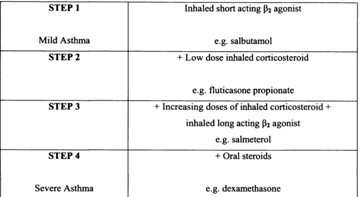
Asthma therapeutics target inflammation, bronchoconstrictions and narrowing of the airways. Both symptom relievers and preventers are clinically available. The drugs used can be broadly classified as either bronchodilators or anti-inflammatories. Treatment is dependent on the severity of the disease, with mild asthma being treated with a short-acting bronchodilator whereas severe asthma is treated with oral steroids. Guidelines for the treatment o f asthma were drawn up by the British Thoracic Society (Wu et al., 2008) (table 2).
Short acting p2-adrenoreceptor agonists are administered to control mild asthma as they are bronchodilators. Beta-agonists are the most effective bronchodilators clinically available as they are effective at protecting against the bronchoconstriction caused by exercise, cold air and allergen (Bames, 1995). The binding of the agonists to the p2 -adrenoreceptor causes activation of the receptor; this leads to the stimulation of the enzyme adenylyl cyclase, which converts adenosine trisphosphate (ATP) into cyclic adenosine monophosphate (cAMP). The formation of cAMP causes relaxation of the bronchial smooth muscle via downstream signals. Two types of p2-adrenoreceptor agonists are available, short acting and long acting. Short acting agonists, such as salbutamol, are used when required for symptom control, whereas long acting agonists, such as salmeterol, cause a prolonged bronchodilation for more than twelve hours
CHAPTER 1 (Boulet, 2004). However, there have been studies that cast doubt on the safety of long acting P2 agonists. It has been suggested that long acting P2 agonists such as salmeterol may increase the risk of asthma deaths (Salpeter et al., 2006). Recently concerns have been reduced as a result of reassuring data (Moore, 2009; Nelson et al., 2009).
STEP 1 Inhaled short acting p2 agonist
Mild Asthma e.g. salbutamol
STEP 2 + Low dose inhaled corticosteroid
e.g. fluticasone propionate
STEP 3 + Increasing doses of inhaled corticosteroid + inhaled long acting P2 agonist
e.g. salmeterol
STEP 4 + Oral steroids
Severe Asthma e.g. dexamethasone
Table 2 - Recom mended guidelines for managing increasingly severe asthma in adults. Adapted from British Thoracic Society (Wu et a l2008).
Another method to control the immediate bronchoconstriction in asthma is to target the muscarinic receptor. In the airways acetylcholine is released from vagus nerves, when this binds with the muscarinic receptor contraction of the smooth muscle and increased secretion o f mucus occurs (Katzung, 2004). Muscarinic receptor antagonists, such as ipratropium bromide, competitively inhibit this acetylcholine effect. Ipratropium bromide is a non-selective muscarinic receptor and therefore blocks the Mi-, M2- and M3 -receptors (Barnes, 2001). However, this is not efficient as the M3-receptor mediates the bronchoconstriction and is therefore the target, whereas the M2-receptor inhibit acetylcholine release and is therefore beneficial (Bames, 2002a). Thus far, M3-selective anatagonists have proved difficult to develop (Maesen et al., 1993). The fact that M3
-CHAPTER 1 receptors can cause mucus secretion suggests that the inhibition of these receptors is beneficial when it comes to controlling airway remodelling.
Phosphodiesterase (PDE) is another target for asthma therapeutics. PDE enzymes decrease intracellular levels o f cyclic adenosine monophosphate (cAMP) by catalysing its degradation to inactive AMP. Inhibiting PDE causes increased intracellular levels of cAMP which has several effects such as relaxing smooth muscle and inhibition of inflammatory cell activity. Methylxanthines, such as theophylline, are non-selective phosphodiesterase inhibitors. Through this inhibition of PDE, levels of cAMP increase and smooth muscle relaxation and anti-inflammatory actions can occur therefore attenuating the early phase bronchoconstriction associated with asthma. It is possible that theophylline works through another mechanism as it can inhibit cell surface adenosine receptors (Katzung, 2004). Adenosine has been shown to cause contraction of airway smooth muscle and to provoke histamine release from mast cells (Katzung, 2004). Therefore, inhibiting adenosine would also attenuate the early bronchoconstriction.
There is increasing evidence that theophylline has anti-inflammatory effects in asthma such as activation of histone deactylase (HDAC), which increases anti-inflammatory gene transcription, (Barnes, 2002a), increased eosinophil apoptosis (Barnes, 2002a), inhibition o f mediator release (Orange et al., 1971) and inhibition of superoxide anion release from neutrophils (Nielson et al., 1988). Whether this action is mediated through cAMP activation, inhibition of adenosine cell surface receptors or through another mechanism is unclear. The side-effects o f theophylline has led to limited usage as the combination of p2-agonists and corticosteroids has less detrimental effects (Barnes, 2002a). Theophylline is a non-selective PDE inhibitor; more recent developments have been the introduction of selective inhibitors o f the PDE subtypes such as the PDE4 inhibitor, roflumilast (see chapter 6 for more information).
Some asthma therapeutics are effective at treating the late phase bronchoconstriction by having anti-inflammatory properties and are therefore also effective against AHR and cellular influx. The most powerful o f this type of drug are the corticosteroids. Corticosteroids are the most commonly used drug in the treatment of asthma, their mechanism of action and effects in asthma are described in chapter 5. Another group o f anti-inflammatory drugs focus on the inhibition of leukotrienes. Leukotrienes cause
CHAPTER 1 airflow obstruction and bronchoconstriction. The leukotriene antagonist, montelukast, blocks leukotriene D4 having a bronchoconstrictor response by binding to the Cysteinyl Leukotriene 1 (CysLTl) receptor on mast cells, smooth muscle and eosinophils, therefore attenuating the early asthmatic response (Finnerty et a l, 1992; Taylor et a l, 1991) and is in fact as effective as short acting p2-agonists (Hui & Barnes, 1991; Reiss et a l, 1997) when administered orally. As well as a bronchoconstriction, CysLTl receptor activation causes eosinophil recruitment, mucus secretion and plasma exudation (Barnes, 2002a). Therefore montelukast is also effective at inhibiting the late asthmatic response (Bames, 2002a).
The expanding knowledge of asthma mechanisms and pathology correlates with an increase in anti-asthma therapeutics. Many o f the recently developed compounds are highly specific to a certain mediator shown to have a role in asthma, examples o f such include tyrosine kinase inhibitors, adhesion molecule inhibitors and nuclear factor kappa-light-chain-enhancer of activated B cells (NF-kB) inhibitors (Bames, 2002a). Another class o f potential anti-asthmatic drugs are inhibitors of inducible nitric oxide synthase (iNOS) which are described in chapter 7. Despite an increasing number of anti-asthmatic therapeutics, a combination o f P2 agonists and corticosteroids is still the main treatment for asthma (Bames, 2002b) and it is believed that the current pharmacological treatment of asthma will not change for the next 10 years (Caramori et a l, 2009).
1.6 MODELS OF ASTHMA
The use o f animal models of asthma is no new phenomenon. Although no animals exhibit asthmatic symptoms spontaneously, certain horses, cats and Basenji-greyhound cross dogs show some features (Bames, 2002a). Consequently the most common method o f developing a model of asthma is using allergen sensitisation and subsequent challenge. Several different allergens can be used to elicit an asthmatic condition, including house dust mite (Cates et a l, 2007), short ragweed extracts (Chapoval et a l, 1999) and ovalbumin (Smith & Broadley, 2007).
Allergen sensitisation and exposure has led to the development of several animal models of asthma in guinea pigs, mice, rats, monkeys and dogs. The similarity between the
CHAPTER 1 pulmonary response and histological findings to antigen exposure in the airways of guinea pigs and humans led to the guinea pig being the model of choice (Nabe et a l, 1997). However, studies that use mice as models of asthma are becoming more common (Kumar & Foster, 2002; McMillan & Lloyd, 2004; Wegmann et al., 2005). This is because mice are low cost, have a well-characterised immune system, knock-out mice are more achievable than other small species and antibodies for molecular studies in inflammatory mediators are readily available.
Several models of asthma concentrate on acute inflammation following antigen exposure (McMillan & Lloyd, 2004). These short-term models develop an inflammatory response which is limited to the proximal airways and as a result is not associated with the chronic pathological changes associated with airway remodelling (Wegmann et a l, 2005). Many studies also use anaesthetised animals (Johnson & Broadley, 1999; Underwood et a l, 1995), however, the possibility exists that the anaesthetic may interfere with the vagal tone or sensory reflex (Toward & Broadley, 2004).
Clearly there are several advantages and disadvantages between species when it comes to using them as a model of asthma. The advantages and disadvantages of guinea pig and mouse models of asthma are discussed in greater detail in chapters 3 and 4, respectively.
Ultimately no animal model can be completely perfect when it comes to representing human asthma; however, several models display many of the characteristics of asthma. Therefore, they have an important role as the first means of testing asthma therapeutics.
CHAPTER 1
1.7 AIMS
The overall aims of this thesis are as follows:
• Develop acute and chronic models of asthma in guinea pigs and mice, which demonstrate the features o f human asthma - early and late asthmatic responses to allergen challenge, airway hyperresponsiveness, airway inflammatory cell influx and changes in lung histology.
• Evaluate the effects o f anti-inflammatory compounds belonging to the steroid, phosphodiesterase type IV inhibitor and inducible nitric oxide synthase inhibitor classes on these acute and chronic guinea pig and mouse models of asthma.
• Assess what effect acute and chronic ovalbumin challenges have on lung oedema in sensitised guinea pigs and whether corticosteroid treatment attenuates this by using magnetic resonance imaging.
CHAPTER 2
Chapter 2
Methods
CHAPTER 2
2.1 MATERIALS AND EQUIPMENT
A full list o f materials and equipment used can be found in Appendix 1. Ovalbumin (OVA) was the allergen o f choice to cause the sensitisation and effector phase of asthma.
2.2 ANIMAL EXPERIMENTS
2.2.1 ANIMAL WELFARE
All animals were obtained through the Joint Animal Service department of Cardiff University from Harlan UK Ltd. Upon arrival the animals were given one week to habituate to their surroundings before any experiments began. They were housed under conventional conditions with a twelve hour light/dark cycle, maintained room temperature of 20°C±2°C and a humidity o f 50%. All experiments were carried out in accordance with the Animal Scientific Procedures Act 1986, under valid Home Office project and personal licences. Experienced technicians were in charge of the welfare of the animals. Before beginning any experiments the animals were exposed to the system that measures their airway function to ensure it was not a novel environment and the animals were habituated when readings were taken. The guinea pigs used in chapter 8 were kept at GlaxoSmithKline, further details can be found in section 8.3.
2.2.1.1 GUINEA PIGS
Male Dunkin-Hartley guinea pigs (200-25Og) were used for all research involving guinea pigs. They received commercial guinea pig pellets, supplemented with ascorbic acid, and drinking water ad libitum. The guinea pigs were housed in grid-floor cages in groups of six and in order to enrich the environment, a cardboard tube and hay was supplied. An n of 6 was used for all experiments.
2.2.1.2 MICE
Male BALB/c mice (20-25g) were the breed used for all mouse studies. They were housed in groups of six in plastic cages, with a sawdust base, and a removable grid roof.
CHAPTER 2 Food and drinking water was supplied ad libtum. For environmental enrichment cardboard tubes were supplied. An n of 6 was used for all experiments.
2.2.2 SENSITISATION PROCESS
2.2.2.1 GUINEA PIGS
Guinea pigs in all studies were sensitised by an intraperitoneal injection of a mixture of OVA (100 pg) and aluminium hydroxide (Al(OH)3) (100 mg) in phosphate-buffered saline (PBS). The mixture was stirred for two hours prior to injection to ensure the OVA and Al(OH) 3 were completely dissolved. Al(OH) 3 was used as an adjuvant to boost the immune response to OVA and promote the development of sensitization. 1 ml o f the suspension was administered bilaterally to the guinea pigs on day 1 and day 5, all procedures then commenced on day 14. This method of sensitization was developed by Smith and Broadley (2007).
2.2.2.2 MICE
The sensitisation process for mice used the same OVA mixture as in guinea pigs. Intraperitoneal injections of 0.25 ml bilaterally were administered on days 1 and 5, with procedures commencing on day 14 (Femandez-Rodriguez et al., 2008).
2.2.3 MEASURING LUNG FUNCTION IN GUINEA PIGS
Throughout all studies non-invasive methods were employed using plethysmography, an airtight chamber, to measure airway function. For respiration to occur there must be a difference in pressure between the mouth or nose and the alveoli, this is known as the pressure difference. If the difference in pressure is divided by the airflow then the outcome is a measure of airway resistance (R aw )- However, airway conductance (Gaw) is regarded as a better measurement of airway function as this incorporates a change in transpulmonary pressure and lung tissue tension (Griffiths-Johnson et al., 1988). In asthma differences in the alveoli pressure occur and therefore changes in the volume of air in the lung, or thoracic gas volume (TGV), occur. Taking this into account a value of Gaw - TGV is used for measuring airway function. This value is known as specific airway conductance
CHAPTER 2 The plethysmograph allows sGaw to be calculated. The pressure in the sealed chamber, box pressure, will decrease and increase in line with the pressure changes between the mouth or nose and alveoli of the subject if the chamber is kept at a constant temperature (Boyle’s Law) (West, 2005). Airflow is measured through a mask placed over the subject’s mouth and nose. The full description of how sGaw is derived, taking the TGV into account is found in Appendix 2.
Guinea pigs are very docile and as a result they can be restrained and have a mask placed over their snout without much complaint. As previously mentioned to measure sGaw in guinea pigs a plethysmograph is used (figure 2.1). This is a perspex box with a removable endplate at one end. The guinea pig was placed in a neck restrainer, this slides into another restrainer that has high sides and therefore stops the guinea pig from moving its body. A perspex mask was attached to the full body restrainer; it was sealed to the snout of the guinea pig by means of a cut balloon. The restrainer was then placed into the plethysmograph and the endplate clamped shut making an airtight environment. The plethysmograph measures two variables, airflow and box pressure. Airflow was measured by using a mesh pneumotachograph that is inside the perspex mask. The mask is connected to a UP1 pressure transducer by means of a plastic tube. Box pressure measures the change in air pressure in the plethysmograph. It is connected to a UP2 pressure transducer by means of a plastic tube.
The pressure transducers are connected to a Biopac data system that converts the airflow and box pressure data into waveforms; this is displayed on the computer by using Acqknowledge software. Acqknowledge allows the analysis of the waveforms by comparing the gradients of airflow and box volume where the flow of the wave tends towards 0. The result is a value of specific airway conductance (sGaw). One reading takes 5 seconds to record and an average of 8 breaths are captured in this time. An example of the waveform data obtained from the plethysmograph is shown in figure 2.1.
C H A PT E R 2
Mask
Restrainer Pneumotachograph
A A A A A A T
Flow pressure transducer Biopac amplifier
□ □□□
PC+Acqknowledge software
Box pressure transducer
Figure 2.1 - A simplified schematic o f the whole body plethysmograph and acquisition packs used to measure specific airways conductance sGaw, in conscious, restrained guinea pigs. Flow rate is measured through a flow pressure transducer which is connected to the pneumotachograph found in the mask that is placed over the snout o f the guinea pig. Changes in box pressure are also measured through a transducer. The two wave forms are captured using a Biopac amplifier, a value o f sGaw is then established using Acqknowledge software by comparing the gradients o f flow rate (red) and box volume (blue) where the flow o f the wave tends towards 0.
CHAPTER 2
2.2.3.1 ACUTE CHALLENGE PROTOCOL
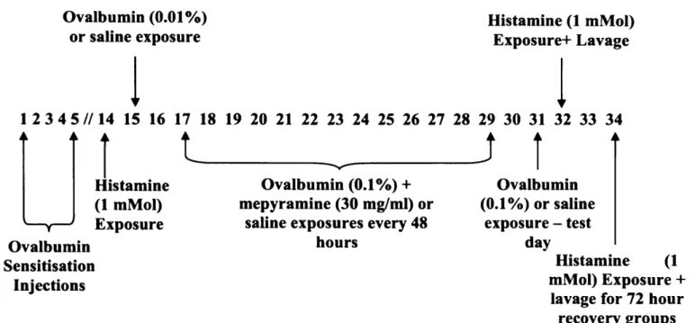
On days 14 and 16 guinea pigs were exposed to histamine to determine whether airway hyperresponsiveness was occurring, this is explained in greater detail in section 2.7.1. The effector phase of asthma was triggered by an allergen challenge on day 15. The guinea pigs were exposed to either OVA (0.01%) or a control solution (saline). Exposure was carried out in a stainless steel exposure chamber (40 cm diameter, 15 cm height) with a Wright nebuliser attached. The nebuliser delivered the OVA or saline at an air pressure of 20 lb p.s.i. and at a rate of 0.3 ml/min. The guinea pigs remained in the chamber for 1 hour, however, if they appeared to be distressed they were immediately removed from the chamber and exposure was considered complete. On day 16 following the second histamine challenge the guinea pigs were killed and a lavage was carried out. This allowed for total and differential cell counts to be obtained, following this the lungs were stored for histological analysis.
An additional group following the same protocol as the antigen-challenged group was run, however, this group was left for 72 hours after the test day before the second histamine challenge and lavage was performed. The purpose of this group was to observe whether any recovery occured in terms o f airway hyperresponsiveness and cellular influx and histology when compared to the antigen-challenged group that had the lavage immediately after the final histamine exposure. Figure 2.2 hig