R E V I E W
A practical approach to goal-directed
echocardiography in the critical care setting
Patricia E Walley
1*, Keith R Walley
2,3, Ben Goodgame
4, Vivek Punjabi
4and Demetrios Sirounis
2Abstract
Urgent cardiac ultrasound examination in the critical care setting is clinically useful. Application of goal-directed echocardiography in this setting is quite distinct from typical exploratory diagnostic comprehensive echocardiography, because the urgent critical care setting mandates a goal-directed approach. Goal-directed echocardiography most frequently aims to rapidly identify and differentiate the cause(s) of hemodynamic instability and/or the cause(s) of acute respiratory failure. Accordingly, this paper highlights 1) indications, 2) an easily memorized differential diagnostic framework for goal-directed echocardiography, 3) clinical questions that must be asked and answered, 4) practical issues to allow optimal image capture, 5) primary echocardiographic views, 6) key issues addressed in each view, and 7) interpretation of findings within the differential diagnostic framework. The most frequent indications for goal-directed echocardiography include 1) the spectrum of hemodynamic instability, shock, and pulseless electrical activity arrest and 2) acute respiratory failure. The differential diagnostic categories for hemodynamic instability can be remembered using the mnemonic‘SHOCK’ (for Septic, Hypovolemic, Obstructive, Cardiogenic, and (K) combinations/other kinds of shock). RESP-F (for exacerbation of chronic Respiratory disease, pulmonary Embolism, ST changes associated with cardiac or pericardial disease, Pneumonia, and heart Failure) can be used for acute respiratory failure. The goals of goal-directed echocardiography in the unstable patient are: assessing global ventricular systolic function, identifying marked right ventricular and left ventricular enlargement, assessing intravascular volume, and the presence of a pericardial effusion. In an urgent or emergent setting, it is recommended to go directly to the best view, which is frequently the subcostal or apical view. The five views are the subcostal four-chamber view, subcostal inferior vena cava view, parasternal long axis view, parasternal short axis view, and the apical four chamber view. Always interpret goal-directed echocardiographic findings in the context of clinically available hemodynamic information. When goal-directed echocardiography is insufficient or when additional abnormalities are appreciated, order a comprehensive echocardiogram. Goal-directed echocardiography and comprehensive echocardiography are not to be used in conflict with each other.
Introduction
Important guidelines have recently been published for the use of echocardiography in the critical care setting [1-6] and a large number of publications have investi-gated important aspects of echocardiography in critically ill patients, in particular the goal-directed approach [1-6]. Here we review the current use of, and a problem-oriented practical guide to, goal-directed echocardiog-raphy in the critical care setting. Effective management of critically ill patients requires rapid assessment and
clear goals. Even the expert practitioner, with knowledge and skills that allow an extensive and comprehensive examination, must focus on the urgent problem at hand and may benefit from a readily remembered diagnostic framework and a goal-directed approach [7,8]. Com-pared with a conventional comprehensive echocardio-gram [9], goal-directed echocardiography has a more limited scope [10] that specifically addresses the poten-tial causes of 1) the spectrum of hemodynamic instabil-ity, shock, and pulseless electrical activity (PEA) arrest and 2) acute respiratory failure. Portability, the time needed to acquire data, ease of use, relatively low cost, and the fact that it is used as an adjunct to the physical examination are major benefits of it [11]. Goal-directed echocardiography can be used instantly for goal-directed * Correspondence:[email protected]
1
Cardiac Echo Laboratory, St Paul’s Hospital, University of British Columbia, Vancouver V6Z 1Y6, Canada
Full list of author information is available at the end of the article
therapy in the setting of a hemodynamically unstable pa-tient [12]. Another major benefit of goal-directed echo-cardiography is that it can be used serially [13] to assess response to interventions in a ‘real-time’ manner and thus guide ongoing therapies [8,14]. Importantly, we emphasize that goal-directed echocardiography should always be interpreted in view of clinically available hemodynamic information (arterial pressures, central venous pressure, use of vasopressor/inotropic drugs, ventilator settings, urine output, and so on) and re-spiratory data (chest radiography, arterial and venous oxygen saturation, shunt fraction, dead space, acid–base status, and so on).
Definition and scope of goal-directed echocardiography
Goal-directed echocardiography is different from the com-prehensive echocardiograms performed by fully trained, experienced, and certified cardiac sonographers, cardiolo-gist echocardiographers, and advanced critical care echo-cardiographers [3-5,9]. Comprehensive echocardiograms are beyond the scope of beginners, who will use only a few echocardiographic views. A key challenge for inten-sivists is to know their (narrow) field of competence while using transthoracic echocardiography in the ICU setting. Advanced practitioners are capable of measur-ing many more parameters designed to assess many additional aspects of cardiac structure and function and requiring more time, additional views, additional tools (intravenous contrast, maneuvers), and a higher level of qualifications and training in the acquisition and inter-pretation of images [3-5,9,15,16].
In conducting goal-directed echocardiography, the most technically difficult patients are imaged in the most challenging situations; hence, diagnostic shortcomings are exaggerated in this population because of, for ex-ample, lung interference, mechanical ventilation, po-sitioning limitations, and the urgency to acquire the information for immediate treatment. Current research suggests that training internists and residents with limited previous ultrasound skills is feasible and can result in ac-curate assessments when using goal-directed echocardiog-raphy [11,17-19]. It is important to recognize, however, when the necessary images cannot be acquired or are in-sufficient for accurate interpretation. It is then necessary to order a comprehensive echocardiogram to further as-sess potential findings and findings that fall outside the scope of goal-directed echocardiography [1] (for example, valvular abnormalities, wall motion abnormalities, dia-stolic dysfunction, abnormal anatomy or other abnormal echocardiographic features). An ‘incidental’ finding of a significantly thickened aortic valve should be noted due to possible implications during resuscitation attempts [20]. When these additional observations are appreciated or
suspected, a full comprehensive echocardiographic exam-ination is indicated as soon as feasible.
The issues of how goal-directed echocardiography is different from conventional echocardiography and when goal-directed echocardiography alone is sufficient are the subject of careful discussion in the recent publication by Spencer and colleagues [1]. The American Society of Echocardiography Expert Consensus Statement specific-ally discusses the role of ‘goal-directed echocardiography when echocardiography is not promptly available’. We address a narrow aspect of these guidelines in detail, specifically the use of goal-directed echocardiography to distinguish between differential diagnostic categories contributing to 1) hemodynamic instability, shock, and PEA arrest requiring immediate therapeutic decisions and interventions - the central indication for goal-directed echocardiography in the critical care setting - and 2) evalu-ation of acute respiratory failure. In every case the operator should consider whether a subsequent comprehensive echocardiographic examination would contribute add-itional information necessary for ongoing care.
Guidelines for training
An expert round table led by the European Society of Intensive Care Medicine has recently developed an inter-national consensus statement on guidelines for train-ing and accreditation which have been widely adopted [3]. Earlier published statements [15,21], the Canadian Society of Echocardiography [22], and the American Society of Echocardiography [1] have also addressed as-pects of training.
While cardiologists and cardiac sonographers are gen-erally the most highly trained and experienced operators, there are emerging important roles for different kinds of providers. Goal-directed echocardiography is often per-formed by physicians who are not trained in acquisition and interpretation of comprehensive echocardiograms and who come from diverse backgrounds and differ-ent clinical experiences. For these physicians guide-lines for goal-directed echocardiography training in the ICU are much more focused and are currently in development [1,15,19,23]. Transesophageal echocardi-ography also plays a role in the ICU but is not ex-plored in this review as it is appropriately performed by skilled and highly knowledgeable operators conduct-ing sufficient examinations every year to maintain com-petency [2]. Training guidelines for transesophageal echocardiography in the critical care setting have re-cently been published [3].
Developing an efficient training program
echocardiography and interpretation takes time, practice, and supervision [11,26,27]. It is imperative to recognize technical and interpretive limitations. The ‘trainees’ are frequently physicians with very busy schedules who are ac-customed to being experts [28]. The recent International Consensus Statement on training standards for advanced critical care echocardiography are clear that a training program should be rigorous and include competence-based testing [3]. Other organizations have endorsed con-cordant views [1]. For example, the American College of Emergency Physicians suggests that didactic training, ex-tensive hands-on experience, and expert review should be included in every case [29] and partnership with a com-prehensive echocardiography laboratory is required [1]. This should be supplemented with a continuing medical education program [2] and there should be a formal certi-fication process to ensure competence in technical skills and interpretation [3,30] as poor training may result in adverse consequences [31].
Indications
Goal-directed echocardiography has now become a reli-able tool and can be performed within minutes [17] and is useful for assessing the spectrum of hemodynamic in-stability, shock, and PEA arrest [2,32-34]. The differen-tial diagnostic categories for hemodynamic instability can be remembered using the mnemonic ‘SHOCK’ (for Septic, Hypovolemic, Obstructive, Cardiogenic, and (‘K’) combinations or other kinds of shock) .‘S’stands for the broad category of septic shock due to infection or simi-lar distributive shock from non-infectious causes. Hypo-volemia (H) is identified by changes in inferior vena cava (IVC) diameter and by small, underfilled ventricles. Ob-structive shock (O) is most frequently due to pericardial tamponade or pulmonary embolism, which manifest dif-ferently (see below). Cardiogenic shock (C) is usually characterized by dilated and/or poorly contractile ventri-cles or marked valvular dysfunction recognized by using color Doppler imaging [5]. When the clinical picture and goal-directed echocardiography do not fit clearly into one of these major categories, other possibilities must be considered - ‘K’, remembered phonetically, for
‘combinations’and other ‘kinds’. That is, shock may be due to combinations of the above etiologies and rarely due to other disease processes such as adrenal insuffi-ciency, neurogenic shock, and so on.
Goal-directed echocardiography can be combined with thoracic ultrasound imaging [35] and improves diagnos-tic accuracy over conventional imaging for patients pre-senting with acute respiratory failure [36]. The most common causes are captured by the mnemonic RESPF -exacerbation of chronic Respiratory disease, pulmonary Embolism, ST changes associated with acute cardiac or pericardial disease, Pneumonia, and heart Failure. Lung
and thoracic imaging (beyond the scope of this review) are an essential component of this examination to additionally identify pleural effusion, pneumothorax, and consolidated or edematous lung [35,36].
Specific goals
The goal of goal-directed echocardiography is to identify the cause (mnemonic SHOCK) of hemodynamic instabil-ity, shock, or PEA arrest and to expedite directed therapies [1,32-34]. To accomplish this, the specific goals are the systematic assessment of 1) left ventricule (LV) systolic function (cardiogenic) [2,17,37], 2) right ventricule (RV) size and function [38] (hypovolemia, obstruction [39,40], cardiogenic), 3) pericardial effusion, potential signs of hemodynamic compromise (obstruction), and 4) size and distensibilty of the IVC for evaluation of volume status (hypovolemia) [14].
A structured approach to accomplishing the goals of goal-directed echocardiography involves specifically ask-ing and addressask-ing key practical questions, as follows. First, does the LV appear significantly dilated or not? Does the LV function appear ‘significantly’ impaired or not? Second, is the RV dilated or not? Third, is there evi-dence of hypovolemia (small LV, narrow IVC (<21 mm) and collapsing >50% with spontaneous respiration). In the more severe cases of hypovolemia, and in a non-mechanically ventilated patient, an IVC diameter of≤1 cm usually indicates preload responsiveness [41]. Fourth, is pericardial effusion present (moderate to large)?
Goal-directed echocardiography should note the pres-ence of additional abnormalities but the diagnostic pur-suit of these additional findings is accomplished by a comprehensive echocardiographic examination. Specific-ally, the presence of significant valvular abnormalities, wall motion abnormalities, LV aneurysm, RV hyper-trophy, cardiac masses, thrombus, diastolic dysfunction, or a dilated ascending aorta with potential dissection should be pursued with a comprehensive echocardio-graphic examination, when recognized [1].
Equipment set up
Transducer
Use a low-frequency transducer for adequate penetration to image cardiac structures. Transducers with a higher frequency have better resolution at the expense of dee-per tissue penetration. Transducers with a lower fre-quency have better penetration at the expense of higher resolution. The ‘footprint’ or size of the low-frequency transducer head is designed to be small enough to better fit between the intercostal spaces.
Initial settings
Decrease‘depth’to 14 to 16 cm for a full screen cardiac image. Continually adjust the‘focus’control to the anat-omy imaged for better resolution. Gain should be ad-justed to see endocardial borders clearly (Figure 1A). Be careful not to ‘over gain’ (too bright, washes out image) or ‘under gain’ (weak echocardiographic images) (Figure 1B,C).
Views
Goal-directed echocardiography comprises four main views. In an urgent or emergent setting,‘go for the gold’. Apical and subcostal views are frequently most useful [42]. That is, if unable to obtain an adequate image in one view, move on to the next view, when necessary. In critical care settings, you may not always have the elec-trocardiogram on the echocardiographic monitor to pro-vide a timing marker. For timing of the cardiac phase, pay attention to whether the mitral/tricuspid valves are open (diastole) or closed (systole). Remember, you must
see endocardium to accurately assess ventricular func-tion for all the views listed.
Subcostal view
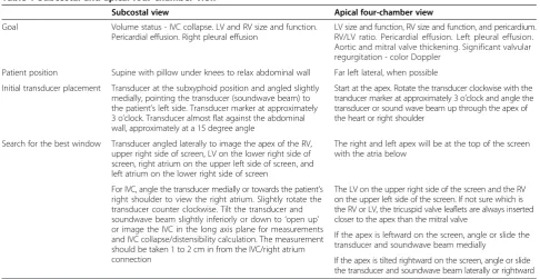
[image:4.595.58.538.345.694.2]The subcostal view (Table 1 and Figure 2) provides more information than any other individual view and, as a re-sult, addresses more of the key questions. In particular, volemia is best addressed in this view and multiple as-pects (IVC collapse [14,43], RV and LV diastolic size) are sometimes evident from this one window. The IVC can be used for estimating central venous pressure, reflective of right atrial pressure [14,43,44]. The ratio of inspira-tory and expirainspira-tory changes in diameter is very useful in evaluating volume status [45-47]. The subcostal view, as with the apical four-chamber (A-4) view, is frequently the best view to appreciate the presence of a pericardial effusion and associated echocardiographic evidence of hemodynamic compromise [2]. This view is frequently used for a pericardiocentesis. It is sometimes difficult
to differentiate pericardial fluid, fat, clot, or thrombus. A small, focal or loculated pericardial effusion may not be visualized nor easily ‘tapped’. When results are in question, order a comprehensive echocardiographic examination.
An incidental finding of ascites or a right pleural effu-sion may be visualized in this view. A left-sided pleural effusion, visualized in other views, is more frequently as-sociated with a pericardial effusion.
Parasternal long-axis view
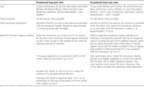
The parasternal long-axis view (Table 2 and Figure 2) is used in the assessment of LV systolic function (normal, decreased, or severely decreased) [17]. This view is often technically easier to obtain due to patient positioning, reliable landmarks, and ease of maintaining a steady transducer position [1]. A small and hyperdynamic LV may indicate hypovolemia. A pericardial effusion may be appreciated. An‘incidental’finding of a left pleural effu-sion may be noted.
Parasternal short-axis view
The parasternal short-axis (PSAX) view (also known as the transverse view; Table 2) is used for the visual assess-ment of LV function at the mid-LV. The mid-LV (Figure 2) is identified when the tips of the papillary muscles are visu-alized as just‘flickers’of the mitral valve apparatus. The LV should appear round, with RV visualization at the mid-LV. The main pulmonary artery with the bifurcation may be visualized.
In the setting of hypovolemia or volume depletion, the LV may appear small and mitral valve leaflets may be visualized. The presence of regional wall motion abnor-malities may be noted, though a comprehensive echocar-diographic examination is indicated for evaluation of regional wall motion abnormalities. The RV may be eval-uated for the presence of abnormal or paradoxical septal motion and the‘D’sign: flattening of the interventricular septum (RV pressure overload). This view is also used for presence of pericardial effusion. Incidental findings of clot/thrombus in the main pulmonary artery may be noted.
Apical four-chamber view
[image:5.595.55.542.96.347.2]Some authors caution about the use of A-4 views since off-axis views lead to erroneous interpretation of the LV and, particularly, RV size and function [1]. This view can be useful for identifying circumferential pericardial effu-sion. Assessment of RV size should be carefully made [38]. The RV/LV ratio may be used for evaluating RV size [48]: a RV/LV ratio <0.6 indicates normal RV size, a RV/LV ratio of 0.6 to 1.0 indicates moderate RV dilata-tion and an RV/LV ratio >1.0 indicates severe diladilata-tion [31,49,50]. The RV apex and LV apex must be at the top of the screen with the atria visualized directly below. An off-axis view may inaccurately cause the RV to appear dilated. This view is used for the assessment of pericar-dial effusion and for evidence of hemodynamic com-promise. Circumferential pericardial effusion is often associated with a large pericardial effusion. Right atrial Table 1 Subcostal and apical four-chamber view
Subcostal view Apical four-chamber view
Goal Volume status - IVC collapse. LV and RV size and function. Pericardial effusion. Right pleural effusion
LV size and function, RV size and function, and pericardium. RV/LV ratio. Pericardial effusion. Left pleural effusion. Aortic and mitral valve thickening. Significant valvular regurgitation - color Doppler
Patient position Supine with pillow under knees to relax abdominal wall Far left lateral, when possible
Initial transducer placement Transducer at the subxyphoid position and angled slightly medially, pointing the transducer (soundwave beam) to the patient’s left side. Transducer marker at approximately 3 o’clock. Transducer almost flat against the abdominal wall, approximately at a 15 degree angle
Start at the apex. Rotate the transducer clockwise with the tranducer marker at approximately 3 o’clock and angle the transducer or sound wave beam up through the apex of the heart or right shoulder
Search for the best window Transducer angled laterally to image the apex of the RV, upper right side of screen, LV on the lower right side of screen, right atrium on the upper left side of screen, and left atrium on the lower right side of screen
The right and left apex will be at the top of the screen with the atria below
For IVC, angle the transducer medially or towards the patient’s right shoulder to view the right atrium. Slightly rotate the transducer counter clockwise. Tilt the transducer and soundwave beam slightly inferiorly or down to‘open up’ or image the IVC in the long axis plane for measurements and IVC collapse/distensibility calculation. The measurement should be taken 1 to 2 cm in from the IVC/right atrium connection
The LV on the upper right side of the screen and the RV on the upper left side of the screen. If not sure which is the RV or LV, the tricuspid valve leaflets are always inserted closer to the apex than the mitral valve
If the apex is leftward on the screen, angle or slide the transducer and soundwave beam medially
If the apex is tilted rightward on the screen, angle or slide the transducer and soundwave beam laterally or rightward
and/or ventricular diastolic collapse/buckling may be seen [2]. An incidental finding of a left pleural effusion may be visualized.
Interpretation
Goal-directed echocardiography should always be inter-preted in the context of clinically available hemodynamic and other clinically relevant information. Accordingly, a goal-directed echocardiography is not an anatomic in-vestigation; it is a physiologic study or hemodynamic as-sessment (Table 3). Full interpretation involves linking goal-directed echocardiography findings to hemodynamic and other clinical information. We provide a practical
‘primer’approach while full discussion of all aspects of hemodynamic instability, shock, and PEA arrest is be-yond the scope of this review [51].
Septic or distributive shock that is less severe or in the earlier stages may show a hyperdynamic LV and a smaller to normal size IVC with >50% collapsibility be-cause septic shock is often initially associated with a de-gree of hypovolemia. More severe distributive shock may demonstrate impaired LV function [31].
[image:6.595.60.539.89.447.2]that cardiac output may increase significantly follow-ing volume infusion [55]. Whether fluid should be given, even when IVC diameter variation is large, is a clinical judgment that depends on other clinical vari-ables [56]. Goal-directed echocardiography only sup-plements other clinical information. Patients who are responsive to volume loading can increase their cardiac output by≥15% [31,54].
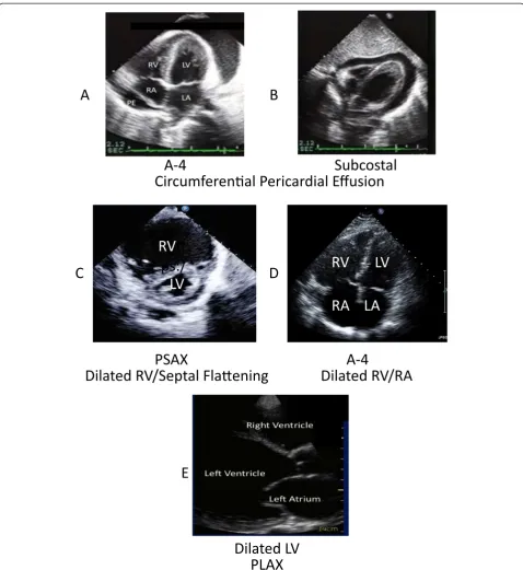
Obstructive shock due to tamponade (evidence of hemodynamic compromise) may be observed in the A-4 view (Figure 3A) and the subcostal view (Figure 3B). These views will demonstrate a significant pericardial effusion (usually encompassing the heart) with right atrial wall or RV diastolic collapse. Delayed RV diastolic expansion may also be noted. These findings may in-dicate increased pericardial pressure [2]. The IVC is dilated indicating high right-sided filling pressures.
‘Swinging heart’ motion always indicates a large amount of pericardial effusion, but is not always associated with hemodynamic compromise. A patient may have tampon-ade physiology at times with a smaller amount of pericar-dial effusion. This is sometimes associated with faster pericardial fluid accumulation. It is beneficial to detect pericardial effusion in patients with PEA and near PEA arrest [32,57]. Pericardial tamponade is a clinical, not echocardiographic, diagnosis [2].
In the subcostal view, where you often image the lar-gest amount of pericardial fluid accumulation, measure maximal echocardiographic-free space in diastole for po-tential pericardial ‘tap’ [58]. It is beneficial to note how many centimeters there are from the skin surface to the pericardium to help prevent inserting the pericardios-centesis needle too deep and close to the myocardium.
[image:7.595.58.537.99.387.2]Obstructive shock due to acute pulmonary embolism will demonstrate a dilated RV (Figure 3C) and an RV/LV Table 2 Parasternal views
Parasternal long-axis view Parasternal short-axis view
Goal LV size and function, RV size, RV wall motion, pericardial effusion, left pleural effusion. Mitral and aortic valve thickening. Significant valvular regurgitation - color Doppler
LV size, wall thickness, and function. RV size and function. Main pulmonary artery (dilated or not). Pericardial effusion. Aortic valve thickening. Significant valvular regurgitation - color Doppler
Patient position Far left lateral, when possible Far left lateral, when possible
Initial transducer placement Second or third ICS, as close to the sternum as possible, with the transducer‘marker’directed at approximately 11 o’clock (towards right shoulder)
Second or third ICS, as close to the sternum as possible. From the PLAX view, rotate the transducer clockwise or to your right until the transducer‘marker’is at approximately 2 o’clock
Search for the best imaging‘window’ Move the transducer up or down an ICS to search for the best view. Structures should appear horizontal on the screen. Image in the middle of the screen by adjusting transducer position
Mid-LV: angle the transducer slightly laterally and inferiorly or towards the patient’s left hip to image the mid-LV. Visualize the tips of the papillary muscles within the LV, below the level of the mitral valve. LV should appear round with RV clearly visualized. If the LV appears ‘egg-shaped’or oblong and the RV is not visualized, slide the transducer‘up’an ICS
If the apex appears to be positioned‘uphill’on the screen, slide the transducer‘up’an ICS
Main pulmonary artery: angle the transducer slightly laterally and slightly superiorly or toward the patient’s left shoulder with a slight clockwise rotation. You may need to move up an ICS, but in doing so may not be able to visualize the pulmonary artery bifurcation
Increase the‘depth’to 20 to 24 cm to image the presence of a possible pleural effusion
Decrease the depth to approximately 14 to 16 cm, depending on the size of the heart, to fill the screen with cardiac structure
ICS, intercostal space; LV, left ventricle; PLAX, parasternal long axis; RV, right ventricle.
Table 3 SHOCK: a practical mnemonic for the differential diagnosis of the spectrum of hemodynamic instability, shock, and pulseless electrical activity arrest
Type of shock Goal-directed echocardiographic features
Septic/distributive Less severe shock - hyperdynamic. Severe shock - can be hypodynamic
Hypovolemic Collapsing IVC, small ventricles, may be hyperdynamic
Obstructive Tamponade: pericardial effusion, tamponade physiology
Pulmonary embolism: large RV, septal shift/diastolic flattening
Cardiogenic Decreased LV (±RV) function, dilated ventricles
K Combinations (for example, septic plus hypovolemic, septic plus cardiogenic, and so on)
Other kinds (adrenal insufficiency, neurogenic, and so on). This broader category is raised by the question‘What does not fit?’
[image:7.595.56.291.559.724.2]diameter ratio >1:1 with decreased RV contractile function [39,40,59]. An RV/LV ratio≤0.6 is normal, an RV/LV ratio of 0.6 to 1.0 indicates moderate RV dilatation and an
RV/LV ratio≥1.0 (Figure 3D) indicates severe dilation [31]. Diastolic flattening of the septum may occur, leading to the
‘D’sign or paradoxical septal motion, indicative of volume Figure 3Examples of pathological findings. (A)Apical four chamber (A-4) view. Circumferential pericardial effusion. Left ventricle (LV), right ventricle (RV), left atrium (LA), right atrium (RA), and pericardium with a circumferential echocardiographic-free space or pericardial effusion (PE).
or pressure overload. The‘D’sign is seen in the PSAX view [38] (Figure 3C). There may also be systolic septal flatten-ing [49]. The RV is sensitive to acute changes in afterload and may dilate under shock conditions [60]. A patient pre-senting with a pulmonary embolism may demonstrate an enlarged RV, a hypokinetic lateral wall, and a hyperdy-namic apex (McConnell’s sign) [61,62].
The sensitivity of these findings to diagnose pulmon-ary embolism is 29% with goal-directed echocardiog-raphy and 51% with a comprehensive echocardiographic examination [2]. Thus, transthoracic echocardiographic examination is not sufficiently sensitive to detect or rule out pulmonary embolism. Goal-directed echocardiography is helpful to identify hemodynamic compromise in a pa-tient having pulmonary embolism, which may be useful in directing the appropriate therapeutic intervention [39,63]. A patient may also have an increased RV/LV ratio in the setting of chronic and acute RV abnormalities such as chronic obstructive pulmonary disease (COPD), pulmon-ary hypertension, and RV myocardial infarction [2].
Cardiogenic shock will demonstrate a dilated LV (Figure 3E; consistent with underlying chronic heart disease) and/or significantly decreased LV function, possibly decreased RV function and a dilated IVC [5]. Additionally, marked valvular regurgitation can cause cardiogenic shock. Color Doppler imaging is useful for identifying significant valvular regurgitation but add-itional training is required for accurate use and interpret-ation [3,5]. LV function is graded as ‘not significantly decreased’or‘significantly decreased’, typically based on a visual estimate of the ejection fraction [64] (ejection fraction = (end-diastolic volume – end-systolic volume)/ end-diastolic volume). These measurements and calcula-tions must be performed by an experienced sonographer, cardiologist, or a physician with advanced echocardio-graphic imaging training and experience. With goal-directed echocardiography, LV function is graded as normal, decreased, or very decreased [37]. A minimal de-crease in ejection fraction is not considered ‘significant’ since it is difficult to call unambiguously and is insufficient, alone, to cause cardiogenic shock.
Correct interpretation must incorporate the impact of preload and surrounding pressures from the lung and chest wall, the effect of afterload, and the effect of medi-cations. For example, a‘normal’number for LV ejection fraction in a hypotensive patient treated with catechol-amine infusions is not normal. Correct interpretation in-cluding these factors as well as decreased afterload and pharmacologically supported contractility may indicate substantially reduced innate LV contractility.
Goal-directed echocardiography can help distinguish between asytole, PEA arrest, and pseudo-PEA [33,34]. In pseudo-PEA there is echocardiographic evidence of ven-tricular contraction with no palpable pulses.
RESP-F
Patients presenting with acute respiratory failure may share etiology and echocardiographic findings of SHOCK conditions. Hemodynamic instability may not yet be manifest in the patient who presents with acute respira-tory failure, due, in part, to the increase in sympathetic tone that generally accompanies severe dyspneic states. There are substantial limitations using goal-directed echocardiography. In the absence of Doppler assessment, LV diastolic properties and filling pressures cannot be properly determined. This may require a more compre-hensive echocardiographic examination.
Patients with acute exacerbation of an obstructive airways disease, such as COPD or asthma, may be hyperinflated so that most echocardiographic views are obstructed and often the heart is best visualized from a subcostal view. Features on goal-directed echocardiog-raphy that support the diagnosis of chronic COPD include evidence of RV hypertrophy and failure. Acute respiratory causes, such as asthma and pneumonia, may have few spe-cific findings on echocardiographic examination. Pulmon-ary embolism leads to evidence of obstruction and right heart failure, as discussed above. Pericardial disease may be associated with ST changes and becomes important when it contributes to obstructive physiology. Acute ventricular dysfunction can result in chest discomfort, dyspnea, and pulmonary edema with hypoxemic re-spiratory failure and even ventilator failure. Myocardial infarction (ST changes) must be considered as a treat-able and potentially very responsive cause. Ventricular dysfunction leading to heart failure is important in the differential diagnosis of acute respiratory failure and may be manifested in a goal-directed echocardiographic examination by the same findings that characterize car-diogenic shock.
Conclusion
The role of goal-directed echocardiography in the crit-ical care setting is continually evolving and becoming ac-cepted as a beneficial modality in the treatment, care, and monitoring of the critically ill patient. It is time to work together as an interdisciplinary group to utilize this modality to the highest level of quality and accuracy for the benefit of our patients. Proper training is essential for the benefit and accurate diagnosis of patients. Poor training may encourage adverse consequences.
Diagnosis and treatment of critically ill patients will likely continue to improve with the use of goal-directed echocardiography with hand carried and portable ultra-sound equipment.
Abbreviations
Competing interests
KRW, BG, VP, and DS declare that they no competing interests. PEW has worked for and was paid as an instructor by the Society of Critical Care Medicine and by CAE Healthcare in the past 5 years.
Acknowledgments
[image:10.595.301.535.130.734.2]We thank Priscilla Peters, BS, RDCS, FASE Cooper Hospital, Camden, NJ, USA, for figures, review, and suggestions; Yanek Beaulieu, MD, Cardiologist and Intensivist at Hospital Sacre’-Coeur of Montreal, PQ, for review and suggestions; and Dave Adams, RDCS, FASE, Duke University Hospital, Raleigh, NC, USA, for Figure 1.
Author details
1Cardiac Echo Laboratory, St Paul’s Hospital, University of British Columbia, Vancouver V6Z 1Y6, Canada.2Intensive Care Unit, St Paul’s Hospital, University of British Columbia, Vancouver V6Z 1Y6, Canada.3Centre for Heart Lung Innovation, St Paul’s Hospital, University of British Columbia, Vancouver V6Z 1Y6, Canada.4Cooper University Hospital, Camden, NJ 08103, USA.
References
1. Spencer KT, Kimura BJ, Korcarz CE, Pellikka PA, Rahko PS, Siegel RJ:Focused cardiac ultrasound: recommendations from the American Society of Echocardiography.J Am Soc Echocardiogr2013,26:567–581. 2. Labovitz AJ, Noble VE, Bierig M, Goldstein SA, Jones R, Kort S, Porter TR,
Spencer KT, Tayal VS, Wei K:Focused cardiac ultrasound in the emergent setting: a consensus statement of the American Society of Echocardiography and American College of Emergency Physicians.
J Am Soc Echocardiogr2010,23:1225–1230.
3. Expert Round Table on Echocardiography in ICU:International consensus statement on training standards for advanced critical care echocardiography.Intensive Care Med2014,40:654–666.
4. Expert Round Table on Ultrasound in ICU:International expert statement on training standards for critical care ultrasonography.Intensive Care Med
2011,37:1077–1083.
5. Mayo PH, Beaulieu Y, Doelken P, Feller-Kopman D, Harrod C, Kaplan A, Oropello J, Vieillard-Baron A, Axler O, Lichtenstein D, Maury E, Slama M, Vignon P:American College of Chest Physicians/La Societe de Reanimation de Langue Francaise statement on competence in critical care ultrasonography.Chest2009,135:1050–1060.
6. Repesse X, Charron C, Vieillard-Baron A:Intensive care ultrasound: V. Goal-directed echocardiography.Ann Am Thorac Soc2014,11:122–128. 7. Rivers E, Nguyen B, Havstad S, Ressler J, Muzzin A, Knoblich B, Peterson E,
Tomlanovich M, Early Goal-Directed Therapy Collaborative Group:Early goal-directed therapy in the treatment of severe sepsis and septic shock.
N Engl J Med2001,345:1368–1377.
8. Boyd JH, Walley KR:The role of echocardiography in hemodynamic monitoring.Curr Opin Crit Care2009,15:239–243.
9. Bierig SM, Ehler D, Knoll ML, Waggoner AD, American Society of Echocardiography:American Society of Echocardiography minimum standards for the cardiac sonographer: a position paper.J Am Soc Echocardiogr2006,19:471–474.
10. Royse CF, Canty DJ, Faris J, Haji DL, Veltman M, Royse A:Core review: physician-performed ultrasound: the time has come for routine use in acute care medicine.Anesth Analg2012,115:1007–1028.
11. Razi R, Estrada JR, Doll J, Spencer KT:Bedside hand-carried ultrasound by internal medicine residents versus traditional clinical assessment for the identification of systolic dysfunction in patients admitted with decompensated heart failure.J Am Soc Echocardiogr2011,24:1319–1324. 12. Gunst M, Ghaemmaghami V, Sperry J, Robinson M, O’Keeffe T, Friese R,
Frankel H:Accuracy of cardiac function and volume status estimates using the bedside echocardiographic assessment in trauma/critical care.
J Trauma2008,65:509–516.
13. Mayron R, Gaudio FE, Plummer D, Asinger R, Elsperger J:Echocardiography performed by emergency physicians: impact on diagnosis and therapy.
Ann Emerg Med1988,17:150–154.
14. Weekes AJ, Tassone HM, Babcock A, Quirke DP, Norton HJ, Jayarama K, Tayal VS:Comparison of serial qualitative and quantitative assessments of caval index and left ventricular systolic function during early fluid
resuscitation of hypotensive emergency department patients.Acad Emerg Med2011,18:912–921.
15. Picard MH, Adams D, Bierig SM, Dent JM, Douglas PS, Gillam LD, Keller AM, Malenka DJ, Masoudi FA, McCulloch M, Pellikka PA, Peters PJ, Stainback RF, Strachan GM, Zoghbi WA, American Society of Echocardiography:
American Society of Echocardiography recommendations for quality echocardiography laboratory operations.J Am Soc Echocardiogr2011,
24:1–10.
16. Quiñones MA, Douglas PS, Foster E, Gorcsan J 3rd, Lewis JF, Pearlman AS, Rychik J, Salcedo EE, Seward JB, Stevenson JG, Thys DM, Weitz HH, Zoghbi WA, Creager MA, Winters WL Jr, Elnicki M, Hirshfeld JW Jr, Lorell BH, Rodgers GP, Tracy CM, Weitz HH, American Society of Echocardiography; Society of Cardiovascular Anesthesiologists; Society of Pediatric Echocardiography:ACC/AHA clinical competence statement on echocardiography: a report of the American College of Cardiology/American Heart Association/American College of Physicians-American Society of Internal Medicine Task Force on clinical competence.J Am Soc Echocardiogr2003,16:379–402. 17. Melamed R, Sprenkle MD, Ulstad VK, Herzog CA, Leatherman JW:
Assessment of left ventricular function by intensivists using hand-held echocardiography.Chest2009,135:1416–1420.
18. Culp BC, Mock JD, Ball TR, Chiles CD, Culp WC Jr:The pocket echocardiograph: a pilot study of its validation and feasibility in intubated patients.Echocardiography2011,28:371–377.
19. Vignon P, Dugard A, Abraham J, Belcour D, Gondran G, Pepino F, Marin B, Francois B, Gastinne H:Focused training for goal-oriented hand-held echocardiography performed by noncardiologist residents in the intensive care unit.Intensive Care Med2007,33:1795–1799.
20. Mijuskovic D, Stamenkovic DM, Borovic S, Karanikolas M:Successful resuscitation from two cardiac arrests in a female patient with critical aortic stenosis, severe mitral regurgitation and coronary artery disease.
Vojnosanit Pregl2012,69:714–716.
21. Pelliccia F, Palmiero P, Maiello M, Losi MA, Italian Chapter of the International Society of Cardiovascular Ultrasound:Italian chapter of the International Society of cardiovascular ultrasound expert consensus document on training requirements for noncardiologists using hand-carried ultrasound devices.Echocardiography2012,29:745–750. 22. Burwash IG, Basmadjian A, Bewick D, Choy JB, Cujec B, Jassal DS, MacKenzie S,
Nair P, Rudski LG, Yu E, Tam JW:Canadian Cardiovascular Society/Canadian Society of Echocardiography guidelines for training and maintenance of competency in adult echocardiography.Can J Cardiol2010,2011(27):862–864. 23. Sicari R, Galderisi M, Voigt JU, Habib G, Zamorano JL, Lancellotti P,
Badano LP:The use of pocket-size imaging devices: a position statement of the European Association of Echocardiography.Eur J Echocardiogr2011,12:85–87.
24. Moore CL, Copel JA:Point-of-care ultrasonography.N Engl J Med2011,
364:749–757.
25. Solomon SD, Saldana F:Point-of-care ultrasound in medical education - stop listening and look.N Engl J Med2014,370:1083–1085.
26. Beaulieu Y:Specific skill set and goals of focused echocardiography for critical care clinicians.Crit Care Med2007,35(5 Suppl):S144–S149. 27. Royse CF, Seah JL, Donelan L, Royse AG:Point of care ultrasound for basic
haemodynamic assessment: novice compared with an expert operator.
Anaesthesia2006,61:849–855.
28. Vignon P, Mücke F, Bellec F, Marin B, Croce J, Brouqui T, Palobart C, Senges P, Truffy C, Wachmann A, Dugard A, Amiel JB:Basic critical care echocardiography: validation of a curriculum dedicated to noncardiologist residents.Crit Care Med2011,39:636–642. 29. American College of Emergency Physicians:Emergency ultrasound
guidelines.Ann Emerg Med2009,53:550–570.
30. Nair P, Siu SC, Sloggett CE, Biclar L, Sidhu RS, Yu EH:The assessment of technical and interpretative proficiency in echocardiography.J Am Soc Echocardiogr2006,19:924–931.
31. Beaulieu Y:Bedside echocardiography in the assessment of the critically ill.Crit Care Med2007,35(5 Suppl):S235–S249.
32. Breitkreutz R, Walcher F, Seeger FH:Focused echocardiographic evaluation in resuscitation management: concept of an advanced life support-conformed algorithm.Crit Care Med2007,35(5 Suppl):S150–S161. 33. Chardoli M, Heidari F, Rabiee H, Sharif-Alhoseini M, Shokoohi H,
pulseless electrical activity cardiac arrest.Chin J Traumatol2012,
15:284–287.
34. Salen P, Melniker L, Chooljian C, Rose JS, Alteveer J, Reed J, Heller M:Does the presence or absence of sonographically identified cardiac activity predict resuscitation outcomes of cardiac arrest patients?Am J Emerg Med2005,23:459–462.
35. Volpicelli G, Elbarbary M, Blaivas M, Lichtenstein DA, Mathis G, Kirkpatrick AW, Melniker L, Gargani L, Noble VE, Via G, Dean A, Tsung JW, Soldati G, Copetti R, Bouhemad B, Reissig A, Agricola E, Rouby JJ, Arbelot C, Liteplo A, Sargsyan A, Silva F, Hoppmann R, Breitkreutz R, Seibel A, Neri L, Storti E, Petrovic T, International Liaison Committee on Lung Ultrasound (ILC-LUS) for International Consensus Conference on Lung Ultrasound (ICC-LUS):International evidence-based recommendations for point-of-care lung ultrasound.Intensive Care Med2012,38:577–591.
36. Silva S, Biendel C, Ruiz J, Olivier M, Bataille B, Geeraerts T, Mari A, Riu B, Fourcade O, Genestal M:Usefulness of cardiothoracic chest ultrasound in the management of acute respiratory failure in critical care practice.
Chest2013,144:859–865.
37. Moore CL, Rose GA, Tayal VS, Sullivan DM, Arrowood JA, Kline JA:
Determination of left ventricular function by emergency physician echocardiography of hypotensive patients.Acad Emerg Med2002,
9:186–193.
38. Rudski LG, Lai WW, Afilalo J, Hua L, Handschumacher MD, Chandrasekaran K, Solomon SD, Louie EK, Schiller NB:Guidelines for the echocardiographic assessment of the right heart in adults: a report from the American Society of Echocardiography endorsed by the European Association of Echocardiography, a registered branch of the European Society of Cardiology, and the Canadian Society of Echocardiography.J Am Soc Echocardiogr2010,23:685–713. quiz 786–688.
39. ten Wolde M, Sohne M, Quak E, Mac Gillavry MR, Buller HR:Prognostic value of echocardiographically assessed right ventricular dysfunction in patients with pulmonary embolism.Arch Internal Med2004,164:1685–1689. 40. Mansencal N, Vieillard-Baron A, Beauchet A, Farcot JC, El Hajjam M, Dufaitre G,
Brun-Ney D, Lacombe P, Jardin F, Dubourg O:Triage patients with suspected pulmonary embolism in the emergency department using a portable ultrasound device.Echocardiography2008,25:451–456.
41. Kaplan A, Mayo PH:Echocardiography performed by the pulmonary/ critical care medicine physician.Chest2009,135:529–535.
42. Maizel J, Salhi A, Tribouilloy C, Massy ZA, Choukroun G, Slama M:The subxiphoid view cannot replace the apical view for transthoracic echocardiographic assessment of hemodynamic status.Crit Care2013,
17:R186.
43. Jue J, Chung W, Schiller NB:Does inferior vena cava size predict right atrial pressures in patients receiving mechanical ventilation?J Am Soc Echocardiogr1992,5:613–619.
44. Brennan JM, Blair JE, Goonewardena S, Ronan A, Shah D, Vasaiwala S, Kirkpatrick JN, Spencer KT:Reappraisal of the use of inferior vena cava for estimating right atrial pressure.J Am Soc Echocardiogr2007,20:857–861. 45. Natori H, Tamaki S, Kira S:Ultrasonographic evaluation of ventilatory
effect on inferior vena caval configuration.Am Rev Respir Dis1979,
120:421–427.
46. Nagdev AD, Merchant RC, Tirado-Gonzalez A, Sisson CA, Murphy MC:
Emergency department bedside ultrasonographic measurement of the caval index for noninvasive determination of low central venous pressure.Ann Emerg Med2010,55:290–295.
47. Kircher BJ, Himelman RB, Schiller NB:Noninvasive estimation of right atrial pressure from the inspiratory collapse of the inferior vena cava.Am J Cardiol1990,66:493–496.
48. Śpiewak M, MałekŁA, Petryka J, MazurkiewiczŁ, Miłosz B, Biernacka EK, Kowalski M, Hoffman P, Demkow M, Miśko J, Rużyłło W:The ratio of right ventricular volume to left ventricular volume reflects the impact of pulmonary regurgitation independently of the method of pulmonary regurgitation quantification.Eur J Radiol2012,81:e977–e981. 49. Jardin F, Dubourg O, Bourdarias JP:Echocardiographic pattern of acute
cor pulmonale.Chest1997,111:209–217.
50. Vieillard-Baron A, Page B, Augarde R, Prin S, Qanadli S, Beauchet A, Dubourg O, Jardin F:Acute cor pulmonale in massive pulmonary embolism: incidence, echocardiographic pattern, clinical implications and recovery rate.Intensive Care Med2001,27:1481–1486.
51. Bet 1: bedside echocardiography for prognosis of emergency department cardiac arrest.Emerg Med J2011,28:990–991.
52. Lang RM, Bierig M, Devereux RB, Flachskampf FA, Foster E, Pellikka PA, Picard MH, Roman MJ, Seward J, Shanewise JS, Solomon SD, Spencer KT, Sutton MS, Stewart WJ, Chamber Quantification Writing Group; American Society of Echocardiography’s Guidelines and Standards Committee; European Association of Echocardiography:Recommendations for chamber quantification: a report from the American Society of Echocardiography's Guidelines and Standards Committee and the Chamber Quantification Writing Group, developed in conjunction with the European Association of Echocardiography, a branch of the European Society of Cardiology.J Am Soc Echocardiogr2005,18:1440–1463. 53. Brennan JM, Ronan A, Goonewardena S, Blair JE, Hammes M, Shah D,
Vasaiwala S, Kirkpatrick JN, Spencer KT:Hand-carried ultrasound measurement of the inferior vena cava for assessment of intravascular volume status in the outpatient hemodialysis clinic.Clin J Am Soc Nephrol
2006,1:749–753.
54. Stawicki SP, Braslow BM, Panebianco NL, Kirkpatrick JN, Gracias VH, Hayden GE, Dean AJ:Intensivist use of hand-carried ultrasonography to measure IVC collapsibility in estimating intravascular volume status: correlations with CVP.J Am College Surg2009,209:55–61.
55. Wetterslev M, Haase N, Johansen RR, Perner A:Predicting fluid responsiveness with transthoracic echocardiography is not yet evidence based.
Acta Anaesthesiol Scand2013,57:692–697.
56. Vincent JL, Rhodes A, Perel A, Martin GS, Della Rocca G, Vallet B, Pinsky MR, Hofer CK, Teboul JL, de Boode WP, Scolletta S, Vieillard-Baron A, De Backer D, Walley KR, Maggiorini M, Singer M:Clinical review: Update on hemodynamic monitoring - a consensus of 16.Crit Care2011,15:229.
57. Tayal VS, Kline JA:Emergency echocardiography to detect pericardial effusion in patients in PEA and near-PEA states.Resuscitation2003,
59:315–318.
58. Callahan JA, Seward JB, Nishimura RA, Miller FA Jr, Reeder GS, Shub C, Callahan MJ, Schattenberg TT, Tajik AJ:Two-dimensional echocardiographically guided pericardiocentesis: experience in 117 consecutive patients.Am J Cardiol1985,55:476–479.
59. Grifoni S, Olivotto I, Cecchini P, Pieralli F, Camaiti A, Santoro G, Pieri A, Toccafondi S, Magazzini S, Berni G, Agnelli G:Utility of an integrated clinical, echocardiographic, and venous ultrasonographic approach for triage of patients with suspected pulmonary embolism.Am J Cardiol
1998,82:1230–1235.
60. Vieillard-Baron A:Assessment of right ventricular function.Curr Opin Crit Care2009,15:254–260.
61. Unluer EE, Senturk GO, Karagoz A, Uyar Y, Bayata S:Red flag in bedside echocardiography for acute pulmonary embolism: remembering McConnell’s sign.Am J Emerg Med2013,31:719–721.
62. Vaid U, Singer E, Marhefka GD, Kraft WK, Baram M:Poor positive predictive value of McConnell’s sign on transthoracic echocardiography for the diagnosis of acute pulmonary embolism.Hosp Pract2013,41:23–27. 63. Kasper W, Konstantinides S, Geibel A, Olschewski M, Heinrich F, Grosser KD,
Rauber K, Iversen S, Redecker M, Kienast J:Management strategies and determinants of outcome in acute major pulmonary embolism: results of a multicenter registry.J Am College Cardiol1997,30:1165–1171.
64. Beaulieu Y, Marik PE:Bedside ultrasonography in the ICU: part 1.Chest2005,
128:881–895.
doi:10.1186/s13054-014-0681-z
Cite this article as:Walleyet al.:A practical approach to goal-directed