R E S E A R C H
Open Access
Infusion of 2.5 meq/min of lactic acid minimally
increases CO
2
production compared to an
isocaloric glucose infusion in healthy
anesthetized, mechanically ventilated pigs
Alberto Zanella
1, Marco Giani
1, Sara Redaelli
1, Paolo Mangili
1, Vittorio Scaravilli
1, Valentina Ormas
1, Marco Costanzi
2,
Mariangela Albertini
2, Giacomo Bellani
1, Nicolò Patroniti
1and Antonio Pesenti
1*Abstract
Introduction:Blood acidification by lactic acid infusion converts bicarbonate to CO2. This effect can be exploited to increase the transmembrane PCO2gradient of an extracorporeal membrane lung, resulting in a significant increase of extracorporeal CO2removal. Lactic acid, however, is an energetic substrate and its metabolism might increase total body CO2production (VCO2), limiting the potential beneficial effects of this technique. The aim of our study was to compare VCO₂during isocaloric infusion of lactic acid or glucose.
Methods:Six pigs (45 ± 5 kg) were sedated and mechanically ventilated. Estimated caloric needs were 2,300–2,400 Kcal/die (95 to 100 Kcal/h). A sequence of two steps lasting four hours each was performed: 1) Glucose, 97 kcal/h were administered as 50% glucose solution, and 2) Lactic Acid, approximately 48.5 kcal/h were administered as lactic acid and approximately 48.5 kcal/h as 50% glucose solution. This sequence was repeated three times with two-hour intervals. Every hour VCO₂, arterial blood gases and lactate were measured. Blood glucose level was kept constant by titrating an insulin infusion, ventilation was adjusted to maintain arterial PCO2at 50 mmHg, a normal value for our animal model.
Results:During Lactic Acid steps VCO2increased less than 5% compared to the Glucose steps (282 vs. 269 ml/min, P<0.05); blood glucose did not differ between the two groups (respectively 101 ± 12 vs. 103 ± 8 mg/dl). Arterial lactate was always lower than 3 mmol/L. Arterial pH was lower during Lactic Acid steps (7.422 vs. 7.445,P<0.05).
Conclusions:Replacing 50% of the caloric input with lactic acid increased total CO2production by less than 5% compared to an equal caloric load provided entirely by a 50% glucose solution.
Introduction
Partial extracorporeal CO2removal (ECCO2R), initially
in-troduced in the late 1970s [1-5], allows ultra-protective mechanical ventilation, and might help limit Ventilation-Induced Lung Injury (VILI) in Acute Respiratory Distress Syndrome (ARDS) patients [6]. Moreover, it may enable awake hypercapnic patients [7,8] or patients awaiting lung transplantation to avoid endotracheal intubation [7,9]. Despite major improvements in extracorporeal technology,
moderate/high extracorporeal blood flow rates (500 to 1,000 ml/minute) [10,11] are still required to remove a sig-nificant fraction (for example, 50%) of the total CO2
pro-duction of an adult patient. This implies the use of large diameter vascular catheters and specific technical require-ments that limit the application of the procedure. The rate of CO2removal is limited by the fact that most of the CO2
in blood (approximately, 90%) is present as bicarbonate ion that cannot cross the artificial lung membrane, which is permeable to gases but not to solutes and water. Acid infu-sion shifts bicarbonate dissociation to the gaseous CO2
* Correspondence:antonio.pesenti@unimib.it 1
Department of Medical Sciences, University of Milano-Bicocca, Ospedale San Gerardo Nuovo dei Tintori, via Donizetti 106, 20900 Milan, Monza, Italy Full list of author information is available at the end of the article
form, dramatically increasing the transmembrane pres-sure gradient and thus increasing extracorporeal CO2
removal. Regional extracorporeal infusion of lactic acid before the artificial lung has proved to effectively increase the efficiency of an extracorporeal CO₂removal system in an experimental setting [12,13]. This technique may allow the removal of more than 100 ml/minute of CO2from an
extracorporeal blood flow as low as 250 ml/minute [14]. Such a low blood flow may be achieved with vascular ac-cesses and technology similar to the ones used for con-tinuous renal replacement therapy in the intensive care unit. Lactic acid, however, is an energetic substrate and its metabolism, producing CO₂, may potentially reduce the clinical benefit of extracorporeal CO₂ removal [15-17]. The present study was designed to assess, in a swine model, the impact of lactic acid infusion on whole body CO₂ production compared to an isocaloric glu-cose infusion.
Materials and methods Experimental setting
Animal care and treatment were conducted in accord-ance with the Institutional Guidelines for the Care and Use of Laboratory Animals (University of Milan) and in compliance with national laws and policies (D.L. n.116 G.U., suppl. 40, 18/02/1992; Circolare n.8, G.U., 14/07/1994), under the supervision of the veterinarian responsible for laboratory animal welfare. The protocol was ap-proved by the University of Milan and MIUR (Minis-tero dell’Istruzione, dell’Università e della Ricerca).
Six female Large White pigs (weight 44.9 ± 5 kg) were initially sedated with an intramuscular injection of medetomidine (Domitor® PFIZER ITALIA Srl (DIV. VET.), Latina, Italy, 0.03 mg/kg) and tiletamine-zolazepam (Zoletil® VIRBAC Srl, Milan, Italy, 4 mg/kg); after catheterization of an ear vein, anesthesia was induced with propofol (Propofol Kabi® Fresenius Kabi Italia S.r.l. Isola della Scala, Verona, 2 to 2.5 mg/kg) to allow endotracheal intubation and mechanical ventilation (Servo 900C, Siemens-Elema AB, Sweden). Anesthesia was then maintained with continuous intravascular infusion of sodium thiopental (Pentothal Sodium® HOSPIRA SpA, Liscate (MI), Italy, 10 to 25 mg/kg/h), fentanyl (Fentanest® Pfizer Italia S.r.l., Latina, Italia, 50 to 200 mcg/h) and rocuronium (Esmeron® MSD Italia S.r.l., Roma, Italia, 30 to 100 mg/h). A Foley urinary catheter (8 to 10 Fr) was inserted and connected to a urine collection bag. Before surgery antibiotic prophylaxis with ceftriaxone (Fidato® Fidia Farmaceutici S.p.A., Abano Terme (PD), Italia, 2 g) and gentamycin (Gentamicina Pensa® Pensa Pharma S.p.A., Milano, Italia, 80 mg) was adminis-tered. The right femoral artery and left internal jugular vein were surgically cannulated for pressure monitor-ing, blood gas analysis and drug infusion. Following
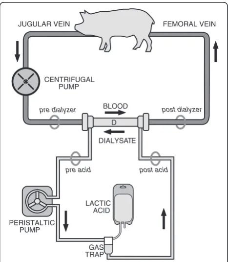
administration of an unfractioned heparin bolus (Epsoclar® Mayne Pharma Srl, Napoli, Italia, 200 U/kg), pigs were connected to a veno-venous extracorporeal circuit after surgical cannulation of the right external jugular vein with a 14-Fr catheter (Medtronic, Minne-apolis, MN, USA) for blood drainage and of the right femoral vein with a 10 to 12-Fr cannula for reinfusion. The extracorporeal circuit, required to infuse a concen-trated (40%) lactic acid solution, consisted of a centrifu-gal pump (Jostra rotaflow, Maquet, Hechingen, Germany), a gas trap, a polysulphone membrane dialyzer (F8HPS, Fresenius, Bad-Homburg, Germany) and 1/4 to 3/16 inch
inner-diameter tubing. No membrane lung was placed in the extracorporeal circuit. The outlet port of the dialyzer was connected to the inlet port to create a closed system with the dialysate flowing countercurrent to blood flow.
The experimental setup is outlined in Figure 1. The extracorporeal blood flow was set to 250 ml/minute and the dialysate flow at 300 ml/minute. A target activated clotting time (Hemocron Jr. Signature, ITC, Edison, NJ, USA) value of 250 to 300 sec was maintained by titrating a continuous infusion of unfractioned heparin.
During the experiment, the tidal volume was adjusted in order to maintain an arterial PCO2 of 50 mmHg,
which is a physiologic value for both our animal model and the chronic hypercapnic patients and corresponds to a nearly neutral pH due to the high bicarbonate con-tent of their blood. The inspiratory oxygen fraction was
pre dialyzer post dialyzer
post acid
BLOOD
DIALYSATE
FEMORAL VEIN JUGULAR VEIN
CENTRIFUGAL PUMP
D
LACTIC ACID
PERISTALTIC PUMP
pre acid
[image:2.595.305.539.439.707.2]GAS TRAP
40%; body temperature was controlled at 38°C through external physical methods. An intravenous infusion of a rapid-acting insulin analog was titrated to keep blood glucose levels constant (target = 100 mg/dl). At the end of the experiment all the pigs were sacrificed with a bolus injection of KCl (40 mEq) in the central venous line.
Study design
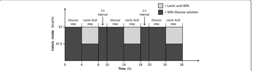
The experiment consisted of two steps, lasting four hours each, which differed only for the caloric source provided:
– Glucose step: 97 kcal/h were administered, through the central venous catheter, as a 50% glucose solution (52 ml/h)
– Lactic Acid step: 48.5 kcal/h were administered (34 ml/h) as a L-(+)-Lactic acid solution approxi-mately 40% in H2O (Sigma-Aldrich Corp. St. Louis, MO, USA) into the gas trap of the recirculating di-alysate [14], 48.5 kcal/h as 50% glucose solution (26 ml/h) were infused through the central venous catheter), for a total caloric load of 97 Kcal/h. The infused lactic acid amounted, therefore, to 2.5 mEq/ minute.
These two steps, always performed in a preset se-quence (Glucose followed by Lactic Acid) were repeated three times with a two-hour interval following the Lactic Acid step to allow a complete clearance of the infused lactate (Figure 2).
The total caloric intake during the whole study period was approximately 2,330 kcal/day, consistent with the estimated basal metabolic rate of anesthetized and cura-rized swine weighing approximately 45 kg [18].
Every hour (except during the two-hour interval after discontinuation of lactic acid infusion) we recorded: total CO2production (VCO2), body temperature, arterial blood
gases, blood glucose and arterial lactate level, hemodynamic parameters (heart rate, systemic arterial pressure, central
venous pressure) and ventilatory parameters (tidal volume, respiratory rate, minute ventilation, airway pressures). All the blood gas analyses were performed with the ABL 800 FLEX (Radiometer, A. De Mori, Milano, Italy) which is equipped for the automatic determination of pH, blood gases, oxymetry, electrolytes, glucose and lactate. Fully au-tomated quality controls are carried out; every four hours a manual calibration was performed. The ABL 800 measures the lactate concentration in the plasma. We calculated the ventilation of the dead space (VD) according to the stand-ard equation: VD = VE * (PaCO2-PeCO2/PaCO2). PeCO2=
partial pressure of expired mixed CO2. Alveolar ventilation
(VA) was calculated as follows: VA = VE - VD.
Total CO2 production (VCO2) was calculated as the
product of expired CO2concentration and minute
venti-lation (VE). The expired CO2 concentration was
mea-sured through an infrared CO2analyzer (WMA-4, GMR
Strumenti SAS, Florence, Italy) sampling at 400 ml/mi-nute from a 20-liter mixing box connected to the ex-haled gas exhaust port of the ventilator. VE was read from the ventilator, which was calibrated against a pneumotachograph (MLT300L, ADInstruments, Sydney, QLD, Australia), whose accuracy was regularly checked with a calibrated syringe.
Statistical analysis
[image:3.595.58.544.579.715.2]Statistical analysis was performed using the SigmaPlot 11.2 statistical software (Systat Software Inc., Chicago, IL, USA). Data are expressed as means ± standard devia-tions. To compare data of Glucose steps versus Lactic Acid steps a paired Student t-test was used. A paired-t analysis was performed to increase the statistical power, eliminating the confounding factor of the between-pig variation; actually a much larger sample would have been required to reject the null hypothesis without a paired analysis. The Shapiro-Wilk test was used to assess normality of the samples. A two-way ANOVA was per-formed to assess whether thetime factor had an impact on VCO2during the different steps.
Results
A total of 16 two-step sequences (Glucose - Lactic Acid), were conducted on six animals. No complications re-lated to lactic acid infusion were reported. In two pigs the study was interrupted before the third two-step se-quence, in one case due to abrupt clotting of an extra-corporeal cannula and in the other case due to severe hemodynamic instability.
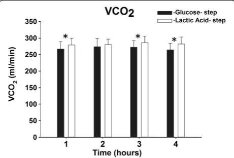
The average total CO2 production was 269 ± 22 and
282 ± 19 ml/minute, respectively, during Glucose and Lactic Acid infusion (P <0.05). The increase in VCO2
during the Lactic Acid step was 4.8%. This difference in VCO2,though quantitatively small, was, however,
statis-tically significant at the first, third and fourth hours of each step (see Figure 3), but not at the 2nd. The time fac-tor did not affect VCO2 (P= 1.00), which remained
stable over time during both the Glucose and Lactic Acid steps.
Arterial lactate was higher (2.6 ± 0.6 vs. 0.4 ± 0.2 mmol/l,
P <0.05), arterial pH was lower (7.422 ± 0.03 vs. 7.445 ± 0.02,P <0.05) and HCO3- was lower (32.2 ± 2.3 vs. 33.9 ±
1.7 mmol/L,P<0.05) during the Lactic Acid steps. Arterial PCO2(50.1 ± 1.4 vs. 50.3 ± 1.5 mmHg,P= 0.54) and
arter-ial glucose (107 ± 15 and 104 ± 13 mg/dl, P= 0.25) were kept constant; as was the amount of insulin needed to maintain target blood glucose (1.2 ± 0.8 and 1.3 ± 1 U/h during Lactic Acid and Glucose, respectively,P= 0.61). Ar-terial PO2 was 175 ± 30 mmHg during Lactic Acid and
167 ± 34 during Glucose (P= 0.22).
Minute ventilation (8.7 ± 1.2 vs. 8.3 ± 1.2 L/minute,
P <0.05) and alveolar ventilation (4 ± 0.3 vs. 3.8 ± 0.3 L/minute, P <0.05) were slightly (barely 5%) but significantly higher during Lactic Acid steps.
Body temperature was not different during Lactic Acid (38.0 ± 0.1°C) or Glucose (38.1 ± 0.1°C), P= 0.06. At all sample times all hemodynamic and respiratory parame-ters were comparable in both groups, except for a small
increase in tidal volume which was required to compen-sate for the VCO2rise (see Table 1).
Discussion
In the present study, we evaluated the VCO2changes in
anesthetized pigs associated with lactic acid infusion. While maintaining a constant caloric intake, replace-ment of 50% of the caloric input with lactic acid, corre-sponding to an infusion of 2.5 m Eq/minute, caused an increase in total CO2production of less than 5% (13 ml/
minute).
This clinically small but statistically significant differ-ence in total CO2 production may be due to several
causes. First, a small increase in total VCO2during lactic
acid infusion is expected since the complete oxidation of lactate load produces 3.6% more CO2than an amount of
glucose of equal caloric content. Therefore, since in the present study lactic acid infusion provided 50% of the total caloric intake, a VCO2increase of about 1.8% was
expected. A variable proportion of the infused lactic acid may enter gluconeogenesis, and then possibly be stored as glycogen. The net result of this process will also cause a net increase in VCO2.
Another factor contributing to the VCO2increase may
result from the conversion of bicarbonate ions into CO2,
caused by the increased systemic steady state levels of lactate and the resulting very mild metabolic acidosis. Indeed, although lactate levels remained lower than 3 mEq/l, a small reduction in arterial pH (of 0.023 pH units) was recorded. Such a systemic pH drop, promot-ing the «shift» of bicarbonate towards dissolved CO2,
may have caused a transient increase in VCO2, despite a
stable metabolic CO2 production. Failure to reach a
steady state condition during lactic acid infusion might have contributed to the slight increase in VCO2. Overall,
during lactic acid infusion, in order to maintain a con-stant arterial PCO2, a small increase in alveolar
ventila-tion (5%) was required.
The infusion of lactic acid at the inlet of a membrane lung, by shifting the equilibrium towards the dissolved form of carbon dioxide, determines a significant increase of extracorporeal CO2removal [12]. We have previously
[image:4.595.305.539.102.184.2]shown, in a similar animal model [14], that an infusion
Figure 3VCO2during Glucose and Lactic Acid steps over
[image:4.595.58.289.549.705.2]time.*:P<0.05 versus the glucose step at the same time.
Table 1 Main hemodynamic and respiratory parameters
Glucose Lactic acid P
Heart rate, bpm 91 ± 15 87 ± 18 P= 0.19
MAP, mmHg 74 ± 14 71 ± 12 P= 0.12
CVP, mmHg 10 ± 3 10 ± 4 P= 0.32
TV, ml 489 ± 52 513 ± 50 P <0.05
RR, breaths per minute 17.2 ± 1.2 17.2 ± 1.2 P= 0.32
of 2.5 mEq/minute of lactic acid (the same rate as tested in the present study) at the inlet of a pediatric artificial lung (extracorporeal blood flow 0.25 L/minute), could increase extracorporeal CO2 removal by approximately
40 to 45 ml/minute (60 ± 9 vs. 101 ± 16 ml/minute, with-out and with lactic acid infusion, respectively), while the PaCO2 was maintained constant at 50 mmHg as in the
current study. In that experiment we did not maintain a constant caloric intake, (caloric input increased from 43 to 90 Kcal/h when lactic acid was infused), hence, the total VCO2was not evaluated.
In the current study, at variance, we kept the caloric in-put constant and we observed that lactic acid infusion slightly increases the total CO2production (13 ml/minute
with our experimental setting). We may, therefore, specu-late that regional extracorporeal blood acidification at the inlet of the membrane lung may allow a substantial de-crease in the patient’s ventilatory needs; actually, despite increasing the total CO2 production (13 ml/minute
in-crease), it produces a significantly higher increase in extracorporeal CO2 removal (40 to 45 ml/minute
in-crease). This assumption may be true only if, during lactic acid infusion, the total caloric intake is main-tained constant.
The caloric input (around 2,330 kcal/day) applied in the present study, was based on literature and prelim-inary data [14,18]. The infusion of 2.5 mEq/minute of lactic acid (approximately 1,150 Kcal/day), represent-ing around 50% of the daily energy requirement of our model, would provide a conspicuous fraction of the daily caloric needs of an ICU patient and, there-fore, it should always be included in the energy balance.
The actual impact of lactic acid infusion, as part of an extracorporeal CO2 removal technique, on the
nutri-tional balance is still to be determined. Lactate enters the glucose metabolism pathway after conversion to pyruvate; if a patient’s total caloric input is kept con-stant, we can assume that this pyruvate may follow the same metabolic pathway as the pyruvate produced from glucose metabolism. Furthermore, the insulin infusion rate required during the Glucose and Lactic acid steps to maintain a constant blood glucose concentration was similar and very low; hence, it is unlikely that insulin in-fusion may have altered the balance between glucose storage and glucose oxidation.
The choice of lactic acid to achieve blood acidifica-tion has several advantages: it is not toxic [17], it has a very fast metabolism and point-of-care testing of lactate is available in most ICUs. In our previous study we also demonstrated that a 48-hour lactic acid infusion, performed through an extracorporeal dialysis circuit, is safe and does not lead to any injury of red blood cells and major organs [14]. Blood acidification
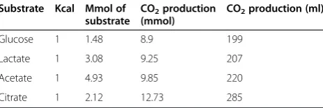
may be achieved by infusion of other metabolizable acids, such as citric or acetic acid; however, their an-ions are difficult to measure in the clinical setting. Moreover, their clearance is less predictable than that of lactate, at least for citric acid [19]. Furthermore, oxi-dation of equal caloric amounts of these acids produces more CO2than lactic acid (see Table 2).
An alternative choice may be the utilization of non-metabolizable acids (for example, hydrochloric acid), which do not provide calories; however, their usage re-quires an extracorporeal removal of the infused anions which may significantly increase the complexity of the extracorporeal system [20,21].
Some limitations of the present study deserve to be discussed. First, our animal model may present some dif-ferences from humans: in a growing pig, even if anesthe-tized and paralyzed, the basal energy requirement and VCO2 per unit weight are far greater than those of an
adult man [22]. Moreover, we cannot exclude the fact that different results would have derived from a critically ill animal model. Second, in our model, the arterial pH and bicarbonate ion concentrations are elevated, and may not closely represent the critical patients, even though the bicarbonate levels may be comparable to the one of chronic hypercapnic patients. Third, we did not investigate the metabolic pathway of infused lactate, which would have required the application of specific techniques (for example, use of isotopic carbon-labeled glucose or lactate) that are beyond the scope of the present investigation.
Conclusions
Administration of 50% of the total caloric input with an infusion of lactic acid (2.5 mEq/minute) only marginally increased (less than 5%) the total CO2production
[image:5.595.306.538.112.190.2]com-pared to an equal caloric load provided entirely by infu-sion of a 50% glucose solution. Since an equal rate of acidification, as shown in a previous study, increased the membrane lung removal by at least 50 ml/minute, we may speculate that blood acidification at the inlet of a membrane lung could be a promising technique to re-duce the ventilatory needs.
Table 2 CO2production from total oxidation of different
substrates to produce 1 Kcal
Substrate Kcal Mmol of
substrate
CO2production
(mmol)
CO2production (ml)
Glucose 1 1.48 8.9 199
Lactate 1 3.08 9.25 207
Acetate 1 4.93 9.85 220
Key messages
Infusion of lactic acid slightly increases the total CO2production compared to an isocaloric infusion of glucose.
An extracorporeal CO2removal technique based on blood acidification with lactic acid infusion could be a promising technique to reduce the ventilatory needs, if the total caloric intake is kept constant.
Abbreviations
ECCO2R:Extracorporeal CO2removal; VA: Alveolar ventilation; VCO2: CO2
production; VD: Ventilation of the dead space; VE: Minute ventilation; VILI: Ventilation-Induced Lung Injury.
Competing interests
Some procedures here described are part of a blood processing technique covered by patents or for which patents are pending: inventors: Antonio Pesenti, Nicolò Patroniti.
1). International patent application: PCT/EP 2008/003661; 7 May 2008 2). Italy: MI2007A000913; 7 May 2007
3). Italy: BO2012A000404; 26 July 2012; Bologna, Italy
Authors’contributions
AZ and MG participated in the design of the study, took part in the experiments, performed the statistical analysis and drafted the manuscript. PM, SR and VS participated in the design of the study and took part in the experiments. MC and MA took part in the experiments. VO took part in the experiments and data analysis. GB helped with the revision of the paper. NP and AP conceived of the study, and participated in its design and coordination, and helped to review the manuscript. All authors read and approved the final manuscript.
Acknowledgements
Special thanks go to Dr. G. Castagna, Dr. S. Abd El Aziz El Sayed Deab, Dr. D. Ferlicca, Dr. E. Rezoagli, Dr. S. Arrigoni, and Dr. S. Sosio for his help with the management of the experiments. We also thank Dr. G. Grasselli for his time spent in reviewing our manuscript.
The study was funded by Regione Lombardia (Bando Accordo Quadro Con il Sistema Universitario 1 Luglio 2009) and by Università Degli Studi Milano-Bicocca (Departemental Funds).
Author details
1Department of Medical Sciences, University of Milano-Bicocca, Ospedale San
Gerardo Nuovo dei Tintori, via Donizetti 106, 20900 Milan, Monza, Italy.
2Dipartimento di Scienze Veterinarie e Sanità Pubblica, Università degli Studi
di Milano, Via Celoria, 10, 20133, Milan, Italy.
Received: 27 March 2013 Accepted: 22 October 2013 Published: 11 November 2013
References
1. Pesenti A, Pelizzola A, Mascheroni D, Uziel L, Pirovano E, Fox U, Gattinoni L, Kolobow T:Low frequency positive pressure ventilation with
extracorporeal CO2 removal (LEPPV-ECCO2R) in acute respiratory failure (ARF): technique.Trans Am Soc Artif Intern Organs1981,27:263–266. 2. Gattinoni L, Kolobow T, Agostoni A, Damia G, Pelizzola A, Rossi GP, Langer
M, Solca M, Citterio R, Pesenti A, Fox U, Uziel L:Clinical application of low frequency positive pressure ventilation with extracorporeal CO2 removal (LFPPV-ECCO2R) in treatment of adult respiratory distress syndrome (ARDS).Int J Artif Organs1979,2:282–283.
3. Kolobow T, Gattinoni L, Tomlinson TA, Pierce JE:Control of breathing using an extracorporeal membrane lung.Anesthesiology1977,46:138–141. 4. Marcolin R, Mascheroni D, Pesenti A, Bombino M, Gattinoni L:Ventilatory
impact of partial extracorporeal CO2 removal (PECOR) in ARF patients.
ASAIO Trans1986,32:508–510.
5. Pesenti A, Rossi GP, Pelosi P, Brazzi L, Gattinoni L:Percutaneous extracorporeal CO2 removal in a patient with bullous emphysema with
recurrent bilateral pneumothoraces and respiratory failure.Anesthesiology
1990,72:571–573.
6. Terragni PP, Del Sorbo L, Mascia L, Urbino R, Martin EL, Birocco A, Faggiano C, Quintel M, Gattinoni L, Ranieri VM:Tidal volume lower than 6 ml/kg enhances lung protection: role of extracorporeal carbon dioxide removal.Anesthesiology2009,111:826–835.
7. Fuehner T, Kuehn C, Hadem J, Wiesner O, Gottlieb J, Tudorache I, Olsson KM, Greer M, Sommer W, Welte T, Haverich A, Hoeper MM, Warnecke G: Extracorporeal membrane oxygenation in awake patients as bridge to lung transplantation.Am J Respir Crit Care Med2012,185:763–768.
8. Kluge S, Braune SA, Engel M, Nierhaus A, Frings D, Ebelt H, Uhrig A, Metschke M, Wegscheider K, Suttorp N, Rousseau S:Avoiding invasive mechanical ventilation by extracorporeal carbon dioxide removal in patients failing noninvasive ventilation.Intensive Care Med2012,38:1632. 9. Javidfar J, Brodie D, Iribarne A, Jurado J, Lavelle M, Brenner K, Arcasoy S,
Sonett J, Bacchetta M:Extracorporeal membrane oxygenation as a bridge to lung transplantation and recovery.J Thorac Cardiovasc Surg2012, 144:716–721.
10. Batchinsky AI, Jordan BS, Regn D, Necsoiu C, Federspiel WJ, Morris MJ, Cancio LC:Respiratory dialysis: reduction in dependence on mechanical ventilation by venovenous extracorporeal CO2 removal.Crit Care Med
2011,39:1382–1387.
11. Crotti S, Lissoni A, Tubiolo D, Azzari S, Tarsia P, Caspani L, Gattinoni L: Artificial lung as an alternative to mechanical ventilation in COPD exacerbation.Eur Respir J2012,39:212–215.
12. Zanella A, Patroniti N, Isgro S, Albertini M, Costanzi M, Pirrone F, Scaravilli V, Vergnano B, Pesenti A:Blood acidification enhances carbon dioxide removal of membrane lung: an experimental study.Intensive Care Med
2009,35:1484–1487.
13. Snider MT, Chaudhari SN, Richard RB, Whitcomb DR, Russell GB: Augmentation of CO2 transfer in membrane lungs by the infusion of a metabolizable organic acid.ASAIO Trans1987,33:345–351.
14. Zanella A, Mangili P, Redaelli S, Ferlicca D, Patroniti N, Pesenti A:Blood acidification enhances extracorporeal carbon dioxide removal: long term animal study.Am J Respir Crit Care Med2012,185:A6020.
15. Gladden LB:A lactatic perspective on metabolism.Med Sci Sports Exerc
2008,40:477–485.
16. Gladden LB:Lactate metabolism: a new paradigm for the third millennium.J Physiol2004,558:5–30.
17. Leverve XM:Lactate in the intensive care unit: pyromaniac, sentinel or fireman?Crit Care2005,9:622–623.
18. Noblet J, Shi XS, Dubois S:Effect of body weight on net energy value of feeds for growing pigs.J Anim Sci1994,72:648–657.
19. Revelly JP, Tappy L, Martinez A, Bollmann M, Cayeux MC, Berger MM, Chiolero RL:Lactate and glucose metabolism in severe sepsis and cardiogenic shock.Crit Care Med2005,33:2235–2240.
20. Nolte SH, Benfer RH, Grau J:Extracorporeal CO2 removal with
hemodialysis (ECBicCO2R): how to make up for the bicarbonate loss?Int J Artif Organs1991,14:759–764.
21. Chang BS, Garella S:Complete extracorporeal removal of metabolic carbon dioxide by alkali administration and dialysis in apnea.Int J Artif Organs1983,6:295–298.
22. Pestana D, Garcia-de-Lorenzo A, Madero R:Metabolic pattern and lipid oxidation during abdominal surgery: midazolam versus propofol.
Anesth Analg1996,83:837–843.
doi:10.1186/cc13098
Cite this article as:Zanellaet al.:Infusion of 2.5 meq/min of lactic acid minimally increases CO2production compared to an isocaloric glucose