HERBAL BIOSYNTHESIS OF ZINC NANOPARTICLES AND EVALUATION OF THEIR ANTIFUNGAL
AND ANTIBACTERIAL EFFECT FOR BUFFALOES SKIN
*,1
Hassan A. Atef.,
2Hanan K.
1
Department of Mycology and Mycotoxins
2
Buffalo Diseases Research Department
3
Department Biochemistry, Animal Health Research Institute,
4Central Laboratory of Elemental and Isoto
ARTICLE INFO ABSTRACT
Herbal biosynthesis of
evaluation of their antimicrobial potential against some fungal and bacterial causes of skin affection in buffaloes were investigated. Out of 100 buffaloe cases that s
Trichophyton verucosum, T. mentagrophytes and C.albicans
and from (65% ; 37% and 13%) of skin scales samples of affected animals, respecti animals had comparatively lower rate of infection that ranged from 0.3
Dermatophilus congolensis
14% of skin scales samples of the affected animals, respectively. Whereas, in case of apparently healthy animals, all cases were not affected by
were affected by
plant, identified and characterized by visual inspection; in a UV transmission electron micro
size and the purity of the prepared powder. The particle size of prepared ZnO nanoparticles was 50 nm. The antimicrobial potential of prepared ZnONPs was evaluated by broth
microbial species from skin affection of buffaloes.
and turbidity of treated spore suspension decreased and reached 100% transmittance and cl MIC of ZnONPs. The
mg/ml and it was 17 and 14 mg/ml for
D.congolenesis,
treated fungal and bacterial cells were subjected to SEM, the damage and rupture of their cell wall membrane damage
Further studies are needed to investigate the efficacy of pharmaceutical preparation of ZnONPs as ointments, skin lotions and synergistic therapy with other traditional antibiotics in the treatment of animal ski
Copyright © 2015. Hassan A. Atef et al. This is an open access article distributed under the Creative Commons Att use, distribution, and reproduction in any medium, provided the original work is properly cited.
1. INTRODUCTION
The bacterial and fungal skin affections of animals cause many economic loses among affected animals and may cause zoonotic infection to the contact human. Dermatophytosis or ringworm infection and bacterial skin affection are considered as the most common cutaneous conditions all over the world, these infections are related to the stratum corneum of the
*Corresponding author: Hassan A. Atef.,
Department of Mycology and Mycotoxins Animal Health Research Institute, Agriculture Research Center, Dokki, Cair
ISSN: 0975-833X
Vol.
Article History:
Received 02nd September, 2015
Received in revised form
29th October, 2015
Accepted 25th November, 2015
Published online 30th December,2015
Citation: Hassan A. Atef., Hanan K. Mahmoud
biosynthesis of zinc Nanoparticles and evaluation of their antifungal and antibacterial effect for buffaloes skin affections
Journal of Current Research, 7, (12), 24338-24349 Key words:
Herbal biosynthesis,
Trichophyton verucosum, T. mentagrophytes, C.albicans, D.congolenesis,
Buffaloes, Nanotechnology, Antimicrobial, ZnONPs.
RESEARCH ARTICLE
HERBAL BIOSYNTHESIS OF ZINC NANOPARTICLES AND EVALUATION OF THEIR ANTIFUNGAL
AND ANTIBACTERIAL EFFECT FOR BUFFALOES SKIN AFFECTIONS
Hanan K. Mahmoud,
3Taha Hesham,
1Rasha M.H. Sayed El
and
4Mahmoud, H. H.
Department of Mycology and Mycotoxins, Animal Health Research Institute, Agriculture Research Center,
Dokki, Cairo, Egypt
Research Department, Animal Health Research Institute,
Agriculture Research Center,
Dokki, Cairo, Egypt
, Animal Health Research Institute, Agriculture Research Center,
Central Laboratory of Elemental and Isotopic Analysis, Nuclear Research Centre,
Atomic Energy Authority, Egypt
ABSTRACT
Herbal biosynthesis of zinc oxide nanmoparticles (ZnONPs) using extract of
evaluation of their antimicrobial potential against some fungal and bacterial causes of skin affection in buffaloes were investigated. Out of 100 buffaloe cases that showed skin lesions and 30 apparently healthy cases, the fungi of
Trichophyton verucosum, T. mentagrophytes and C.albicans were recovered from (50%, 31% and 15%) of hair
and from (65% ; 37% and 13%) of skin scales samples of affected animals, respecti animals had comparatively lower rate of infection that ranged from 0.3-1%. On the other hand,
Dermatophilus congolensis and S. aureus were recovered from 15% and 13% of hair samples and from 17% and
14% of skin scales samples of the affected animals, respectively. Whereas, in case of apparently healthy animals, all cases were not affected by Dermatophilus congolensis and only 1.8% of hair and 6 % of skin sc
were affected by S. aureus, respectively. The ZnONPs was synthesized using
identified and characterized by visual inspection; in a UV-visible spectrophotometer and Scanning by transmission electron microscope (TEM) and scanning electron microscope (SEM) for detection of their particle size and the purity of the prepared powder. The particle size of prepared ZnO nanoparticles was 50 nm. The antimicrobial potential of prepared ZnONPs was evaluated by broth microdilution methods against recovered microbial species from skin affection of buffaloes. As the concentrations of
and turbidity of treated spore suspension decreased and reached 100% transmittance and cl MIC of ZnONPs. The MIC that inhibited 50% of the growth of T.verucosum and T.mentagrophytes mg/ml and it was 17 and 14 mg/ml for MIC 100%, respectively. The MIC that inhibited
D.congolenesis, S. aureus species was 12.5-25 ug/ml and it was 150 ug
treated fungal and bacterial cells were subjected to SEM, the damage and rupture of their cell wall
membrane damage and some pits that have been caused leakage in inter cellular components and finally cell death. Further studies are needed to investigate the efficacy of pharmaceutical preparation of ZnONPs as ointments, skin lotions and synergistic therapy with other traditional antibiotics in the treatment of animal ski
is an open access article distributed under the Creative Commons Attribution License, which use, distribution, and reproduction in any medium, provided the original work is properly cited.
The bacterial and fungal skin affections of animals cause many economic loses among affected animals and may cause zoonotic infection to the contact human. Dermatophytosis or ringworm infection and bacterial skin affection are considered cutaneous conditions all over the world, these infections are related to the stratum corneum of the
Department of Mycology and Mycotoxins Animal Health Research Institute, Agriculture Research Center, Dokki, Cairo, Egypt.
epidermis and keratinized tissues such as skin, hair and nails of humans and animals (Popoola
Hassan et al., 2015). The diseases are caused by keratinophilic filamentous fungi, called dermatophytes, belonging to the genera Trichophyton, Microsporum, and Epidermophyton (Deacon, 1988). In cattle, dermatophytosis is most often caused by T. verrucosum called ringworm which
entity characterized by annular skin lesions
and Stenwig, 1985). It is transmitted easily from one animal to another and its spores may survive in the environment for 2 to 3 years and calves or purchased animals int
herd are likely to contract infection. According to
International Journal of Current Research
Vol. 7, Issue, 12, pp.24338-24349, December, 2015
INTERNATIONAL
Hanan K. Mahmoud, Taha Hesham, Rasha M.H. Sayed El-Ahl and Mahmoud, H. H. valuation of their antifungal and antibacterial effect for buffaloes skin affections
24349.
HERBAL BIOSYNTHESIS OF ZINC NANOPARTICLES AND EVALUATION OF THEIR ANTIFUNGAL
AFFECTIONS
Rasha M.H. Sayed El-Ahl
Agriculture Research Center,
Agriculture Research Center,
Agriculture Research Center, Dokki, Cairo, Egypt
pic Analysis, Nuclear Research Centre,
ZnONPs) using extract of Corriandrum sativum plant leafs and evaluation of their antimicrobial potential against some fungal and bacterial causes of skin affection in buffaloes howed skin lesions and 30 apparently healthy cases, the fungi of were recovered from (50%, 31% and 15%) of hair and from (65% ; 37% and 13%) of skin scales samples of affected animals, respectively. The apparently healthy
1%. On the other hand, bacterial species of 15% and 13% of hair samples and from 17% and 14% of skin scales samples of the affected animals, respectively. Whereas, in case of apparently healthy animals, and only 1.8% of hair and 6 % of skin scraping samples The ZnONPs was synthesized using extract of Corriandrum sativum visible spectrophotometer and Scanning by scope (TEM) and scanning electron microscope (SEM) for detection of their particle size and the purity of the prepared powder. The particle size of prepared ZnO nanoparticles was 50 nm. The microdilution methods against recovered As the concentrations of ZnONPs increased, the optical density and turbidity of treated spore suspension decreased and reached 100% transmittance and clear medium at the
T.verucosum and T.mentagrophytes was 11 -12
MIC that inhibited 50% of the growth of 25 ug/ml and it was 150 ug/ml for MIC 100%, respectively. The treated fungal and bacterial cells were subjected to SEM, the damage and rupture of their cell wall was detected or cellular components and finally cell death. Further studies are needed to investigate the efficacy of pharmaceutical preparation of ZnONPs as ointments, skin lotions and synergistic therapy with other traditional antibiotics in the treatment of animal skin diseases.
ribution License, which permits unrestricted
epidermis and keratinized tissues such as skin, hair and nails of (Popoola et al., 2006 ; Ameen, 2010 and The diseases are caused by keratinophilic filamentous fungi, called dermatophytes, belonging to the Microsporum, and Epidermophyton In cattle, dermatophytosis is most often caused called ringworm which denotes the clinical entity characterized by annular skin lesions (Weiss et al., 1979 It is transmitted easily from one animal to another and its spores may survive in the environment for 2 to 3 years and calves or purchased animals introduced into the herd are likely to contract infection. According to Hay (2003)
INTERNATIONAL JOURNAL OF CURRENT RESEARCH
and Havlickova et al. (2008), a number of factors including geographic location, prevailing climate as (temperature, humidity, wind, water activity etc.), overcrowding, health care,
immigration, environmental hygiene culture and
socioeconomic dispositions have great implication for the proliferation of dermatophytosis. The inflammation of the skin is associated with unthriftiness and general discomfort in affected animals. Dermatophytosis also has important economic significance, as apparently healed skin lesions re-appear after the tanning process (Haab, 1991).
On the other hand, the bacterial skin affections of buffaloes particularly with the dermatophilus species, can cause skin disease of animal (Radositis et al., 1994) and man (Harman et al., 2001). This disease in cattle caused by agram-positive actinomycetes, Dermatophilus congolensis is characterized by acute or chronic, local or progressive and sometimes fatal exudative dermatitis; it starts as an erythema, progressing through serous exudation and drying to form characteristic matting of the hair (Harman et al., 2001; Ambroso et al., 1999; Abdullahi, 2001 and Loria et al., 2005)
There is definitely a need for effective prophylaxis against the disease as hygienic and other preventive measures often fail. Consequently, a national vaccination program was advocated by the Swedish hide industry and restrictions on sale of breeding animals and on the access of livestock to common pastures are imposed on affected herds. Treatment of clinical ringworm in cattle is expensive and time-consuming (Shams-Ghahfarokhi, 2009).
The emerging infectious diseases and the development of drug resistance in the pathogenic bacteria and fungi at an alarming rate is a matter of serious concern. Despite the increased knowledge of microbial pathogenesis and application of modern therapeutics, the morbidity and mortality associated with the microbial infections still remains high (Kolar et al., 2001). Therefore, there is an urgent demand to discover novel strategies and identify new antimicrobial agents from natural and inorganic substances to develop the next generation of drugs or agents to control microbial infections. Prior to the extensive use of chemotherapeutics in modern health care system, inorganic antimicrobials such as silver, zinc and copper were used since ancient times to treat microbial infections (Moghimi, 2005). In the recent times, the advances in the field of nanosciences and nanotechnology has brought to form the nanosized inorganic and organic particles which are used for many applications as amendments in industrial, medicine and therapeutics, synthetic textiles and food packaging products (Gajjar et al., 2009).
The plant phytochemical with antioxidant properties is accountable for the preparation of metal nanoparticles. Recently, nanoparticles synthesis was achieved with bacteria, fungi and actinomycetes (Holmes et al., 1995 and Mukherjee et al., 2001) and use of plant extract such as neem, camellia sinensis and Corriandrum and several others which are compatible with the green chemistry principles (Badri et al., 2008; Begum et al., 2009 and Gnanasangeetha and Sarala, 2013). The biosynthetic approache using plant extracts is a compensation of adverse effects of chemical methods such as
easily available to be applied, safe to handle and possess a broad viability of metabolites.
Therefore, the present study was undertaken to study the prevalence of some fungal and bacterial causes of skin affection of buffaloes in some Egyptian farms. The herbal biosynthesis of ZnO nanoparticles and investigation of their antimicrobial potential were also evaluated.
2. MATERIALAND METHODS
2.1. Animals and samples
One hundred Egyptian buffaloes clinically showing skin lesions and 30 apparently healthy animals were selected from different farms at Giza and Cairo governorates for mycological and bacteriological examination. Hair and skin scraping scales samples were collected from all animals. The affected skin areas of the animal body were cleaned with 70% alcohol and skin scraping were taken from the edge of the lesion by scalpel blade and hair as mentioned by Refai et al.(2014 a). The scales and hairs were collected in clean labeled envelops or in clean sterile labeled Petri dishes.
2.2. Antifungal and antibacterial agents
Fluconazol (20 ug) and Entrofloxacin (20%) were purchased from Sigma Chemical Company (USA) and used as a comparable control.
2.3. Mycological and bacteriological examination
2.3.1. Direct microscopical examination
The hair and skin scraping scale specimens from all animals were examined microscopically as described by Refai et al. (2014 a) using KOH wet preparation method.
2.3.2. Isolation of dermatophytes and C.albicans from infected or apparently healthy animals
The samples from clinically affected or apparently healthy animals were cultured as described by Larone (1995) and Refai et al. (2014 a &b) in Petri dishes or test tubes containing Sabouraud Dextrose Agar supplemented with chloramphenicol and cycloheximide. A light inoculum of each hairs and skin scrapings scale specimen was scattered over the surface of the medium and gently pressed down into the agar. The inoculated plates were incubated at 37ºC for 24 hours up to 2-3 weeks. After the establishment of dermatophyte or yeast of candida growth, a subculture was made on SDA with cycloheximide for further identification and preservation of the obtained colonies.
2.3.3. Identification of the dermatophyte and C. albicans isolates
The isolated colonies were identified by macroscopical morphology and microscopical examination using Lactophenol cotton blue (LPCB) wet mount (Refai et al., 2014a and b), to
demonstrate the presence of hyphae, macroconidia,
chlamydospores, pseudohyphe, germ tube and other fungal structure.
2.3.4. Isolation and identification of S. aureus and dermatophilus species
A. Direct Microscopical Examination (Quinn et al., 1994): Small pieces were taken from the underside of the scab and softened in few drops of distilled water on a clean microscope
slide, a smear was made and stained by Loeffler,s methylene
blue and Gimsa or Gram's stains.
B. Culturing Method for isolation and identification of S. aureus : A part of skin scales and hair samples was inoculated into the test tubes containing nutrient broth and were incubated at 37 ˚C for 24 hrs. The subcultures were also made on nutrient agar, 5% sheep blood agar and Baird Parker and incubated at 37 ˚C for overnight. Based on morphological and staining characteristics, hemolytic activities on blood agar, biochemical characteristic and antibiotic sensitivity test, the bacteria were isolated and identified as described by (Quinn et al., 1994; Anon, 2007)
C. Culturing Method for isolation and identification of dermatophilus species (Haalstra, 1965): A small amount of scab material was ground up, placed in a screw capped bottle, moistened with one ml sterilized distilled water and allowed to stand open for 3 and half hours on the bench. Then the opened bottle transferred to candle jar with a candle was burned within
the jar to obtain 10- 20% CO2 tension (so the motile zoospores
were chemo-tactically attracted to the CO enhanced atmosphere and move to the surface of distilled water). After 15 minutes, the bottle was carefully removed and a drop was taken from the water surface with a bacteriological loop and cultivated on brain heart infusion agar plates which were
incubated at 37oC in 20% CO2 tension for 24 to 48 hours. The
suspected colonies were identified as recommended by (Silva et al., 2003; Yardley, 2004; Anon, 2007).
2.4. Herbal biosynthesis and characterization of Zinc oxide nanoparticles
A. Preparation of the leaf extract (Harborne, 1973; Trease and Evans, 1989): Fresh leaves were collected from Corriandrum sativum plants and washed several times with water to remove the dust particles and then sun dried to remove the residual moisture. The used extract for the reduction of zinc ions (Zn2+) to zinc nanoparticles (ZnONPs) was prepared by placing 50 grams of washed dried fine cut leaves in 250 mL glass beaker along with 100 mlof sterile distilled water. The mixture was then boiled for 60 minutes until the color of the aqueous solution changes from watery to light yellow. The extract was cooled to room temperature and filtered using filter paper. The extract was stored in a refrigerator in order to be used for further experiments.
B. Preparation of zinc nanoparticles (Gnanasangeetha and Sarala, 2013): 50 ml of Corriandrum sativum leaves extract
were taken and boiled to 60-80oC using a stirrer-heater. 5
grams of Zinc acetate dehydrate (99%purity) were added under constant stirring for 10 minutes. Then, 2.0 M NaOH was added
to make pH 12 resulted in a formation of pale white aqueous solution. This was then placed in a magnetic stirrer for 2hrs. The pale white precipitate was then taken out and washed several times with distilled water followed by ethanol to get rid of the impurities. Then a pale white powder of ZnONPs nanoparticles was obtained after drying at 60°C in vacuum oven over night. Complete conversion of Zn (OH) 2 into ZnO NPs took place during drying. The obtained powder in the filter
paper was dried in hot oven at 50-60 oC.
C. Characterization of zinc nanoparticles (Awodugba and Ilyas, 2013): The prepared ZnO nanoparticles were characterized for their optical and structural properties by using a UV-Vis spectrophotometer (Lamda-25; PerkinElmer; Waltham, Massachusetts) and the particle sizes and morphology were observed and measured under transmission electron micrograph (TEM) HITACHI H-800 (Hitachi) and scanning electron microscope (SEM) (Joe, JSM-5600LV, Japan).
2.5. Measurement of MIC of prepared ZnO nanoparticles against isolated dermatophytes and bacteria from skin affection of buffaloes (CLSI 2008)
The minimum inhibitory concentration (MIC) of ZnO-NPs for the tested isolated was determined by a broth micro-dilution method based on the National Committee for Clinical Laboratory Standards (NCCLS ) for bacteria (Balachandran et al. 2015), for filamentous fungi (CLSI 2008 ) and for yeasts (NCCLS, M27-A2 2002). In sterile 12- x 75-mm plastic test tubes, 900 ul of Roswell Park Memorial Institute broth medium (RPMI 1640) or Sabouraud dextrose broth medium (SD broth) (for fungi) or nutrient broth (for bacteria) was inoculated separately, then, 100 ul of spore suspension to adjust the
inoculums of dermatophytes spp and S. aureus to (2.5 х 103
cells/ml) and dermatophytes to (5 х 104 spores/ml). 100 ul of
zinc oxide nanoparticles concentrations (250, 200, 150, 100, 50, 25 , 12.5 and 6.25 ug/ml) for bacteria and (10, 11,12, 13, 14, 15, 16, 17 & 18 mg/ml) for fungi, were added . The antifungal agent Fluconazole 20 ug and antibacterial agent Entrofluxacin 20% were included in the assays as positive controls. For fungi, the tubes were incubated for 48 hr–14 days
at 30-37o C and for bacteria the tubes were incubated for 24-48
h at 37o C. The experiment was repeated twice. The MIC for
fungi and bacteria was defined as the lowest zinc oxide nanoparticles concentration that showing no visible fungal or bacterial growth after incubation time. After end of incubation period, 5 ul of tested broth were inoculated on the sterile nutrient agar plates for bacteria and SDA plate for fungi and
incubated at 37o C for 24 hr- 2 weeks. The MIC was
determined as the lowest concentration of ZnONPs inhibiting the visual growth of the test cultures on the agar plate. The turbidity of the growth in tubes was observed every 24 hrs. The growth was assayed by measurement of optical density and transmittance percentage of each tubes content at 405 nm using spectrophotometer.
2.6. Scanning Electron Microscopy (SEM) (Gong et al., 2007)
The morphological changes of T.mentagrophytes,
S. aureus which were treated by zinc oxide -NPs were observed with a scanning electron microscope (SEM). All the content of tubes were centrifugated and the sediments of each tube were dehydrated separately through a graded series of ethanol (30, 50, 60, 70, 80, 90, 95, and 100%), each level was applied twice for 15 min each time, then the ethanol :isoamyl acetate (3:1, 1:1, 1:3) and 100% isoamyl acetate applied twice for 30 min). The solutions in wells were dried with a critical-point drier
using liquid CO2 and coated with gold-coater for 5 min. The
coated samples were observed under SEM, JSM-5600LV with accelerating voltage of 10 kV.
RESULTS AND DISSCUSSION
Recently , the prevalence of the skin affection has significantly reduced in many developed nations of the world compared to the developing countries due to improved social, economic, health care and hygiene practice factors evident in the former (Loria et al., 2005 ; Havlickova et al., 2008 and Ilkit, 2010). Numerous medical and mycological literatures reported the incidence of dermatophytic infections around the globe with high prevalence rate recorded in many developing countries within the tropical and subtropical regions of the world. The disease can be epizootic and can result in considerable economic losses as a result of lost production, public health concern, premature culling and treatment costs and down grading of hides and skins (Wabacha et al., 1998; Nweze, 2010 and Hassan et al., 2015). The clinical disease has been recognized in several African countries (Efuntoye and Fashanu, 2002) and the distribution of dermatophytosis consequently reflected the distribution of the causative agents in these countries (Rahbar et al., 2010) with countries variations in dominant etiological species. Dermatophytic infections may be symptomatic or asymptomatic pose public health problems in many parts of the world causing cutaneous disfigurement (Havlickova et al., 2008). On the other hand, the fungal dermatophytosis infection in buffaloes (ringworm) and bacterial dermatophilosis are important skin infections which have received major consideration not only for economical losses in the animal breeding industry but also in regards to their zoonotic transmission to humans which represent the widest spread and most prevalent diseases of man and animal (Loria et al., 2005; Hassan et al., 2008 and 2015). The occurrence of these serious skin affections have public health problem. Despite aggressive treatment with new or more established licensed antimicrobial agents, these infections are important causes of morbidity and mortality in animals, Espinel-Ingroff (2009). The prevalence of dermatophytes infection in buffaloes was investigated by (Hassan et al., 2008) who recovered T.verrucosum and T.mentagrophytes from cases of suspected ringworm in cattle at incidence rates of 70% and 30% in skin scraping samples, respectively. Whereas, Shams-Ghahfarokhi (2009) reported that the most frequent dermatophyte isolated from cases of cattle ringworm was Trichophyton verrucosum (99% of total isolates) which was obtained from all cultures of positive cases except five cases (1.0%) infected with Trichophyton mentagrophytes.
Recently, Hassan et al. (2015), investigated the microbial causes of 120 cases of cattle suffering from obvious skin lesions. The recovered fungal species from samples included
Trichophyton verucosum and T. mentagrophytes and bacterial species of Dermatophilus congolensis. In this work, 100 cases of Egyptian buffaloes clinically showed skin lesions and 30 apparently healthy animals were selected from different farms in Giza and Cairo governorates for mycological and bacteriological examination. Hair and skin scraping scale samples were collected from all animals (Table 1). The fungi of Trichophyton verucosum, T. mentagrophytes and C.albicans were recovered from (50%, 31% and 15%) of hair and from (65%; 37% and 13%) of skin scales samples of affected animals, respectively. While, the apparently healthy animal had comparatively lower rate of infection, ranged from 0.3-1%.
Table 1. Prevalence of dermatophytes in affected and apparently healthy buffaloes
Type of fungi
Apparently healthy (30 cases)
Affected with skin lesion (100 cases)
Hair Skin scales Hair Skin scales
No. % No. % No. % No. %
T. verrocosum 2 0.6 3 1 50 50 65 65
T. mentagrophytes 1 0.3 1 0.3 31 31 37 37
C.albicans 3 1 1 0.3 15 15 13 13
Currently, the isolation of Trichophytone verucosum, T. mentagrophytes and C.albicans from cases of buffaloes
ringworm indicated that these fungi are the most common dermatophytes affecting animals in Egypt (Refai et al., 2014 a; Hassan et al., 2015). The primary dermatophyte isolation in this work was achieved by using a commercial Sabouraud-cycloheximide medium and direct microscopy for any given specimen type as recommended by Kane et al. (1997). The dermatophytes are able to survive in skin scales of infected animals for up to several months in moist and dark places where they can be easily transmitted to human and other animals (Weitzman and Summerbell, 1995). The zoophilic fungi, T. verucosum and T. mentagrophytes isolated in this study may be associated with frequent contacts of the buffaloes with rats and pet animals like dogs and cats as it was reported in cattle cases from traditional type farms, Moretti et al. (1998).
Regarding, the bacterial skin affection of buffaloes, several factors are involved in the pathogenesis of dermatophilosis and staphylococcus infections, among them are the mechanical injury to the skin, rainfall, tick infestation, concurrent diseases and or stresses that compromise the hosts immune system (Amabrose, 1996).
Hassan et al. (2014) isolated 6 species of bacteria namely, Staphylococcus species., Escherichia coli species, Klebsiella species, Coryne bacterium species, Enterococcus species and Streptococcus species from diseased buffaloe. While, Hassan et al. (2015) recovered D. congolensis from cattle cases suffered from skin affection.
The infection occurs when the integrity of the skin is impaired, as in case of long exposure to rain or traumatic injuries resulting from arthropod bites which serve as mechanical transmitters of microorganisms into epidermal layers, where germination zoospores takes places (Zaria 1993). The high relative humidity has a significant influence on the maturation and motility of the infective zoospores and it has been claimed
to be a major predisposing factor in the spread and epizootiology of dermatophilosis and other bacteria (Zaria, 1993) and latently infected animals may serve as the major reservoir of infection (Stewart, 1972). The organism persists in dry scabs and crusts and can survive in the environment for long periods (up to 42 months) (Oduyeo, 1989; Martinez and Prior, 1991). It may be transmitted by direct contact with a carrier host, by contaminated fomites or by biting and non- biting flies and is also encountered in a wide range of hosts such as buffalo, rabbit and sheep and rarely in man (Pal, 1989 and 1995 and Nagpal et al., 2012).
[image:5.595.307.561.408.518.2]The current results in Table (2) revealed that the Dermatophilus congolensis and S. aureus were recovered from 15% and 13% of hair samples and from 17% and 14% of skin scales of the affected animal, respectively. Whereas, in case of apparently healthy animal, all cases were not affected by Dermatophilus congolensis and only 1.8% of hair and 6 % of skin scraping samples were affected by S.aureus, respectively . Similar results were obtained by (Hassan et al., 2015).
Table 2. Prevalence of some bacterial infection in buffaloes with or without skin affection
Type of Bacteria
Apparently healthy (30 cases)
Affected with skin lesion (100 cases)
Hair Skin scales Haire Skin scales
No. % No. % No. % No. No.
Dermatophilus congolensis
0 0 0 0 15 15 17 17
S.aureus 6 1.8 20 6 13 13 14 14
Several studies indicated that there was significant prevalence of Dermatophilus infection in cattle and buffaloes that infested with ticks. This may be due to the fact that toxins present in saliva of ticks result in immunosuppression of the animals. In cattle, it causes high economic losses as body weight loss, decreased milk yield and the acceptance of live animals in the market would also be reduced (Hassan et al., 2015).
The alarming rate for antimicrobial resistance is growing poses great challenge, especially by bacteria and fungi which is one of the major concerns to public health and scientific communities worldwide (Dismukes, 2006 and Goossens, 2005). One promising approach is the application of nanotechnology in the battle against microorganisms and is being applied not only to the treatment of infectious diseases but also to diagnostics of infections and to prophylaxis against its effect (Klabunde et al., 1996). In addition, Zinc oxide nanoparticles had been utilized in patients for both diagnosis and therapy, leading to more effective medication with less unfavorable effects (Richards et al., 2000).
In the chemical synthesis of nanoparticles, high temperature and/or high pressure, inert atmosphere protection are required
for initiating the reaction or using toxic matters such as H2S
and metallic precursors which lead to non-eco-friendly byproducts Garima et al., 2011; Manish et al., 2012). Hence, the growing need of environmental friendly biosynthesis methods of metal nanoparticles interested the researchers for using green methods for pharmaceutical applications and biological approaches using microorganisms and plant extracts
have been suggested as valuable alternatives to chemical methods (Akl et al., 2012). Several biological systems including bacteria, fungi and yeast have been used in synthesis of nanoparticles (Alagumuthu and Kirubha, 2012) and the use of herbal methods in the synthesis of zinc oxide nanoparticles has increasingly become a topic of interests (Cynthia et al, 2012). In addition, the use of environmentally benign plant leaf extract (Rajasekharreddy et al., 2010) for the synthesis of zinc oxide nanoparticle offers copious profit of eco-friendliness, where toxic chemicals are not used (Vijayaraghavan and Nalini, 2010).
In the present study, the green synthesis of ZnONPs using aqueous leaf extracts of Corriandrum sativum plant was undertaken. The prepared ZnONPs were identified and characterized by visual inspection; in a UV-visible spectrophotometer and Scanning by transmission electron microscope (TEM) and scanning electron microscope (SEM) for detection of their particle size and the purity of the prepared powder. The particle size of prepared ZnO nanoparticles was 50 nm (Fig.1). It was reported that the characterized absorption peak of ZnONPs is detected at wave length of 350 nm due to electron transition from valence band to conduction band (Fig 2). The results revealed that ZnONPs with spherical and granular morphology had uniform distribution. Because of their unique properties and large number of applications, zinc oxide nanostructures are one of the main subjects of the nowadays research. ZnO nanoparticles are durable, free from affecting the soil fertility in comparison to traditional antifungal agents (Lopes et al., 2002 and Hassan et al., 2012 and 2013 a & b).
Fig.1. The TEM image of thesize and distribution of the particles
of zinc oxide (50 nm) (x 20 000)
Fig. 2. The UV-VIS absorbance spectra ofZNO-NPS
(the optimal W.L was 350 nm)
Several studies evaluated the antimicrobial activity of metal oxide NPs particularly ZnO powder against fungi in culture
0 0.5 1 1.5
0 200 400 600 800
A
b
so
rb
an
ce
(O
D
)
[image:5.595.310.558.554.707.2]media. Metal oxides nanoparticles are characterized by very high surface area to volume particularly ZnONPs which is of the least hazards to the environment (Sawai and Yoshikawa, 2004; Hassan et al., 2013 b and Nabawy, 2015). Zinc oxide is one of five zinc compounds that are currently listed as generally recognized as safe (GRAS) by the U.S. Food and Drug Administration. Zinc salt has been used for the treatment of zinc deficiency (Lopes et al., 2002).
Recently, the antifungal activities of ZnONPs was evaluated, the yeast of C. albicans was more sensitive for relatively lower concentrations (100 ug/ml). While, A. niger and A. ochraceus required higher concentration of ZnONPs to inhibit their growth. The diameters of of inhibition zones of ZnONPs (MIC) against A. flavus and A. ochraceus were 7 and 15 mm at the concentration of 300 ug/ml using well diffusion test, whereas, A. niger required relatively lower concentration (200 ug/ml) of nanoparticles to inhibit their growth (Hassan et al., 2014). Other study by Hosseini et al. (2011) reported that the MIC of ZnONPs against Aspergillus spp. and C. albicans was 1.013-296 μg/ml and for SDS and of Fluconazole were 0.001-0.56 and 0.062-128 μg/ml, respectively. They added that lower concentrations of ZnONPs were most effective as antifungal and antibacterial agent. Furthermore, different studies conducted in different laboratories showed that the antimicrobial activity is influenced by not only nanoparticles concentration but also by the size of the ZnO particles (Violeta et al., 2011 and Shawky et al., 2014).
On the other hand, Hassan et al. (2013 b) evaluated the inhibition of aflatoxigenic mold growth and aflatoxins production on yeast extract sucrose medium by zinc oxide NPs and they detected that a lower concentration of (8 ug/ml) was required to obtain effective inhibition. While, Hassan et al. (2013 c) investigated the inhibitory effect of prepared iron oxide nanoparticles against C.neoformance. They detected that
the MIC of Fe2O3-NPs by the use of disc diffusion test was
efficient than well diffusion test, where the MIC of Fe2O3-NPs
for C.neoformance was 50 ppm but in case of well diffusion test the MIC was 100 ppm or more. However, EL-Diasty et al. (2013) evaluated the antifungal activity of zinc oxide nanoparticles against species of Trichophyton mentagrophyte, Microsporum canis, Candida albicans and Aspergillus fumigatus that were isolated from diseased cases. They detected that the largest inhibition of the germination of all the tested fungi was observed at largest ZnO nanoparticles concentration (40 mg/ml).
Nabawy et al. (2014) evaluated the antifungal potential of ZnO nanoparticles in comparison with commercial antifungal feed additives. The authors revealed that the mean of growth inhibition zone diameters for non-aflatoxigenic strains ranged from (11.0±0.5 mm) to (21.3±0.96 mm) when the concentration of ZnO nanoparticles elevated from 25-250 ug/ml using well diffusion test. Whereas, at the same concentrations of ZnO nanoparticles, the mean growth inhibition zone diameters of aflatoxigenic strains were relatively smaller (ranged from 8.2±0.4 to 19.3±0.94 mm).
Whereas, nano-silver exhibited potent activity against clinical isolates and ATCC strains of Trichophyton mentagrophytes and
Candida species (IC80, 1-7 μg/ml). The activity of nano-Ag was comparable to that of amphotericin B and fluconazole. The antifungal activity of nano-Ag is attributed to its effects on the fungal mycelia (Kim et al., 2009; Hassan et al., 2013 c). There are also reports of the application of nanosilver having antifungal activity in bio-stabilization of footwear materials, wherein, 1% solution inhibited the growth of the majority of yeast-like fungal and mold strains (Falkiewicz and Macura, 2008).
In the present study, the antimicrobial potential of prepared ZnONPs was evaluated by broth micro dilution method as recommended by (CLSI, 2008). The inoculum size, turbidity and transmittance of treated spore suspension of dermatophytes and bacteria was determined spectrophotometrically to provides target percent of transmittance (T%) readings. It is difficult to hold microtiter tests longer than 72 h due to dehydration. Many of the less frequently encountered fungi as dermatophytes may require as long as 120–144 hours before growth is detected in the drug-free growth control well. For this reason, isolates that are known to be slow growers should be tested via the macro broth method (CLSI, 2008). The endpoint determination depend on a reduction in turbidity which typically easy to visualize. Reading the MIC endpoint determined at the lowest concentration that prevents discernable growth, the first clear well (Ghannoum et al., 2004). The dermatophytes are a specialized group of fungi, which infect the keratinized tissues of humans and animals, such as skin, hair, and nails, commonly causing superficial infections. While, there have been several studies involving yeasts which have been used to develop the Clinical and Laboratory Standards Institute (CLSI) reference method and the antifungal susceptibility of dermatophytes has only recently been addressed in the standard for filamentous fungi (CLSI, 2008).
Currently, the tabulated results in Table (3), illustrated that the antifungal potential of ZnONPs against T.verucosum and T.mentagrophyte species was concentration dependant, when the concentrations of ZnONPs increased up to 20 mg/ml , the optical density and turbidity of treated spore suspension were decreased till reach 100% transmittance and clear medium. The inhibitory concentration of ZnONPs that inhibited 50% of the growth of T.verrucosum and T.mentagrophytes was 11, 12 mg/ml and it was 17 , 14 mg/ml for IC 100%, respectively. The transmittance percentage and clearance of spore turbidity were confirmed by re-cultivation of inoculums from treated tubes on the specific medium SDA for dermatophyte and NA for bacteria. The accordance of all previous parameters was repeated 2 times to pooled data (Table, 3 and Fig. 3 & 4).
In other study, Hassan et al. (2015) evaluated the antimicrobial
effect of prepared Fe2O3 NPs against isolated Dermatophytes
and Dermatophilus species that recovered from skin affection of cattle and it had an inhibitory effect against the growth of T. verrucosum at concentrations of 3 mg /ml and 4 mg /ml, respectively (using well diffusion test). While, in case of T.mentagrophytes, iron oxide NPs revealed an inhibitory effect at concentration of 1, 2, 3, 4 and 5 mg/ml by well diffusion test.
The treatment by Fe2O3 NPs had no effect on the growth of
Dermatophilus sp. at the concentration ranged from 1- 3 mg/ml
using disc diffusion test. While, the treatment by 4 mg/ml or more resulted in inhibition of bacterial growth.
Table 3. Optical density and degree of turbidity of treated Dermatophytes recovered from skin affection of buffaloes at
gradual concentration of ZnONPs
Gradual doses of ZnONPS mg/ml
Dermatophyte Sp
T.verrucosum T.mentagrophytes
O.D T % D.T.&G.T. OD T% D.T.&G.T.
10 0.60 25.1 4+ 0.75 17.7 4+
11 0.56 27.5 3+ 0.52 30.2 3+
12 0.30 50.1 2+ 0.18 66 2+
13 0.20 63.1 1+ 0.05 89.1 1+
14 0.17 67.6 1+ 0.04 91.2 1+
15 0.11 77.7 1+ 0.00 100 0-
16 0.05 89.1 0- 0.00 100 0-
17 0.00 100 0- 0.00 100 0-
18 0.00 100 0- 0.00 100 0-
Control antifungal: Fluconazol 20 ug (Its OD: 00 and turbidity: 00)- OD : Optical density of treated spores at wave length ; 405 nm.
- DT : Degree of turbidity of treated suspension. - G.T.: Growth after Treatement.
T%: Transmitance %
Fig. 3. Transmittance % of treated T.verucosum at gradual concentration of ZnO NPs
Fig.4. Transmittance % of treated T.mentagrophyte at gradual concentration of ZnO NPs
Several studies showed that nanoparticls as zinc homeostasis is regulated through a number of specific and nonspecific membrane-bound uptake and efflux pumps (Abou-Mohamed et al., 1998) and prevents sulfhydryl groups from oxidation (Lim et al., 2004). The antimicrobial activity of ZnONPs have
been studied against the food related bacteria Bacillussubtilis,
Escherichia coli and Pseudomonas fluorescens (Jiang et al., 2009). Zinc oxide nanoparticles could potentially be used as an effective antibacterial agent to protect agricultural and food safety from food borne pathogens, especially E. coli O157:H7 (Zhang et al., 2007). Also, ZnONPs possess antimicrobial
activities against Listeria monocytogenes, Salmonella
enteritidis and E. coli O157:H7 in culture media (Jin et al., 2009).There are also other studies confirming the strong antimicrobial activity of ZnO nanoparticles where, the nanoparticles could completely lyse the food-borne bacteria
Salmonella typhimurium and Staphylococcus aureus (Liu
et al., 2009 and Hassan et al., 2014). In another study, ZnO nanoparticles (12 nm) inhibited the growth of E. coli by disintegrating the cell membrane and increasing the membrane permeability (Jiang et al., 2009). The above findings suggest that ZnO nanoparticles can find applications in food systems and can be used to inhibitgrowth of pathogenic bacteria.
[image:7.595.37.292.474.653.2]Regarding, the antibacterial potential of prepared ZnONPs in this study, the current data in Table (4) and figures (5, 6), showed the antifungal potential of ZnO-NPs against D.congolenesis and S.aureas species. As the concentrations of ZnONPs increased up to 250 ug/ml, the optical density and turbidity of treated cell suspension were decreased till reach 100% transmittance and clear medium at the IC80-100 of ZnONPs for treated bacteria. The IC that inhibited 50% of the growth of D.congolenesis and S.aureus species was 12.5-25 ug/ml and it was 150 ug/ml for MIC80-100%, respectively. The transmittance % and clearance of cells turbidity were confirmed by re-cultivation of inoculums from treated wells on the nutrient agar medium for D.congolenesis and S.aureus species. The accordance of all previous parameters were repeated 2 times to pooled data (Table, 3 and Fig. 5 & 6).
Table 4. Optical density and degree of turbidity of treated Dermatophilus and Staphylococci bacteria recovered from skin
affection of buffaloes at gradual concentration of ZnO NPs
Gradual doses of ZnO NPS
ug/ml
Bacterial species
D. congolenesis S.aureus
O.D T% D.T.&G.T.. OD T% D.T.&G.T.
0 1.27 5.4 4+ 0.80 15.1 4+
50 0.40 39.8 3+ 0.75 17.7 3+
100 0.20 63.1 2+ 0.17 67.6 2+
200 0.10 79.4 1+ 0.11 77.6 2+
300 0.03 93.3 1+ 0.08 83.2 1+
500 0.00 100 0- 0.00 100 0-
1000 0.00 100 0- 0.00 100 0-
2000 0.00 100 0- 0.00 100 0-
3000 0.00 100 0- 0.00 100 0-
Control antibacterial as Entrofloxacin 20 ug (Its OD: 00 and turbidity: 00)- OD : Optical density of treated spores at wave length ; 405 nm. - DT : Degree of turbidity of treated suspension.- G.T.: Growth after Treatment.
T%: Transmittance%
On the other hand, the antimicrobial effect of ZnONPs was
reported to occur by 2 ways. The first is the formation of H2O2
on the surface of ZnONPs due to the possible formation of hydrogen bond between hydroxyl group of cellulose molecules of fungi with oxygen atom of ZnONPs leading to inhibition of
the microbial growth, while the second is the release of Zn2+
that causes damages of cell membrane and interacts with intraocular contents (Moraru et al., 2003). Several natural and
engineered nanomaterials have demonstrated strong
0 20 40 60 80 100 120
0 5 10 15 20
T%
o
f
tr
e
at
e
d
s
p
o
re
su
sp
e
n
si
o
n
o
f
T.
V
e
ru
co
su
m
CONCENTRATIONS OF ZnO NPs (mg/ml)
0 20 40 60 80 100 120
0 5 10 15 20
T
%
.
O
F
T
R
EA
T
ED
S
U
SP
EN
SI
O
N
O
F
T
.m
e
n
ta
gr
o
p
h
yt
e
[image:7.595.298.564.487.612.2]antimicrobial properties through diverse mechanisms including photocatalytic production of reactive oxygen species that damage cell components and viruses (as ZnO
[image:8.595.41.287.131.297.2]the cell envelope (e.g. peptides, chitosan carboxyfullerene, carbon nanotubes, ZnO and interruption of energy transduction (Matei et al., 2010; Violeta et al., 2011).
Fig.5. Transmittance % of treated D.congolenesis concentration of ZnO NPs
Fig.6. Transmittance % of treated S.aureas concentration of ZnO NPs
There are several mechanisms, which have been proposed to explain the antibacterial activity of ZnONPs.
hydrogen peroxide from the surface of ZnO is considered as an effective mean for the inhibition of bacterial growth (Yamamoto, 2001). It is presumed that with decreasing particle size, the number of ZnO powder particles per unit
powder slurry increases resulting in increased surface area and increased generation of hydrogen peroxide.
mechanism for ZnO antibacterial activity is the
ions which cause the damage of cell membrane and interact with intracellular contents (Brayner et al., 2006).
In the present work, it was proved that the antifungal and antibacterial effects of metal nanoparticles was probably due to the damage of the cell wall of the microbial cells leading to leakage of the cell contents and finally cell death.
are confirmed when the treated dermatophytes, dermatophilus species and S.aureas were subjected to SEM, the damage and rupture of their cell wall were detected in the area surrounding
0 20 40 60 80 100 120
0 100 200
T
%
O
F
T
R
EA
T
ED
S
P
O
R
E
SU
SP
EN
SI
O
N
O
F
D
.
C
o
n
go
lo
n
e
si
s
CONCENTRATIONS OF ZnO NPs (ug/ml)
0 20 40 60 80 100 120
0 100 200
T%
O
F
T
R
EA
TE
D
S
P
O
R
E
SU
SP
EN
SI
O
N
O
F
S.
a
u
re
as
CONCENTRATIONS OF ZnO NPS (ug/ml)
24345 Hassan A. Atef et al. Herbal biosynthesis of zinc Nanoparticles and evaluation of their antif
antimicrobial properties through diverse mechanisms including photocatalytic production of reactive oxygen species that components and viruses (as ZnO), compromising pe (e.g. peptides, chitosan carboxyfullerene, carbon nanotubes, ZnO and interruption of energy transduction
D.congolenesis at gradual
S.aureas at gradual
There are several mechanisms, which have been proposed to explain the antibacterial activity of ZnONPs. The generation of hydrogen peroxide from the surface of ZnO is considered as an effective mean for the inhibition of bacterial growth ). It is presumed that with decreasing particle size, the number of ZnO powder particles per unit volume of powder slurry increases resulting in increased surface area and ration of hydrogen peroxide. Another possible mechanism for ZnO antibacterial activity is the release of Zn2+ the damage of cell membrane and interact
., 2006).
that the antifungal and antibacterial effects of metal nanoparticles was probably due to the damage of the cell wall of the microbial cells leading to leakage of the cell contents and finally cell death. These effects tophytes, dermatophilus were subjected to SEM, the damage and rupture of their cell wall were detected in the area surrounding
growth. The effect of high concentration of ZnO nanoparticles on the treated fungi and bacteria was observ
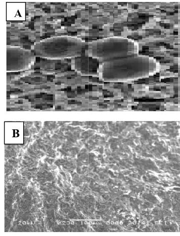
damage of cells and some pits that have been caused leakage in inter cellular components and finally cell death (Fig.7&8). Similar findings were also reported by
Hassan et al., 2014; Hassan et al 2014).
Fig, 7. (A) Scanning Electron Microscopy (SEM) of normal
T. verrucosum (B): Scanning Electron Microscopy (SEM) of
T. verrucosumafter treatement with
Fig. 8. (A) Scanning Electron Microscopy (SEM) of normal
D. congolensis bacteria (B): Scanning Electron Microscopy
(SEM) of D. congolensis after treatement
200 300
200 300
CONCENTRATIONS OF ZnO NPS (ug/ml)
A
B
A
B
Herbal biosynthesis of zinc Nanoparticles and evaluation of their antifungal and antibacterial effect for buffaloes skin affections
growth. The effect of high concentration of ZnO nanoparticles on the treated fungi and bacteria was observed as membrane damage of cells and some pits that have been caused leakage in components and finally cell death (Fig.7&8). Similar findings were also reported by (Violeta et al., 2011, et al., 2014, 2015; Shawky et al.,
) Scanning Electron Microscopy (SEM) of normal ): Scanning Electron Microscopy (SEM) of
after treatement with ZnO NPS
[image:8.595.341.523.145.384.2] [image:8.595.35.293.328.500.2] [image:8.595.330.536.427.718.2]Conclusion
From the present study it is concluded that the periodical examination of human and animals for skin affections pathogens should be undertaken. All environmental factors which predispose for these affections must be under hygeinic measures and good control. The rapid biosynthesis of zinc oxide nanoparticles using aqueous leaf extract of Corriandrum sativum plant provides an environmental friendly, simple and efficient route. The use of plant extracts avoids the usage of harmful and toxic reducing and stabilizing agents. In addition, the antimicrobial effects of metal nanoparticles are due to the damage of the cell wall of the microbial cells leading to leakage of the cell contents and finally cell death. Further studies are needed to investigate the efficacy of pharmaceutical preparation of ointments, skin lotions and synergistic therapy with other traditional antibiotics in the treatment of animal diseases.
Acknowledgement
The authors are gratefully acknowledged to Prof. Dr. M.K. Refai for his kind assistance and advice in initiating the achievement of this work and proof revision of this paper for publication.
REFERENCES
Abdullahi, U.S. 2001. Chemotherareumatic and chemoprophy-lactic control of bovine Dermatophilosis Ph.D thesis. Ahmadu Bello Univ, Zaria, Nigeria pp. 66-68.
Abou-Mohamed, G.; Papapetropoulos, A.; Catrava, J.D. and Caldwell, R.W. 1998. Zn2+ inhibit nitric oxide formation in response to lipopolysaccharides :implication in its anti- inflammatory activity. Eur. J. Pharmaco., 341,265-272. Akl M. Awwad1, Nidà M. Salem, Amany O. Abdeen 2012.
Biosynthesis of Silver Nanoparticles using Olea europaea Leaves Extract and its Antibacterial Activity. Nanoscience and Nanotechnology, 2, 6, 164-170.
Alagumuthu, G. and R.Kirubha 2012. Green synthesis of silver nanoparticles using Cissus quadrangularis plant extract and their antibacterial activity, International Journal of Nanomaterials and Biostructures, Vol. 2, No. 3, pp. 30-33
Amabrose, N.C. 1996. The pathogenesis of Dermatophilosis. Trop.Anim. Health-peod., 26: 295-375.
Ambroso, N., Lioyd, D. and Millard, C. 1999. Immune
response to Dermatophilus congolensis infections,
Parasital. Today, 15; 295-300.
Ameen M. 2010. Epidemiology of superficial fungal infections. Clin. Dermatol. 28:197 -201.
Anon, S.K. 2007. Dermatophilus dermatitis with clinical observations and laboratory finding. J. Clin. Microbiol., 9, 7-21.
Awodugba, A.O. and Ilyas, A. O. 2013. Synthesis and characterization of Zn-O nanoparticles with zinc chloride as zinc source. Asian J. Nature Appl. Sci., 2 : 41-44.
Badri S., Narayanan K., Sakthivel N. 2008. Coriander leaf mediated biosynthesis of gold nanoparticles, Mat Lett., 62, 4588-4600.
Balachandran, C., P. Saravana Kumar; Y. Arun, V. Duraipandiyan, R. Lakshmi Sundaram, A. Vijayakumar; K. Balakrishna, S. Ignacimuthu, N. A. Al-Dhabi and P. T. Perumal, 2015. Antimicrobial, antioxidant, cytotoxic and molecular docking properties of N- benzyl-2,2,2-trifluoroacetamide. Appl Nanosci., 5:207–216.
Begum N.A, Mondal S. Basu, R.A. Laskar, D. Mandal, 2009. Colloids and Surface B: Bio interfaces. D. Philip, Spectrochimica Acta: Part A, 73, 374-390.
Brayner R, Ferrari-Iliou R, Brivois N, Djediat S, Benedetti MF, Fievet F. 2006. Toxicological impact studies based on Escherichia coli bacteria in ultrafine ZnO nanoparticles colloidal medium. Nano Lett., 2006;6:866–870.
Clinical and Laboratory Standards Institute (CLSI) 2008.
Reference method for Broth dilution antifungal
susceptibility testing of filamentous fungi; Approved standard second—Edition CLSI document M38-A2 (ISBN 1-56238-668-9) (2008) Clinical and Laboratory Standards Institute, 940, West valley Road, Suite 1400, Wayne, Pennsylvania 19087-1898 USA
Cynthia Mason, Singaravelu Vivekanandhan, Manjusri Misra, Amar Kumar Mohanty, 2012. Switchgrass (Panicum virgatum) Extract Mediated Green Synthesis of Silver Nanoparticles, World Journal of Nano Science and Engineering, Vol. 2, pp.47-52
Deacon JW. 1988. Introduction to modern mycology, 2nd ed. Oxford: Blackwell Scientific Publications, 1988: 1-239. Dismukes WE. 2006. Antifungal therapy: lessons learned over
the past 27 years. Clin Infect Dis 42:1289–1296
Efuntoye, M. O. and Fashanu, S. O. 2002. Fungi isolated from skins and pens of healthy animals in Nigeria. Mycopathologia, 153(1): 21-32.
EL-Diasty, E.M.; Ahmed, M.A.; Okasha, N.; Mansour, S.F.; EL- Dek, S.I.; Abd EL-Khalek, H.M. and Youssif, M.H. 2013. Antifungal activity of zinc oxide nanoparticles against dermatophytic lesions of cattle. Romanian J.
Biophys., 23, (3).191 -202.
Espinel-Ingroff, A. 2009 Novel antifungal agents, target or therapeutic strategies for the treatment of invasive fungal diseases: A review of the literature (2005-2009) J. Rev Iberoam Micol. 26(1):1-22
Falkiewicz-Dulik M, Macura AB. 2008. Nanosilver as substance biostabilising footwear materials in the foot mycosis prophylaxis. Mikologia Lekarska. 2008; 15:145-150.
Gajjar P, Pettee B, Britt DW, Huang W, Johnson WP, Anderson J. 2009. Antimicrobial activities of commercial nanoparticles against an environmental soil microbe, Pseudomonas putida KT2440. Journal of Biological Engineering, 3 :9-22.
Garima Singhal, Riju Bhavesh, Kunal Kasariya, Ashish Ranjan Sharma, Rajendra Pal Singh, 2011. Biosynthesis of silver nanoparticles using Ocimum sanctum (Tulsi) leaf extract and screening its antimicrobial activity, Journal of Nanoparticle Research, Vol. 13, pp.2981–2988.
Gnanasangeetha D.1 and SaralaThambavani D. 2013. One Pot Synthesis of Zinc Oxide Nanoparticles via Chemical and Green Method. Res. J. Material Sci., Vol. 1(7), 1-8. Gong, P.; H. Li ; X. He ; K. Wang ;. J. Hu ; S. Zhang and X.
Yang, 2007. Preparation and antibacterial activity of Fe3O4@Ag na-noparticles.Nanotechnology, 18, No. 28, 1000-1010
Goossens, H. 2005. European status of resistance in nosocomial infections. Chemotherapy, 51:177–189.
Haab C. 1991. Epidemiologie der Trichophytie beim Mastkalb (Inaugural-Dissertation). Switzerland, Zurich: University of Zurich, 77, p.
Haalstra, R. 1965. Isolation of Dermatophilus congolensis from skin lesions in the diagnosis of streptothricosis. Vet. Rec., 77: 824-834.
Harborne J.B. 1973. Phytochemical methods, London, Chapman and Hall Ltd, 2nd ed. 49-188.
Harman M., Sekin S, and Akdeniz S. 2001. Human Dermatophilosis mimicking ringworm. Br. Dermatol., 145(1): 170-171.
Hassan, A, A.; Mogda, K. Mansour, B. and H.H. Mahmoud 2013 a. Biosynthesis of silver nanoparticles (Ag-Nps) (a model of metals) by Candida albicans and its antifungal activity on some fungal pathogens (Trichophyton mentagrophytes and Candida albicans). New York Science Journal. 6, 27-34.
Hassan, A. Atef ; Howayda , M. El-Shafei and Mahmoud , H.H.( 013 b. Effect of Zinc Oxid Nanoparticles on The Growth of Some Mycotoxigenic Moulds J. Studies in Chemical Process Technology (SCPT), American Society of Science and Engineering, 1 (3) :16-25
Hassan, A. Atef., Noha H. Oraby., El-Dahshan, E.M. E. and Ali, M.A. 2015. Antimicrobial Potential of Iron Oxide Nanoparticles in Control of Some Causes of Microbial Skin Affection in Cattle. European Journal of Academic Essays, 2(6): 20-31.
Hassan, A.A.; El Shorbagy, M.M.; El-Barawy, A.M. and Hassan, M.A. 2008. Study the availability of using buckthorn (Rhamnus cathartica) plant extract in laboratory
control of some bacterial and fungal diseases. The 5th
Scientific Congress, Minufiya Vet. J., 5, 27–39.
Hassan, A.A.; Howayda, M. El Shafei; Noha, H. Oraby; Rasha, M.H. Sayed El Ahl and Mogeda, K. Mansour, 2012.
Studies on mycosis and mycotoxicosis in cattle. 1st Conf. of
An. Health Res. Inst. Assoc., December 2012. pp. 216 – 227
Hassan, A.A.; M.A. Rashid ; Noha H. Oraby; S. El-Araby and M.M. Minshawy 2013 c. Using of molecular biology techniques for detection of Cryptococcus neoformans in respiratory disorders in cows with references to its control by nanoparticles of iron oxide (Fe2O3). Egypt. J. of Appl. Sci., 28,(6): 433-448.
Hassan, A.A.; Noha, H. Oraby; Aliaa.A. E. Mohamed and Mahmoud H.H. 2014. The possibility of using Zinc Oxide nanoparticles in controlling some fungal and bacterial strains isolated from buffaloes. Egypt. J. of Appl. Sci., 29 (3): 58-83.
Havlickova B, Czaika VA, Friedrich M. 2008. Epidemiological trends in skin mycoses worldwide. Mycoses 51:2-15. Hay RJ. 2003. Fungal infections (mycoses). In: Warrell DA,
Cox TM, Firth JD and Benz EJ Jr (Eds.). Oxford Textbook
of Medicine 4th Ed. Oxford. Oxford University Press Section, 7.12.1
Holmes J.D, Smith P.R, Evans. Growing R, Richardson D.J., Russel D.A and Sodeau J.R. 1995. Energy-dispersive X-ray analysis of the extracellular cadmium sulfide crystallites of Klebsiella aerogenes. Arch. Microbial., 163, 143-150. Hosseini, S.S.; Roudbar Mohammadi Sh.; Joshaghani HR.
and Eskandari M (MSc) 2011. Antifungal effect of Sodium Dodecil Sulfate and Nano particle ZnO on growth inhibition of standard strain of Candida albicans. Journal of Gorgan University of Medical Sciences, Vol. 12,(4), 200-205.
Ilkit, M. 2010. Favus of the scalp: an overview and update. Mycopathologia., 170: 143-154.
Jiang W, Mashayekhi H, Xing B. 2009. Bacterial toxicity comparison between nano- and micro-scaled oxide particles. Environ.Pollut., 157:1619–1625.
Jin T, Sun D, Su Y, Zhang H, Sue HJ. 2009. Antimicrobial efficacy of zinc oxide quantum dots against Listeria monocytogenes, Salmonella enteritidis and Escherichia coli O157:H7. J. Food Sci., 2009;74:46–52.
Kane, J.; Summerbell, R.C.; Sigler, L.; Krajden, S. and Land, G. 1997. Laboratory handbook of dermatophytes. Belmont CA, USA: Star Publishers.
Kim KJ, Sung WS, Suh BK, Moon SK, Choi JS, Kim JG. 2009. Antifungal activity and mode of action of silver nano-particles on Candida albicans. Biometals, 22:235-242. Klabunde KJ, Stark J, Koper O, Mohs C, Park D, Decker S,
Jiang Y, Lagadic I, Zhang D. 1996. Nanocrystals as Stoichiometric Reagents with Unique Surface Chemistry. J. Phys. Chem., 1996;100:12142-12153.
Kolar M, Urbanek K, Latal T. 2001. Antibiotic selective pressure and development of bacterial resistance. Int J Antimicrob., 17:357–363.
Larone, D.H. 1995. Medically important fungi: a guide to identification, 3rd edition, ASM Press, Washington DC. Lim, Y.; Levy, M. and Bray, T.M. 2004. Dietary zinc alters
early inflammatory responses during cutaneous wound healing in weanling mice. J. Nutri., 134, 811 –816.
Liu Y, He L, Mustapha A, Li H, Hu ZQ, Lin M. 2009. Antibacterial activities of zinc oxide nanoparticles against Escherichia coli O157:H7. J.Appl.Microbiol., 107:1193– 1201.
Lopes, de Romana, and D.; Brown, K.H. and Guinard, J.X. 2002. Sensory trial to assess the accept-ability of zinc fortificants added to iron-fortified wheat products. J Food Sci., 67: 461–465.
Loria, G.R., E; La Barber, V; Monteverde, O.A; Sparangano and Caracappa, S. 2005. Dermatophilosis in goats in Sicily. Vet. Rec., 156: 120-121.
Manish Hudlikar, Shreeram Joglekar, Mayur Dhaygude and Kisan Kodam 2012. Latex-mediated synthesis of ZnS nanoparticles, Journal of Nanoparticle Research, Vol.14, pp. 865
Martinez, D. and Prior, P. 1991. Survival of Dermatophilus congolensis in tropical clay soils submitted to different water potenials. Vet. Microbial., 29, 135-145
Matei, E.; Enculescu, I.; Vasilache, V. and Teodorescu, C.M. 2010. Cobalt doped ZnO prepared by electrochemistry: chemistry, orphology, and magnetism. Physica Status Solidi, 207 (11), 2517-2522