ONLAY PREPARATION TECHNIQUES
*Dr. Rubeena. A.
Dept. Conservative Dentistry &
ARTICLE INFO ABSTRACT
Aim:
regarding appropriate materials and techniques for onlay restorations. Background:
compromised posterior teeth. These restorations are bonded directly to the tooth using resin cement and can actually increase the strength of a tooth by up to 75%. Many techniques have been suggested for the preparation of onlays. Advances in adhesive
composite resins and ceramics have enabled clinicians to use conservative preparations to place restorations that also reinforce the remaining tooth structures. In addition, these restorations satisfy the incre
developed systems with fiber
optimism for the future application of these restorations to the dail
Copyright©2017, Dr. Rubeena. A. Azeem and Dr. Nivedhitha Malli Sureshbabu
Attribution License, which permits unrestricted use, distribution, and reproduction in any medium, provided the original work is properly cited.
INTRODUCTION
The most commonly placed partial coverage
restoration would be an onlay where weakened tooth structure can be protected without further extensive tooth
common indication for an onlay would be a root posterior tooth where cuspal protection is required. Root canal treatment in molars and premolars is usually the result of caries and restorative procedures and, as such, these teeth are extensively broken down and have weakened cusps. The access cavity for a root treatment removes the roof of the pulp chamber, weakening the tooth further, and can leave a limited amount of buccal and lingual tooth tissue which might be completely removed if prepared for a crown. Preservation of some part of the buccal and lingual tooth helps to retain the core and reduces the need to consider a post, especially in premolar teeth (Christensen, 2012 and
Bonding materials (including gold) to a tooth, using adhesive cements, reduces some of the need for conventional principles of retention. Onlays can be considered when there is no or little intracoronal shape to the preparation and where retention is poor. Despite the retention provided by adhesive cement, conventional concepts of tooth preparation for retention should not be ignored and, where possible, should
into the design of the preparation; routine use of adhesive cements to achieve retention poses problems if retrieval is ever
*Corresponding author: Dr. Rubeena. A. Azeem,
Dept. Conservative Dentistry & Endodontics, Saveetha Dental College & University, Chennai, India.
ISSN: 0975-833X
Article History:
Received 08th February, 2017 Received in revised form 10th March, 2017 Accepted 29th April, 2017 Published online 23rd May,2017
Key words:
Collection Development of Libraries, Evaluative, Comparative.
Citation: Dr. Rubeena. A. Azeem and Dr. Nivedhitha Malli Sureshbabu
International Journal of Current Research, 9, (05), 50646
RESEARCH ARTICLE
ONLAY PREPARATION TECHNIQUES - CLINICAL PRACTICE GUIDELINES
A. Azeem and Dr. Nivedhitha Malli Sureshbabu
Dept. Conservative Dentistry & Endodontics, Saveetha Dental College & University, Chennai, India
ABSTRACT
Aim: The aim of this clinical practice guideline is to help dental practitioners make decisions regarding appropriate materials and techniques for onlay restorations.
Background: Onlay restorations are an excellent choice for the clinicians to restore structur compromised posterior teeth. These restorations are bonded directly to the tooth using resin cement and can actually increase the strength of a tooth by up to 75%. Many techniques have been suggested for the preparation of onlays. Advances in adhesive system and esthetic dental materials such as composite resins and ceramics have enabled clinicians to use conservative preparations to place restorations that also reinforce the remaining tooth structures. In addition, these restorations satisfy the increasing patient expectations for a natural or enhanced appearance. More technologically developed systems with fiber - reinforced materials which can be placed in a single
optimism for the future application of these restorations to the dail
Dr. Rubeena. A. Azeem and Dr. Nivedhitha Malli Sureshbabu.This is an open access article distributed under the Creative Commons use, distribution, and reproduction in any medium, provided the original work is properly cited.
coverage extracoronal restoration would be an onlay where weakened tooth structure can be protected without further extensive tooth removal. A common indication for an onlay would be a root-filled posterior tooth where cuspal protection is required. Root canal atment in molars and premolars is usually the result of caries and restorative procedures and, as such, these teeth are extensively broken down and have weakened cusps. The access cavity for a root treatment removes the roof of the pulp the tooth further, and can leave a limited amount of buccal and lingual tooth tissue which might be completely removed if prepared for a crown. Preservation of some part of the buccal and lingual tooth helps to retain the der a post, especially in
, 2012 and Jackson, 1994). Bonding materials (including gold) to a tooth, using adhesive
cements, reduces some of the need for conventional principles when there is no or little intracoronal shape to the preparation and where retention is poor. Despite the retention provided by adhesive cement, conventional concepts of tooth preparation for retention should not be ignored and, where possible, should be incorporated into the design of the preparation; routine use of adhesive cements to achieve retention poses problems if retrieval is ever
,
Dept. Conservative Dentistry & Endodontics, Saveetha Dental
necessary (Christensen, 2012 and
resin onlay restorations have gained popularity since 1980s. The direct composite onlay restoration is formed in the cavity; after an initial cure, it is remo
postcured in a heat-and-light oven. Improved mechanical and physical properties are expected compared with direct light cured-only composite, mainly due to the overall increase in conversion. Higher stress relaxation and improved ma adaptation is also expected. Shrinkage is limited to a thin luting composite resin layer
laboratory-processed composite onlay restorations also have gained increased popularity over the last decade. Heat, pressure, and nitrogen atmospheric treatment may be combined to form a relatively void-free well
an attempt to improve the wear resistance of composite resin.
However, the basic chemistry remains similar to that of the direct materials (Swift, 2001)
composite has been introduced as a dental restorative composite resin. These single-visit restorations are intended to be used in stress bearing areas since it has the ability to form a strong and reinforcing sub-structure.
restoration of cavities where inlays and onlays would be indicated (Garoushi, 2013; Garoushi
2008). Long-term clinical studies reported no difference in clinical and mechanical properties between direct and d heat-treated composite resin inlay/onlay restorations 2000).
International Journal of Current Research
Vol. 9, Issue, 05, pp.50646-50650, May, 2017
INTERNATIONAL
OF CURRENT RESEARCH
Dr. Rubeena. A. Azeem and Dr. Nivedhitha Malli Sureshbabu. 2017. “Onlay preparation techniques
50646-50650.
CLINICAL PRACTICE GUIDELINES
Dr. Nivedhitha Malli Sureshbabu
Endodontics, Saveetha Dental College & University, Chennai, India
The aim of this clinical practice guideline is to help dental practitioners make decisions regarding appropriate materials and techniques for onlay restorations.
Onlay restorations are an excellent choice for the clinicians to restore structurally compromised posterior teeth. These restorations are bonded directly to the tooth using resin cement and can actually increase the strength of a tooth by up to 75%. Many techniques have been suggested system and esthetic dental materials such as composite resins and ceramics have enabled clinicians to use conservative preparations to place restorations that also reinforce the remaining tooth structures. In addition, these restorations satisfy asing patient expectations for a natural or enhanced appearance. More technologically reinforced materials which can be placed in a single - visit provides optimism for the future application of these restorations to the daily clinical practice.
is an open access article distributed under the Creative Commons use, distribution, and reproduction in any medium, provided the original work is properly cited.
, 2012 and Jackson, 1994).Composite resin onlay restorations have gained popularity since 1980s. The direct composite onlay restoration is formed in the cavity; after an initial cure, it is removed from the cavity and light oven. Improved mechanical and physical properties are expected compared with direct
light-only composite, mainly due to the overall increase in conversion. Higher stress relaxation and improved marginal adaptation is also expected. Shrinkage is limited to a thin luting composite resin layer (Wendt, 2012). Indirect processed composite onlay restorations also have gained increased popularity over the last decade. Heat, en atmospheric treatment may be combined free well-polymerized resin matrix, in an attempt to improve the wear resistance of composite resin.
However, the basic chemistry remains similar to that of the 001).Recently, fiber-reinforced composite has been introduced as a dental restorative visit restorations are intended to be used in stress bearing areas since it has the ability to form a structure. It can be used as a for the restoration of cavities where inlays and onlays would be Garoushi, 2007 and Garoushi, term clinical studies reported no difference in clinical and mechanical properties between direct and direct treated composite resin inlay/onlay restorations (Wassell,
INTERNATIONAL JOURNAL OF CURRENT RESEARCH
Overview of various onlay preparation techniques
Tooth preparation for the onlay casting
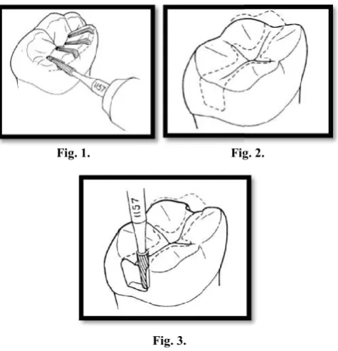
This technique was given by Dr. Jack G. Seymour in 1987. Most posterior teeth can be restored conservatively with a type II gold by using a minimal number of instruments in an organized manner. The instruments needed are No. 1157, No. 17OL, and No. 282-010 carbide burs and a ½ inch fine emery disk with No. 80-7-14 and No. 10-7-14 gingival marginal trimmers (Seymour, 1987).
Place depth penetration grooves on the occlusal surface, develop axial surfaces, and establish the traditional outline form with a No. 1157 carbide bur. Remove existing restorations and excavate caries. This may modify the conventional outline. (Fig. 1, Fig. 2, Fig. 3)
[image:2.595.309.553.158.303.2]Fig. 1. Fig. 2
Fig. 3.
[image:2.595.43.281.260.507.2] Place the retentive grooves in ¾ crowns and prepare proximal boxes and occlusal offset for onlays with the use of a No. 170 L carbide bur. (Fig.4)
Fig. 4.
Prepare the gingival bevel, buccal and lingual flares, and occlusal offset with the No. 282-010 bur. (Fig. 5)
Use a fine emery disk on the proximal aspect of the preparation to finish the flares and slight
develop during use of the No. 282-010 bur.
Define the proximal box and refine the reverse gingival bench with hand instruments No. 80-7
50647 Dr. Rubeena. A. Azeem and Dr. Nivedhitha Malli Sureshbabu,
Overview of various onlay preparation techniques
This technique was given by Dr. Jack G. Seymour in 1987. conservatively with a type II gold by using a minimal number of instruments in an organized manner. The instruments needed are No. 1157, No. 010 carbide burs and a ½ inch fine emery 14 gingival marginal
Place depth penetration grooves on the occlusal surface, develop axial surfaces, and establish the traditional outline form with a No. 1157 carbide bur. Remove existing restorations and excavate caries. This may ntional outline. (Fig. 1, Fig. 2, Fig. 3)
Fig. 2.
Place the retentive grooves in ¾ crowns and prepare proximal boxes and occlusal offset for onlays with the
No. 170 L carbide bur. (Fig.4)
Prepare the gingival bevel, buccal and lingual flares, 010 bur. (Fig. 5) Use a fine emery disk on the proximal aspect of the preparation to finish the flares and slight hollowing that
010 bur.
Define the proximal box and refine the reverse gingival 7-014 and 10-7-14.
Make impressions and pour casts of the prepared onlay. (Fig. 6)
Try the castings on the
contacts, evaluate occlusal relationship, and cement the casting. Use one or two 5/8 inch “metal center” fine garnets to finish the exposed margins. After the complete set of the cement, review the occlusal surface again (Seymour, 1987).
Fig. 5.
Tooth preparation for esthetic onlay
[image:2.595.323.543.463.652.2]The principles of cavity preparation for esthetic inlays or onlays differ from those for gold restorations. For esthetic inlay or onlay restorations, bevels and retention forms are not needed. Resistance form is generally not necessary but may be required in very large onlay restorations. Cavity walls are flared 5 degrees to 15 degrees in total (10 degrees to 12 degrees ideal), and the gingival floor can be prepared with a butt joint. The internal line angles are rounded, the minimum isthmus width is 2 mm, and the minimum depth thickness is 1.5 mm (Christensen, 2012 and
Fig. 7.
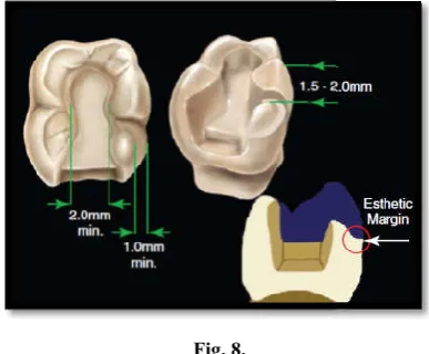
For onlay restorations, nonworking
covered with at least 1.5 mm and 2 mm of material, respectively. If the cusp to be onlayed shows in the patient’s smile, a more esthetic blended margin is achieved by a further to 2-mm reduction with a
1-and Jackson, 1994). When the occlusal aspect of the cavity is prepared, undercuts should not be eliminated by removing healthy tooth structure, which compromises the conservatism of this approach. The objective is to establish divergence in the enamel, then block out all undercuts. This is possible using bonded resin or a resin-modified glass ionomer. For cemented castings it is generally best to overlay a working cusp when the
Dr. Rubeena. A. Azeem and Dr. Nivedhitha Malli Sureshbabu, Onlay preparation techniques - Clinical practice guidelines
Make impressions and pour casts of the prepared onlay.
Try the castings on the tooth, adjust the proximal contacts, evaluate occlusal relationship, and cement the casting. Use one or two 5/8 inch “metal center” fine garnets to finish the exposed margins. After the complete set of the cement, review the occlusal surface
Fig. 6
preparation for esthetic onlay
The principles of cavity preparation for esthetic inlays or onlays differ from those for gold restorations. For esthetic onlay restorations, bevels and retention forms are not tance form is generally not necessary but may be required in very large onlay restorations. Cavity walls are flared 5 degrees to 15 degrees in total (10 degrees to 12 the gingival floor can be prepared with a butt joint. The internal line angles are rounded, the minimum isthmus width is 2 mm, and the minimum depth thickness is
, 2012 and Jackson, 1994).
Fig. 7.
For onlay restorations, nonworking and working cusps are covered with at least 1.5 mm and 2 mm of material, respectively. If the cusp to be onlayed shows in the patient’s smile, a more esthetic blended margin is achieved by a further -mm chamfer (Christensen, 2012 When the occlusal aspect of the cavity is prepared, undercuts should not be eliminated by removing healthy tooth structure, which compromises the conservatism of this approach. The objective is to establish divergence in the then block out all undercuts. This is possible using modified glass ionomer. For cemented castings it is generally best to overlay a working cusp when the
cavosurface margin is more than 50% up the incline of the cusp. The cavosurface margin can extend up to 75% up the cuspal incline of a nonworking cusp before overlaying of the cusp is considered. Studies have investigated the use of bonded inlay or onlay restorations for this area, but no clinical consensus on when to remove a cusp has been reached. Because these restorations reinforce the remaining tooth structure, the traditional guidelines for overlay
[image:3.595.63.257.170.330.2]cast gold onlays have been modified.
Fig. 8.
When there is no dentin support directly underneath the cusp tip, onlay restorations can be done. The palatal or working cusp is onlayed, even with dentin support if the margin is within 1 mm of the cusp tip. When the margin is beyond 1 mm from the cusp tip, the cusp gains dentin support and bond strength increases. The horizontal lines depict the direction of the enamel rods. At the cusp tip the enamel rods are almost vertical and etching would be on their sides. As the margin moves away from the cusp tip the ends become etched, which has been shown to increase bond strength (
and Jackson, 1994). The non-working or buccal cusp is not onlayed in this diagram even when the margin is at the cusp tip. If the posterior teeth are discluded in lateral jaw movements, there are no forces applied to this cusp.
uncommon to find cracks on the pulpal floor under cusps when removing amalgams that have been in place for some time, particularly moderate-sized ones. Whether the teeth exhibit pain on chewing (e.g., cracked tooth syndrome) or are asymptomatic, these cusps should be overlayed.
technique, called immediate dentin sealing
described by Paul and Scharer in 1997, this technique has been clinically popularized by Dr Pascal Magne. The technique is based on the logic that the strongest dentin bond is achieved when dentin is bonded immediately after being cut and before becoming contaminated, such as occurs during the provisional phase. Besides the pulpal protection afforded by this procedure, the patient has more comfort while th
is in place. Finally, early data show that the ultimate bond of the restoration and the marginal integrity over time are improved.
This technique requires placement of a self
followed immediately after curing by a very thin layer of low-viscosity flowable composite resin. Any under
blocked out simultaneously with the flowable resin. After curing, it is necessary to remove the air-inhibited layer. This can be done by wiping the surface with a cotton pledget soaked in alcohol. An alternative technique is to cover the surface with a glycerin product and light curing again. After washing and drying, the vertical enamel walls are prepared again with a cavosurface margin is more than 50% up the incline of the ace margin can extend up to 75% up the cusp before overlaying of the cusp is considered. Studies have investigated the use of bonded inlay or onlay restorations for this area, but no clinical p has been reached. Because these restorations reinforce the remaining tooth structure, the traditional guidelines for overlaying a cusp as in
When there is no dentin support directly underneath the cusp tip, onlay restorations can be done. The palatal or working cusp is onlayed, even with dentin support if the margin is within 1 mm of the cusp tip. When the margin is beyond 1 mm from the cusp tip, the cusp gains dentin support and bond The horizontal lines depict the direction of the enamel rods. At the cusp tip the enamel rods are almost vertical and etching would be on their sides. As the margin moves away from the cusp tip the ends become etched, which (Christensen, 2012 working or buccal cusp is not onlayed in this diagram even when the margin is at the cusp tip. If the posterior teeth are discluded in lateral jaw movements, there are no forces applied to this cusp. It is not uncommon to find cracks on the pulpal floor under cusps when removing amalgams that have been in place for some time, sized ones. Whether the teeth exhibit pain on chewing (e.g., cracked tooth syndrome) or are , these cusps should be overlayed. A popular
immediate dentin sealing (IDS), first
described by Paul and Scharer in 1997, this technique has been clinically popularized by Dr Pascal Magne. The technique is ngest dentin bond is achieved diately after being cut and before becoming contaminated, such as occurs during the provisional tion afforded by this procedure, the patient has more comfort while the provisional is in place. Finally, early data show that the ultimate bond of the restoration and the marginal integrity over time are
This technique requires placement of a self-etching adhesive followed immediately after curing by a very thin layer of
very-viscosity flowable composite resin. Any undercuts are blocked out simultaneously with the flowable resin. After inhibited layer. This can be done by wiping the surface with a cotton pledget soaked hol. An alternative technique is to cover the surface with a glycerin product and light curing again. After washing and drying, the vertical enamel walls are prepared again with a
finishing bur to remove any adhesive that may have flowed onto these surfaces. After preparation, an impression is obtained using an accurate re-pourable material. This is sent to the laboratory with any additional models, records, or information needed to fabricate the restoration. The level of esthetics achieved with this resto
to the level of communication between the clinician and laboratory technician. Consequently, the color prescription must contain the occlusal base shade of the restoration, the gradient of shade from central fossa to cavo
degree and color of the desired pit and fissure stains, and any maverick highlights present. The shade is taken before preparation to avoid the misleading effects produced in a desiccated tooth. Once this diagnostic information has been obtained, a direct provisional restoration is placed while the definitive restorations are fabricated in the laboratory.
A good deal of science is documented in studies over the past 20 years. Significant evidence details the effectiveness of the enamel bonds in terms of both bond strength and durability. For esthetic inlays or onlays, evidence supports the effectiveness of these enamel bonds with regard to tooth reinforcement. The literature lists tooth reinforcement numbers that indicate that when there
surfaces, tooth reinforcement is achieved, even up to 70% to 80% of the original strength of the tooth. Clinical evidence also supports the longevity of these restorations. A significant number of patients show longevity greate
(Christensen, 2012 and Jackson
intra- and extra-coronal restorations follows the similar concepts as used for indirect restorations. The preparation should avoid undercuts between opposing walls within the cavity. All-ceramic restorations depend upon the luting cement for most of the retention, therefore a slightly over
cavity is acceptable provided there are no undercuts.
gold restorations gain most of their retention from the cavity shape and are therefore more near parallel preparations can be done for these restorations. Inlay and onlay restorations preserves tooth tissue to retain the core. If existing cavities contain undercuts they can be blocked out with composite or glass ionomer cements to provide the necessary cavity shape.Tapered burs provide the most convenient shape to prepare onlays and reduce the chance of creating undercuts. If an onlay preparation is to be cut, occlusal clearance/ reduction will be required consistent with th
marginal configuration (shoulder, chamfer or deep chamfer) depends on the material planned
Jackson, 1994).
Technique for placing tooth-colored onlays
Anesthetize the tooth if necessary
Clean the tooth prepara water on a rubber cup
Selectively place phosphoric acid gel on just the enamel portions of the margins and perform a standard acid etch of the enamel; this is the so
technique
Seat the onlay with a sel
Minimally cure the resin cement residue around the margins and remove it before making the final light cure
Floss the contact areas before providing a full light cure of the restoration to avoid difficulty in clearing cement from the contact areas
finishing bur to remove any adhesive that may have flowed After preparation, an impression is pourable material. This is sent to the laboratory with any additional models, records, or information needed to fabricate the restoration. The level of esthetics achieved with this restoration is directly proportional to the level of communication between the clinician and laboratory technician. Consequently, the color prescription must contain the occlusal base shade of the restoration, the gradient of shade from central fossa to cavosurface margin, the degree and color of the desired pit and fissure stains, and any maverick highlights present. The shade is taken before preparation to avoid the misleading effects produced in a desiccated tooth. Once this diagnostic information has been obtained, a direct provisional restoration is placed while the definitive restorations are fabricated in the laboratory.
A good deal of science is documented in studies over the past 20 years. Significant evidence details the effectiveness of the bonds in terms of both bond strength and durability. For esthetic inlays or onlays, evidence supports the effectiveness of these enamel bonds with regard to tooth reinforcement. The literature lists tooth reinforcement numbers that indicate that when there are significant enamel bond surfaces, tooth reinforcement is achieved, even up to 70% to 80% of the original strength of the tooth. Clinical evidence also supports the longevity of these restorations. A significant number of patients show longevity greater than 10 years Jackson, 1994).Tooth preparation for coronal restorations follows the similar concepts as used for indirect restorations. The preparation should avoid undercuts between opposing walls within the ceramic restorations depend upon the luting cement for most of the retention, therefore a slightly over-tapered cavity is acceptable provided there are no undercuts.However, gold restorations gain most of their retention from the cavity and are therefore more near parallel preparations can be done for these restorations. Inlay and onlay restorations preserves tooth tissue to retain the core. If existing cavities contain undercuts they can be blocked out with composite or ents to provide the necessary cavity shape.Tapered burs provide the most convenient shape to prepare onlays and reduce the chance of creating undercuts. If an onlay preparation is to be cut, occlusal clearance/ reduction will be required consistent with the material chosen. The marginal configuration (shoulder, chamfer or deep chamfer) depends on the material planned (Christensen, 2012 and
colored onlays
Anesthetize the tooth if necessary
Clean the tooth preparation with flour of pumice and
Selectively place phosphoric acid gel on just the enamel portions of the margins and perform a standard acid etch of the enamel; this is the so-called selective etching
Seat the onlay with a self-etching resin cement
Minimally cure the resin cement residue around the margins and remove it before making the final light
Finish and polish the margins and correct any high occlusal areas
RECENT ADVANCES
Direct fiber-reinforced composite onlay
ecently, short fiber reinforced composite resin was introduced as a dental restorative composite resin. These direct composite restorations are now intended to be used in high stress bearing areas especially in molars. The results of the laboratory mechanical tests revealed substantial improvements in the load bearing capacity, the flexural strength and fracture toughness of dental composite resin reinforced with short E glass fiber fillers in comparison with conventional particulate filler composite resin. The short fiber composite resin has shown control of the polymerization shrinkage stress by fiber orientation and, thus, marginal micro leakage was reduced compared with conventional particulate filler composite resins (Garoushi, 2012; Garoushi,f, 2007 and Garoushi, 2008) For direct composite restorations, quadrant isolation is done using rubber dam sheet. The tooth to be restored is cleaned with pumice-water slurry in a rubber cup to remove salivary pellicle and any remaining dental plaque.
Onlay preparation is done using high-speed burs. Caries removed with low-speed burs and spoon excavator leaving discolored but hard dentin at the cavity floor. The preparations are done according to the principles of minimally invasive dentistry. In cases where the cavity is deep, MTA Plus can be given (PREVEST Denpro). The prepared tooth is restored using sectional matrix system (Triodent) that is stabilized using anatomical wedges. The bonding procedure begins with the application of self-etch adhesive (Gaenial Bond, GC) to the prepared walls. The application and placement of bonding agent is done according to manufacturer’s instructions. The fiber-reinforced composite (EverX Posterior, GC) is placed and light cured according to an incremental technique. A restorations are fully covered with a layer (1
composite resin (Gaenial Posterior, GC). Occlusion is carefully adjusted using articulating paper. Finishing and polishing procedures are carried at same visit after occlusal adjustment
50649 Dr. Rubeena. A. Azeem and Dr. Nivedhitha Malli Sureshbabu,
Finish and polish the margins and correct any high
reinforced composite onlay
ecently, short fiber reinforced composite resin was introduced as a dental restorative composite resin. These direct composite restorations are now intended to be used in high stress bearing areas especially in molars. The results of the laboratory cal tests revealed substantial improvements in the load bearing capacity, the flexural strength and fracture toughness of dental composite resin reinforced with short E glass fiber fillers in comparison with conventional particulate filler The short fiber composite resin has shown control of the polymerization shrinkage stress by fiber orientation and, thus, marginal micro leakage was reduced compared with conventional particulate filler composite resins
d Garoushi, 2008). For direct composite restorations, quadrant isolation is done using rubber dam sheet. The tooth to be restored is cleaned water slurry in a rubber cup to remove salivary
speed burs. Caries speed burs and spoon excavator leaving discolored but hard dentin at the cavity floor. The preparations are done according to the principles of minimally invasive where the cavity is deep, MTA Plus can be given (PREVEST Denpro). The prepared tooth is restored using sectional matrix system (Triodent) that is stabilized using anatomical wedges. The bonding procedure begins with the aenial Bond, GC) to the prepared walls. The application and placement of bonding agent is done according to manufacturer’s instructions. The reinforced composite (EverX Posterior, GC) is placed and light cured according to an incremental technique. All restorations are fully covered with a layer (1-2 mm) of hybrid composite resin (Gaenial Posterior, GC). Occlusion is carefully adjusted using articulating paper. Finishing and polishing procedures are carried at same visit after occlusal adjustment
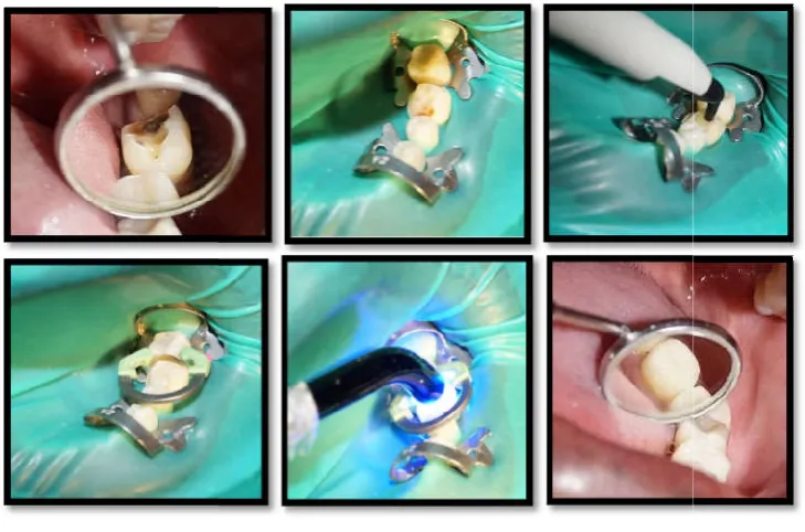
using composite finishing and polishing kit. Hence, these direct restorations can be placed in a single visit and allows for maximum preservation of tooth structure as well as strengthens remaining tooth structure. (Fig. 9
fiber-reinforced composite onlay restorations with 3 year follow-up – by Dr. Rubeena)
CAD-CAM approach
The computer-assisted design and computer
turing (CAD-CAM) approach is a valid procedure for fabricating esthetic inlays or onlays. Many of the ceramic inlays or onlays ordered by dentists today are fabricated in the laboratory using milling machines. The two machines available in the marketplace today are the CEREC (Sirona Dental Systems, Charlotte, North Carolina) and the
Technologies, Richardson, Texas). The quality of the restorations that can be fabricated with these milling machines in the dental office today is as good as that of laboratory fabricated indirect resin or ceramic restorations with respect to fit and function. Both approaches depend on the com
and skill of the operator. This can be the dentist or a dental auxiliary who actually does the design and operates the milling equipment.
Summary and conclusion
Advances in tooth-colored materials and adhesive technology have expanded the scope of restorative dentistry.
more conservative restorative option than are crowns. The results of research are positive regarding onlays’ service potential. Numerous well-proven, as well as some new, materials such as ceramics and fiber
make the use of tooth-colored onlays a viable procedure.
REFERENCES
Christensen, G.J. 2012. The case for onlays versus tooth colored crowns. The Journal of the American Dental
Association. 2012 Oct 1;143(10):1141
Frankenberger, R., Petschelt,
reinforced glass ceramic inlays and onlays after six years. Part I: Clinical behavior, Oper Dent
[image:4.595.116.481.342.583.2]Dr. Rubeena. A. Azeem and Dr. Nivedhitha Malli Sureshbabu, Onlay preparation techniques - Clinical practice guidelines
Fig. 9 – 14.
g composite finishing and polishing kit. Hence, these direct restorations can be placed in a single visit and allows for maximum preservation of tooth structure as well as strengthens remaining tooth structure. (Fig. 9-14: Clinical pictures of direct reinforced composite onlay restorations with 3 year
CAM approach
assisted design and computer-assisted manufac-CAM) approach is a valid procedure for
or onlays. Many of the ceramic inlays or onlays ordered by dentists today are fabricated in the laboratory using milling machines. The two machines available in the marketplace today are the CEREC (Sirona Dental Systems, Charlotte, North Carolina) and the E4D (D4D Technologies, Richardson, Texas). The quality of the restorations that can be fabricated with these milling machines in the dental office today is as good as that of laboratory-fabricated indirect resin or ceramic restorations with respect to
and function. Both approaches depend on the commitment and skill of the operator. This can be the dentist or a dental auxiliary who actually does the design and operates the milling
colored materials and adhesive technology have expanded the scope of restorative dentistry. Onlays are a more conservative restorative option than are crowns. The results of research are positive regarding onlays’ service proven, as well as some new, materials such as ceramics and fiber – reinforced composite
colored onlays a viable procedure.
The case for onlays versus tooth-The Journal of the American Dental 2012 Oct 1;143(10):1141–4.
A., Krämer, N. 2000. Leucite-reinforced glass ceramic inlays and onlays after six years.
Oper Dent 25:459-465.
Garoushi, S., Vallittu, P.K., Lassila, L.V.J. 2007. Short glass fiber reinforced restorative composite resin with semi-inter penetrating polymer network matrix. Dent Mater. 2007 Nov;23(11):1356–62.
Garoushi, S., Vallittu, P.K., Watts, D.C., Lassila, L.V.J. Polymerization shrinkage of experimental short glass fiber-reinforced composite with semi-inter penetrating polymer network matrix. Dent Mater. 2008 Feb;24(2):211–5. Garoushi, S., Mangoush, E., Vallittu, M., & Lassila, L. Short
Fiber Reinforced Composite: a New Alternative for Direct Onlay Restorations. The Open Dentistry Journal 2013; 7: 181–185.
Jackson, R. 1999. Indirect resin inlay and onlay restorations: a comprehensive clinical overview, Pract Periodontics Aesthet Dent 11(8):891-900.
Jackson, R.D. 1994. Esthetic inlays and onlays. Curr Opin Cosmet Dent. 30–9.
Kramer, N., Frankenberger, R. 2005. Clinical performance of bonded leucite-reinforced glass ceramic inlays and onlays after eight years, Dent Mater 21(3):262-271.
Kramer, N., Taschner, M., Lohbauer, U., et al. 2008. Totally bonded Ceramic inlays and onlays after eight years, J Adhes Dent. 10:307-314.
Lehner, C., Studer, S., Brodbeck, U., Scharer, P. 1998. Six-year clinical results of leucite-reinforced glass ceramic inlays and onlays, Acta Medica Dentistry Switzerland 3:137-146, 1998.
Otto, T., De Nisco, S. 2002. Computer-aided direct ceramic restorations: 10-year prospective clinical study of CEREC CAD/CAM inlays and onlays, Int J Prosthodont 15:122-128.
Paul, S.J., Schärer, P. 1997. The dual bonding technique: a modified method to improve adhesive luting procedures, Int J Periodontics Restorative Dent 17:536-545.
Schulte, A.G., Vocker, A., Reinhardt, R. 2005. Longevity of ceramic inlays and onlays luted with a solely light curing composite resin, J Dent 33:433-442.
Seymour, J.G. 1987. A simplified technique for the tooth preparation of onlay castings. J Prosthet Dent., Jan;57(1):9–11.
Swift, E.J. 2001. Processed Composites. Journal of Esthetic and Restorative Dentistry. Sep 1;13(5):284–95.
Van Dijken, J.W.V. 2000. Direct resin composite inlays or onlays: an 11 year follow-up, J Dent 28(5):299-306. Wassell, R.W., Walls, A.W., McCabe, J.F. 2000. Direct
composite inlays versus conventional composite restorations: 5-year follow-up. J Dent., 28:375-82.
Wendt, S.L., Leinfelder, K.F. 1992. Clinical evaluation of a heat-treated resin composite inlay: 3-year results. Am J Dent. Oct;5(5):258–62.