EFFECT OF DIAZEPAM ON THE TESTICULAR HISTOLOGY AND
GENE EXPRESSION IN RATS
*Dr. Mohammed A. Taher and Dr. Zainab N. H. Anber
Assistant Professor; College of Pharmacy, University of Baghdad.
ABSTRACT
Steroid biosynthesis is initiated with transportation of cholesterol along
the steroidogenic acute regulatory protein (StAR) into the
mitochondria. It has been reported that Ca2+ channel blockers suppress
the biosynthesis of androgens. Diazepam is one of the most common
benzodiazepines used for anxiety. Recent studies found that diazepam
acts as Ca channels antagonist as it can produce a complete inhibition
of voltage-dependent Ca uptake. The purpose of the present study was
to investigate whether treatment of male rats with diazepam interferes
with steroidogenesis. Fifty male rats were allocated into five groups.
Control (D.W.) (n=10) and test groups that received (2, 5 and 10
mg/kg/day) of diazepam by oral gavage, each group (n=10) and
sulfasalazine (500mg/kg) for 8 weeks. Animals were kept in standard conditions. At the end
of the treatment; animals were sacrificed and the epididymis was removed. Sperms were
collected and the sperm count, motility, viability and morphology were determined. The testis
were also removed and the effect of diazepam on the steroidogenic acute regulatory protein
(StAR) mRNA expression was assessed by using reverse transcription (Reverse-
transcriprase polymerase chain reaction) analysis. Testicular histological analysis was also
achieved. The results of the present study showed a significant decrease in sperm count,
motility and viability with an increase in sperms abnormalities. Also showed that diazepam
significantly inhibits the (StAR) mRNA expression. Histological analyses showed dose
dependent anomalies of the testis. From the data; it can be concluded that diazepam, in a dose
dependent pattern, was effective in attenuating steroidogenesis production through an
inhibitory effects on StAR protein gene expression in rats.
KEYWORDS: steroidogenic, Ca channels antagonist, Ca uptake and StAR.
7105 – 2277 ISSN Research Article . 266 -3 24 Volume 4, Issue 11,
Article Received on 09 Sep 2015,
Revised on 29 Sep 2015, Accepted on 19 Oct 2015
*Correspondence for Author
Dr. Mohammed A. Taher
INTRODUCTION
Calcium ion is implicated in diverse cellular functions in both germ cells and somatic cells in
the testis, particularly, mediating the responses to endocrine hormones and local regulators in
genital tracts.[1,2] A common belief is that the Ca2+ influx and efflux should be tightly
regulated to maintain the intracellular Ca2+ homeostasis, and an alteration in the Ca2+
transport across the cell membrane could result in a drastic impact on spermatogenesis and
steroidogenesis.[3, 4] Leydig cell production of testicular androgens is tightly controlled by
endocrine interactions among the pituitary gland and the testis, as well as through the
paracrine and autocrine regulation within the testis.[5, 6, 7] Leydig cells secrete testosterone
responsible for the onset of both spermatogenesis and male sexual development. Endocrine
control of Leydig cell steroidogenic activity by luteinizing hormone (LH), follicle-releasing
hormone (FSH) or human chorionic gonadotropin (hCG) has been exerted through their
Ca2+- mediated signaling pathway.[8, 9, 10]
Recent studies suggest that Ca2+ affects the transfer of the substrate cholesterol to the inner
mitochondrial membrane, the rate-limiting step in steroidogenesis.[11, 12] This was confirmed
by a study reporting Ca2+ induced increase in steroidogenic acute regulatory (StAR)
protein[13], which is critical for the cholesterol transfer to the inner mitochondrial membrane
to initiate steroidogenesis.[14,15,16] Diazepam is a benzodiazepine that is widely used as an
anxiolytic, anticonvulsant, hypnotic, sedative & skeletal muscle relaxant. Recent studies
found that benzodiazepines can produce a complete inhibition of voltage-dependent Ca2+
uptake. Also, they indicate that benzodiazepines are acting as Ca2+ channels antagonists.[17]
MATERIALS AND METHODS Animals
Sprague-Dawly male rats weighing 250-300 gm and 8 weeks old were obtained from the
animal house of the College of Pharmacy- University of Baghdad. The animals were
maintained on normal conditions of temperature, humidity and light/dark cycles. They were
fed standard rodent pellet and they have free access to water.
The study design
Fifty male rats were used in the present study, the study groups were divided into 5 groups:
First group (control): 10 rats were administered distilled water for 8 weeks by oral gavage.
Second group: 10 rats were used for the study of the infertility activity of diazepam in which
Third group: 10 rats were used for the study of the possible infertility activity of diazepam in
rat model. In this group (5mg/kg BW) dose of diazepam were used for 8 weeks by oral
gavage.
Fourth group: 10 rats were used for the study of the possible infertility activity of (10mg/kg
BW) diazepam was used for 8 weeks by oral gavage.
Fifth group: 10 rats were given a dose of (500mg/kg BW) of sulfasalazine for 8 weeks by oral
gavage as a positive control (in this group, sulfasalazine represents standard infertility agent.
Epididymal tail suspension preparation
At the end of treatment; the cauda epididymis was quickly removed into a petridish that
contains 10 ml of warm normal saline at 37°C and it was cut longitudinally with a pair of
fine pointed scissors and compressed with forceps. The sperms were released by mincing the
cauda epididymis into pieces (at least 200 cuts) to perform the following microscopical
examination on sperm characters.[18]
Determination of sperm concentration
Sperm count was determined using the haemocytometer under light microscope. A cover slip
was placed on the haemocytometer before a drop of the epididymal sperm solution was
loaded under the cover slip. Sperm count was done by counting 5 RBC small squares.
Sperm count was determined using the following formula:
Sperm count= total no. of sperms in 5 squares ×50,000×100 (cells/ml).[19]
Determination of sperm motility
Sperm motility was assessed by placing a drop of the sperm suspension over a clean dry slide
and covered with a cover slip and then the slide was placed under light microscope. The data
were tabulated in the form of percentage using the formula:
Percentage of motile sperms =no. of motile sperms ×100%/total no. of sperms (motile and
immotile).[20]
Determination of sperm viability
In this analysis; a drop from the sperm suspension used before was mixed with one drop of
eosin and then after 30 seconds, a drop of nigrosin was added and mixed. Then a smear was
sperms showed pink color of the head while the viable sperm showed colorless or whitish
head based on the degree of membrane permeability, then the data were tabulated in the form
of percentage using the following formula.
Percentage of viable sperms =no. of viable sperms×100%/ total no. of dead and viable
sperms.[18]
Determination of sperms abnormalities
In this analysis; the same sperm smears made for sperm viability were observed under light
microscope. The smears were examined for abnormal morphology of the head, neck and tail.
Then data were tabulated using the following formula.
Percentage of abnormal sperms =no. of abnormal sperms×100%/ total no. of normal and
abnormal sperms.[21]
Histological evaluation of tissues
After the end of treatment; the testes were excised and cleared off the attached fat and
connective tissues. Histological sections were prepared according to Luna (1968) methods for
histological evaluation.[22]
C DNA synthesis and purification of the StAR cDNA using reverse transcriptase – polymerase chain reaction (RT- qPCR).
Extraction of RNA from tissue
Total RNA was extracted from fresh tissue (testes) using the Geneaid total RNA mini kit
which was designed specifically for purifying total RNA from a variety of fresh and paraffin
– embedded tissues.[23]
Determination of RNA yield and purity
The most common and easiest technique to determine RNA yield and purity is absorbance
using nanodrop instrument which is highly sensitive and directly provides the concentration
of RNA in ng/ml. the measurement depended on the ratios of absorbance at 260 and 280 nm,
i.e A260/A 280 ratio.
Conversion of RNA into cDNA using RT-PCR
The RNA extracted previously from tissues is converted into cDNA using AccuPower®
ready to use lyophilized master mix containing all components for first strand cDNA
synthesis from a purified total RNA template.
The reaction is performed under the following conditions:
Table (1): Conditions for conversion of RNA into cDNA.
Step Temperature Time
cDNA synthesis 42-70°C 1 hr
Heat inactivation 95 °C 5 min
The cDNA formed previously was checked using agarose gel electrophoresis.
The agarose gel electrophoresis was done according to Harisha method.[24] Preparation of the
agarose gel was according to Lee method[25] and the ethidium bromide staining was done
according to Robinson and Lafleche method.[26]
Agarose gel was visualized in a UV transilluminator and photos were captured.
Real time PCR (qPCR)
The cDNA was amplified using Accupower®Green StarTM qPCR premix kit from Bioneer.
Accupower®Green StarTM qPCR premix is a ready to use reagent containing all components
for real- time PCR reaction just by the addition of a specific primer and target gene into tubes
provides results with high sensitivity and specificity. There are three major steps at different
temperatures in a PCR which are repeated for 30 or 45 cycles. Double stranded target DNA is
heat denaturated (denaturation step), the two primers complementary to the target segment
are annealed at low temperature (annealing step), and the annealed primers are then extended
at an intermediate temperature (extension step) with a DNA polymerase. As the target copy
number doubles each cycle, PCR can thereby amplify DNA fragments up to 10 8 –fold in a
short period.[27]
The PCR products are detected with SYBR Green dye. SYBR Green fluorescence is
enormously increased upon binding to double- stranded DNA. During the extension phase,
more and more SYBR Green will bind to the PCR product resulting in an increased
fluorescence. Consequently, during each subsequent PCR cycle, more fluorescence will be
detected.
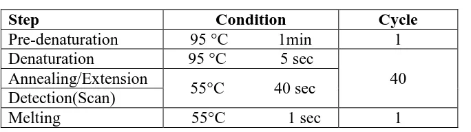
Table (2) Conditions for real-time PCR.
Step Condition Cycle
Pre-denaturation 95 °C 1min 1
Denaturation 95 °C 5 sec
40 Annealing/Extension
55°C 40 sec Detection(Scan)
Melting 55°C 1 sec 1
The oligonucleotide primer sequence used for PCR amplification of StAR gene
(Genebank:Access no.BC060970) is shown in table (3).
Table (3) primer sequence for the StAR gene.
Primer Sequences
Forward primer LP5´-GAC CTT GAA AGG CTC AGG AAG AAC-3´
Reverse primer RP5´-TAG CTG AAG ATG GAC AGA CTT GC-3´
The oligonucleotide primer sequence used for PCR amplification of ß-actin gene
(Genebank:Access no.NM007393) is shown in table (4).
Table (4) primer sequence for the ß-actin gene.
Primer Sequences
Forward primer LP5´- ATGCCCACTGCCGCATCCTCTTCC -3´
Reverse primer RP5´- CACGATGGAGGGGCCGGACTCATC -3´
The data results of qRT –PCR for the target (StAR) and housekeeping gene (ß-actin) were
analyzed by the relative quantification gene expression levels (fold change) Livak method
that described by.[28]
In which the following formula was applicated:
Relative expression ratio= 2 – Ct
Statistical analysis
Analysis of data was carried out using the available statistical package of SPSS-21 (Statistical
Packages for Social Sciences version -21). Student t-test was used for testing the significance
of difference between two groups and ANOVA between three. Statistical significance was
[image:6.595.131.473.86.182.2]RESULTS
Sperm concentration
The data referring to sperm concentration in epididymal suspension of control and treated
groups are shown in table (5).
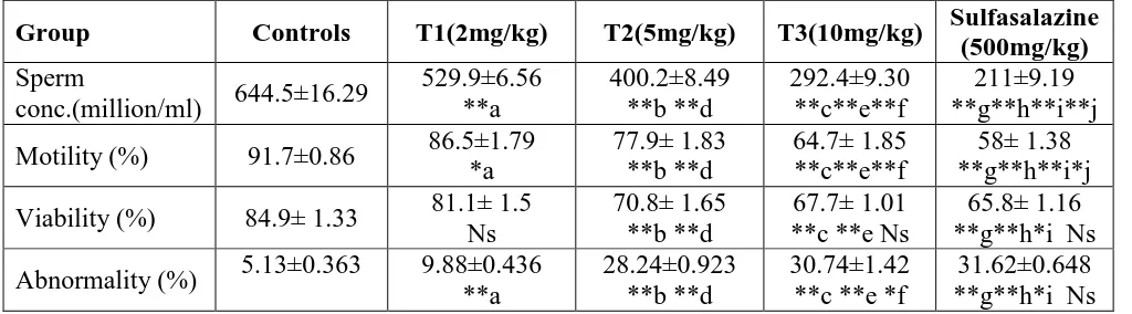
Table (5): Effect of different oral doses of diazepam suspension and sulfasalazine on the sperm characteristics of male rats.
Group Controls T1(2mg/kg) T2(5mg/kg) T3(10mg/kg) Sulfasalazine
(500mg/kg)
Sperm
conc.(million/ml) 644.5±16.29
529.9±6.56 **a 400.2±8.49 **b **d 292.4±9.30 **c**e**f 211±9.19 **g**h**i**j
Motility (%) 91.7±0.86 86.5±1.79
*a 77.9± 1.83 **b **d 64.7± 1.85 **c**e**f 58± 1.38 **g**h**i*j
Viability (%) 84.9± 1.33 81.1± 1.5 Ns
70.8± 1.65 **b **d
67.7± 1.01 **c **e Ns
65.8± 1.16 **g**h*i Ns
Abnormality (%) 5.13±0.363 9.88±0.436 **a
28.24±0.923 **b **d
30.74±1.42 **c **e *f
31.62±0.648 **g**h*i Ns
Data are expressed as mean (±SE); n=10 rats/group; a: t-test between control and T1, b: t-test
between control and T2, c: test between control and T3, d: test between T1 and T2, e:
t-test between T1 and T3, f: t-t-test between T2 and T3, g: t-t-test between control and
sulfasalazine, h: t-test between T1 and sulfasalazine, i: t-test between T2 and sulfasalazine,
j:t-test between T3 and sulfasalazine, *:significant (p<0.05) difference, **:highly significant
(p<0.001) difference, NS: no significant difference.
Table (5) showed that the sperm concentration, motility and viability decreased highly
significantly (p< 0.001) in the T1 (2mg/kg), T2 (5mg/kg), T3 (10mg/kg) groups and in the
sulfasalazine group compared to the control group. While the percentage of sperms
abnormalities increased also highly significantly in all treatment doses and the sulfasalazine
group than in the control group.
The histological changes in testicular tissues
The microscopic study of rat´s testes treated with diazepam suspension for 8 weeks revealed
variable degrees of alteration according to the dose of treatment compared to the control
group. Many histological changes in semniferous tubules, spermatogenesis and number of
[image:7.595.45.557.210.352.2]Figure (1) show sections of the control rat testis with normal structure appearance of
semniferous tubules and full maturation of spermatogonia cells and sperms present inside the
lumen (normal spermatogenesis).
Figure (1) Cross section of normal rat´s testes (control group). (H& E ×20).
Figure (2) show sections of rat´s testis treated with 2mg/kg of diazepam suspension in which
certain semniferous tubules are empty from the production of sperms.
Figurev (2) Cross section of rat´s testes treated with 2 mg/Kg of diazepam suspension. (H&E ×20), black arrows showing few sperms.
Figure (3) show sections of rat´s testis treated with 5mg/kg of diazepam suspension in which
certain tubules show damage of maturity of sperm production cells (primary spermatocytes,
[image:8.595.134.461.146.341.2]Figure (3) Cross section of rat´s testis treated with 5 mg/Kg of diazepam suspension. (H&E ×200), black arrows showing no sperms.
Figure (4) show sections of rat´s testis treated with 10mg/kg of diazepam suspension in
which there is some severe necrosis of products with certain necrosis of spermatogonic cells,
other show no products of sperm.
Figure (4) Cross section of rat´s testis treated with 10 mg/Kg of diazepam suspension. (H&E ×40), black arrows showing spermatogonic cell necrosis.
Figure (5) show sections of rat´s testis treated with sulfasalazine showing immaturation of
[image:9.595.137.456.70.266.2] [image:9.595.126.468.407.650.2]Figure (5): Cross section of rat´s testis treated with sulfasalazine. (H&E×20).
Quantitative Reverse transcriptase Real – time PCR
Quantitative Reverse transcriptase Real – time PCR (RT-q PCR) was performed for
measurement of relative quantification (gene expression analysis) for StAR gene expression
level normalized by housekeeping gene expression (ß-actin). RT-q PCR quantification
method in real-time PCR system was dependent on the values threshold cycle numbers (CT)
of amplification plot of target gene and housekeeping gene.
[image:10.595.134.458.73.273.2] [image:10.595.125.470.457.677.2]Figure (7) amplification plot for ß-actin gene in the control.
Figure (8) amplification plot for StAR gene in 2mg/kg dose of diazepam.
[image:11.595.127.473.72.266.2]Figure (10) amplification plot for StAR gene in 5mg/kg dose of diazepam.
Figure (11) amplification plot for ß-actin gene in 5mg/kg dose of diazepam.
Figure (13) amplification plot for ß-actin gene in 10mg/kg dose of diazepam.
[image:13.595.127.468.73.330.2] [image:13.595.129.464.376.630.2]Figure (15) amplification plot for ß-actin gene in sulfasalazine treatment.
Table (6) relative gene expression between groups using RT-qPCR.
Group Control T1(2mg/kg) T2(5mg/kg) T3(10mg/kg) sulfasalazine
Relative StAR gene expression
1 1.305±0.312 0.888±0.069
**a NS
0.66±0.038 **b*d Ns
0.45±0.086 **c*e*f NS
Data are expressed as mean (±SE); n=3 rats/group; a: t-test between T1 and T2, b: t-test
between T1 and T3, c: t-test between T1 and sulfasalazine, d: t-test between T2 and T3, e:
t-test between T2 and sulfasalazine, f: t-test between control and sulfasalazine, *:significant
(p<0.05) difference, **:highly significant (p<0.01) difference, NS: no significant difference.
Relative gene expression
The relative expression of the target gene (StAR) in male rats testes was calculated by using
Livak method (2 -CT) that is dependent on normalization of RT-qPCR(CT values)of the
target gene with housekeeping gene (ß-actin) as reference gene in control and treatment
groups. Our results of the relative gene expression in StAR gene showed highly significant
(p<0.001) difference in fold change of the gene expression levels between control and
treatment groups. It showed that the relative gene expression of the StAR gene of the T2
(5mg/kg), T3 (10 mg/kg) and the sulfasalazine group; the results were (0.888±0.069;
0.66±0.038 and 0.45±0.086) respectively; were highly significantly (p<0.001) decreased than
[image:14.595.129.465.71.344.2](10mg/kg); (0.66±0.038) and sulfasalazine (0.45±0.086) were significantly (p<0.05)
decreased than the T2 (5mg/kg); (0.888±0.069) group. And the relative gene expression of
the sulfasalazine (0.45±0.086) group was significantly (p<0.05) decreased than the control
group which is equal to 1 fold change of gene expression levels. while there was no
significant difference between both the T2 (5mg/kg); (0.888±0.069) and T3 (10mg/kg);
(0.66±0.038) groups compared to the control (1) group and no significant difference between
the T3(10mg/kg); ( 0.66±0.038) and the sulfasalazine (0.45±0.086)groups.
1.305
0.888
0.66
0.45
1 0
0 0 0 0.2 0.4 0.6 0.8 1 1.2 1.4 f o ld c h an ge m R N A t ra n sc ri p t le ve
l
Relative StAR gene expression
Figure (16) gene expression of StAR by 2 -CT Livak method.
DISCUSSION
In table (5); the sperm count was observed to have reduced significantly, which is an
indication that diazepam suspension had inhibited the spermatogenesis process.[29]
Spermatogenesis is influenced by the hypothalamic- pituitary- testicular axis relating
gonadotropin releasing hormone, LH, FSH and androgens. Thus, the effects evoked by
diazepam on sperm concentration might be strongly linked with status of LH and FSH
hormones which are also reduced and greatly affect sertoli cells functions in the testes
specially sperm production.[30] This implies that the decrease in sperm count caused by the
drug in the treated rats was as a result of a decrease in plasma level of testosterone, because
this hormone has been reported to be important in initiation and maintenance of
[image:15.595.101.494.233.462.2]Table (5) showed also a significant decrease in the sperm motility. This suggests that the drug
was able to permeate the blood- testis barrier with a resultant alteration in the
microenvironment of the semniferous tubules[34] and thus, creating a different
microenvironment in the inner wall of the semniferous tubules from that in the outer parts.[35]
It is known that the structure and function of the epididymis are dependent on androgens.[36]
In this study, a dose related suppression of the epididymis sperm motility in treated rats
suggests an under supply of testosterone to epididymis and therefore an impaired epididymal
function. The impaired epididymal function may also be due to reduced activity of the testes
which affects the normal passage of the testicular fluid into the epididymis.[37, 38, 39] This is
also confirmed by the reduced epididymal weight. Furthermore, It has been reported that
androgen binding protein and testosterone produced by the Leydig cells must reach the
epididymis in a sufficient amount through the testicular fluid and maintains the epididymis
testosterone level, since testosterone level affects the functional integrity of testes and
epididymis, sperm concentration, motility and viability of spermatozoa.[40, 41, 42]
Sulfasalazine induced a reduction in the percentage of progressively motile sperms possibly
by suppressed synthesis of sperm membrane proteins located in the acrosomal region of the
sperms head which could lead later to changes in cytoplasmic calcium levels and to a
decrease in sperms motility.[43]
Kato and coworkers (2002) had observed also that treatment with sulfasalazine caused a
remarkable decrease in sperms velocity and an increase in the beat frequency of the sperm
head which were considered as main causes of infertility.[44] This is in consistent with that
observed by Ohashi et al (1995) who indicated that a reduction in acrosome reaction ratio is
one of the mechanisms of induction of decreased fertility by sulfasalazine as well as the
depressed percentage of progressively motile sperms.[45] This study referred to highly
significant decrease in sperm viability with increasing the dose of diazepam (table 5), this is
in consistent with that indicated by Mohana et al. (2013) who found that diazepam decreased
significantly the motility and viability of goat epididymal sperms.[46] They suggested that
diazepam induced oxidative stress in goat epididymal sperms and that it had a significant role
in disturbing the balance between oxidative stress and antioxidant system. They concluded
that the use of diazepam could be a considerable factor in causing infertility in human males.
Table (5) showed also a significant increase in the percentage of morphologically abnormal
semniferous tubules, epididymal functions and testosterone activity on the hypothalamic
release factor and anterior pituitary secretion of gonadotropins which may result in alteration
of spermatogenesis.[47,48] Kar and Das (1983) demonstrated a significant incidence of
abnormalities induced by diazepam on sperms involving both shape and size of the sperm
head and tail.[49] This is in consistent with our study which had shown a significant increase
in sperm abnormalities.
Figure (1) showed normal morphologically and structurally tissue with normal components
of germinal epithelium that lining the semniferous tubules which include normal
spermatogenic lineage cells (spermatogonia, spermatocyte and spermatid); the spermatogonia
are attached to the basal lamina, while the primary spermatocytes are larger than the
spermatogonia and occupies the middle zone of the germinal epithelium, but the secondary
spermatocytes are about half size of the primary spermatocytes and lie nearer the lumen. The
spermatids lies close to the lumen and are spherical or polyglonal cells. And the supporting
cells (Sertoli cells) are surrounded by thin connective tissues.[50]
Figure (2) showed some of the semniferous tubules clear and empty in the 2mg/kg of
diazepam treated rats, compared to the crowded semniferous tubules filled by spermatozoa in
the control group. The above findings are more pronounced with increasing the dose of
diazepam (5 mg/kg); figure (3) in which the spermatogonia in the semniferous epithelium of
the diazepam treated groups were over-populated most of the other spermatogenic cells and
thus, there is a relative preponderance of primitive germ cells on the expense of more mature
ones. The reduction or loss in number of primary spermatocytes and secondary spermatocytes
and spermatids in treated rats with diazepam reflected the non-avaibility of androgen binding
protein (ABP) from sertoli cells.[51] ABP is required to maintain intra- testicular androgen
concentration and transformation of advance stages of germ cells. Meiotic and post- meiotic
germ cells were highly sensitive to androgen concentration.[52, 53]
Figure (4) showed a cross section of rat´s testis treated with 10mg/kg of diazepam, it showed
degeneration and necrosis among spermatogenic cells in the semniferous tubules and
decreasing in number of sertoli cells also the semniferous tubules were shrink distorted
containing detached spermatogenic cells and few numbers of sperms and leydig cells. The
atrophic state of Leydig cells in the testes of treated animals may be due to declined LH
spermatogenesis as sertoli cells provide all or most nutritional and physical support for the
developing germ cells.[56]
Figure (5) showed cross sections of rat´s testis treated with sulfasalazine; showed different
damage of the semniferous tubules such as disorders of the systemic arrangement of the
stages of spermatogenesis, loss of one or more stages of spermatogenesis and few numbers of
leydig cells with few numbers of sperms. The RT-qPCR analysis showed by this study
indicated that the StAR mRNA expression was decreased with a corresponding increase in
the dose of diazepam, (table 5).
StAR is necessary for the transfer of cholesterol from the outer mitochondrial membrane to
the inner mitochondrial membrane in Leydig cells and this step is the rate-limiting regulated
step in steroidogenesis.[57, 58] The inhibition of StAR expression results in a dramatic decrease
in steroid biosynthesis.[59] In this study, expression levels of StAR were significantly
decreased following exposure to 5 or 10 mg diazepam/kg. This reduction may affect
cholesterol delivery across the mitochondrial membrane and thus contribute to the decrease in
testosterone synthesis.[60]
Several mechanisms underlying the reduction in mRNA expression of StAR gene caused by
the drug closely correlated with the fact that Ca affects the transfer of cholesterol to the inner
mitochondrial membrane, the rate – limiting step in steroidogenesis.[61,62] This was confirmed
by a study reporting a Ca2+ induced increase in StAR protein[13], which is critical for the
cholesterol transfer to the inner mitochondrial membrane to initiate steroidogenesis.[63,64,65]
As Ca2+ ions are required for several steps of steroidgenesis, diazepam as Ca2+ channel
antagonist would be expected to have an effect on StAR protein expression.
StAR expression is stimulated via LH binding to its receptor (LHR). Therefore, LH also plays
an indirect role in the delivery of cholesterol to the inner mitochondrial membrane.[66] This
study revealed a significant reduction in serum LH levels in rats exposed to 5mg and 10
mg/kg. These results suggest that a reduction in LH results in decreased StAR expression at
levels of 5mg and 10 mg of diazepam/kg and therefore, diazepam affects the transport of
CONCLUSION
Diazepam in a dose dependent pattern was effective in attenuating steroidogenesis and
testosterone production through its inhibitory effects on StAR protein gene expression in rats.
REFERENCES
1. Steele, G. L., Leung, P. C. Intragonadal signalling mechanisms in the control of steroid
hormone production. J Steroid Biochem Mol Biol., 1992; 41: 515–22.
2. Berridge, M.J., Bootman, M.D., Roderick, H.L. Calcium signalling: dynamics,
homeostasis and remodeling. Nat Rev Mol Cell Biol., 2003; 4: 517–29.
3. Li, L.H., Wine, R.N., Miller, D.S., Reece, J.M., Smith, M., Chapin, R.E. (1947)
Protection against methoxyacetic-acid-induced spermatocyte apoptosis with calcium
channel blockers in cultured rat seminiferous tubules: possible mechanisms. Toxicol Appl
Pharmacol, 1997; 144: 105–19.
4. Yamaguchi, M. Role of regucalcin in maintaining cell homeostasis and function. Int J
Mol Med., 2005; 15: 371–89.
5. Saez JM. Leydig cells: endocrine, paracrine and autocrine regulation. Endocr Rev, 1994;
15: 574–626.
6. Chen YC, Nagpal ML, Stocco DM, Lin T. Effects of genistein, resveratrol and quercetin
on steroidogenesis and proliferation of MA-10 mouse Leydig tumor cells. JEndocrinol,
2007; 192: 527–37.
7. Medelson C, Dufau ML, Catt KJ. Gonadotropin binding and stimulation of cAMP and
testosterone production inm isolated Leydig cells. J Biol Chem., 1975; 250: 8818–23.
8. 8-Sullivan MH, Cooke BA, The role of Ca 2+ in steroidogenesis in Leydig cells:
stimulation of intracellular free Ca2+ by lutropin (LH), luliberin (LHRH) agonist and
cyclic AMP. Biochem J. 1986; 236: 45–51.
9. Tomić M, Dufau ML, Catt KJ, Stojilkovic SS. Calcium signalling in single rat Leydig
cells. Endocrinology, 1995; 136: 3422–29.
10.Taranta A, Morena AR, Barbacci E, D’Agostino A. Omega- Conotoxin-sensitive Ca2+
voltage-gated channels modulate protein secretion in cultured rat Sertoli cells. Mol Cell
Endocrinol. 1997.
11.Hall PF, Osawa S & Mrotek J The influence of calmodulin on steroid synthesis in leydig
12.Meikle AW, Liu XH & Stringham JD Extracellular calcium and luteinizing hormone
effects on 22-hydroxycholesterol used for testosterone production in mouse Leydig cells.
Journal of Andrology, 1991; 12: 148–151.
13.Manna PR, Pakarinen P, El-Hefnawy T & Huhtaniemi IT Functional assessment of the
calcium messenger system in cultured mouse Leydig tumor cells: regulation of human
chorionic gonadotropin-induced expression of the steroidogenic acute regulatory protein.
Endocrinology, 1999; 140: 1739–1751.
14.Clark BJ, Wells J, King SR & Stocco DM The purification, cloning, and expression of a
novel luteinizing hormone-induced mitochondrial protein in MA-10 mouse Leydig tumor
cells. Characterization of the steroidogenic acute regulatory protein (STAR). Journal of
Biological Chemistry, 1994; 269: 28314–28322.
15.Lin D, Sugawara T, Strauss JF III, Clark BJ, Stocco DM, Saenger P, Rogol A & Miller
WL Role of steroidogenic acute regulatory protein in adrenal and gonadal
steroidogenesis. Science, 1995; 267: 1828–1831.
16.Wang XJ, Liu Z, Eimerl S, Timberg R, Weiss AM, Orly J & Stocco DM Effect of
truncated forms of the steroidogenic acute regulatory protein on intramitochondrial
cholesterol transfer. Endocrinology, 1998; 139: 3903–3912.
17.William C.T. and De Lorenzo R.J. Micromolar-affinity benzodiazepine receptors regulate
voltage-sensitive calcium channels in nerve terminal preparations. Proceedings of the
National Academy of Sciences of the United States of America, 1984; 81(10): 3118–22.
18.Saadat P., Maryam J.Y., Mohammad A.D., Effect of Phaleria macrocarpa on sperm
characteristics in adult rats, 2013; 3(2): 345-52.
19.Rathje TA, Johnson RK, Lunstra DD. Sperm production in boars after nine generations of
selection for increased weight of testis. J Anim Sci., 1995; 73(8): 2177–85.
20.20-Chemineau P., Cogine Y., Guerin Y., Orgeure P. and Valtet J.C. Training Manual
on Artificial Insemination in sheep and goats. FAO, Animal Production and Health, 1991;
83.
21.Siegmund O.H. Reproductive and urinary system. In: Merk Veterinary manual. Siegmund
O.H. and Fraser C.M. USA, Merck and co. Inc. Rahway N.J., 1979; 794-890.
22.Luna L.G. Manual of histology staining. Methods of armed forces Institute of pathology.
3rd edition, New York and London, Mc Graw-Hill Book Company, 1968.
23.Vogelstein B. and Gillespie D. Proc. Natl. Acad. Sci. USA, 1979; 76: 615.
24.Harisha S: Biotechnology procedures and experiments handbook. Infinity science press
25.Lee P.Y., Costumbrado J. and Hsu C.Y. Agarose gel electrophoresis for the separation of
RNA fragements. J. Vis. Exp, 2012; 3923.
26.Robinson D.H. and Lafleche G.L.: nucleic acid electrophoresis in agarose gel. Essential
Molecular Biology, 2nd edition, TA Brawn, 2000; 1: 5.
27.Yoon D.S., Kim Y.H., Jung H.S., Paik S. and Lee J.W., Brain Korea 21 Project for
Medical Science, Yonsei University, Seoul, South Korea and Department of Orthopaedic
Surgery, Yonsei University College of Medicine, Seoul, South Korea Cell Prolif, 2011;
44: 428–40.
28.Livak, K.J. and Schmittgen, T.D. Analysis of relative gene expression data using real-
time quantitative PCR and the 2 (-Delta Delta CT) method. Methods, 2001; 25: 402-10.
29.Reddy C.M., Murthy D.R. and Patil S.B. Antispermatogenic and androgenic activities of
various extracts of Hibiscus rosa sinensis in albino mice, Ind. J. Exp. Biol, 1997; 35:
1170-74.
30.Steinberger E. Hormonal control of mammalian spermatogenesis. Physiological Reviews,
1971; 51: 1-22.
31.Swerdloff R. and Bhasin S. Male Reproductive abnormalities Chapter 7. In: Endocrine
pathophysiology. Hershman J.M. (ed), Philadeliphia, Lea and Febiger, 1988.
32.Behre H.M., Nashan D., Hubert W. and Nieschlag E. Gonadotropin releasing hormone
agonist blunts the androgen induced suppression of spermatogenesis in a clinical trial of
male contraception. Journal of Clinical Endocrinology and metabolism, 1992; 74(1):
84-90.
33.Christensen A.C. Leydig cell: In: Handbook of physiology, P.O. Greep and E.B.
Astwood. Washington DC American physiological Society, 1975.
34.Baldessarini R.J. Indrugs and the treatment of psychiatric disorders. The pharmacological
basis of therapeutics Ed. By Goodman and Gilman. Macmillan Pub. Co. Inc., 1980;
301-417.
35.Bloom W. and Fawcett D.W. Male reproductive system. In the textbook of Histology,
Saunders company, Philadelphia, 1975.
36.Cooper T.G. The epididymis as a site of contraceptive attack. In: spermatogenesis
fertilization, contraception, Nieschlag E. and Habenicbt U.F. (Eds.), Berlin, Springer,
1992; 419-60.
37.Lohiya N.K. Goyal R.B, Jayaprakash, D.; Sharma, S.; Kumer M. and Ansari, A.S.
38.Gupat G., Srivastava A.K. and Setty B.S. Androgenic regulation of glycoltic and HMP
pathway in epididymis and vas deferens of rhesus monkey. Ind. J. Exp. Biol, 1993; 31:
305-11.
39.Ansari A.S., Kumar S., Srivastava S. and Lohiya N.K. Longterm sequelae of tolnidamine
on male reproduction and general body metabolism in rabbits. Contraception, 1998; 57:
271-79.
40.Hou J.W., Collins D.C, and Schliecher R.L Source of cholesterol for testosterone
biosynthesis in murine Leydig cells. Endocrinology, 1990; 127(5): 2047-55.
41.Sandeep G., Dheeraj A., Sharma N.K., Jhade D. and Bharti A. Effect of plumbagin free
alcohol extract of Plumbago zeylanica Linn. root on reproductive system of female
Wistar rats. Asian pacific Journal of tropical medicine, 2011; 4(12): 978-84.
42.Meena R., Misro M.M., Ghosh D. and Nandan D. Extended intervention time and
evaluation of sperm suppression by dienogest plus testosterone undecanoate in male rat.
Contraception, 2012; 85(1): 113-21.
43.Tamio F., Masashi K., Tetsuya A., Yoshimasa H. and Masao H. Effects of sulfasalazine
on sperms acrosome reaction and gene expression in the male reproductive organs of rats,
toxicological sciences, 2005; 85: 675-682.
44.Kato M., Makino S., Kimura H. and Hirano K. In vitro evaluation of acrosomal status and
motility in rat epididymal spermatozoa treated with a-chlorohydrin for predicting their
fertilizing capacity. J. Reprod. Develop, 2002; 48: 461-68.
45.Ohashi K., Saji F., Kato M. and Tomiyama T. Acrobeads test: A new diagnostic test for
assessment of the fertilizing capacity of human spermatozoa. Fertil. Steril, 1995; 63:
625-30.
46.Mohana K.M., Asha K. and Mathura P.P. Diazepam: a peripheral benzodiazepines
receptor ligand, inhibits mitochondrial F1-F0 ATPase and induces oxidative stress in goat
epididymal sperm in vitro. International journal of Scientific & engineering research,
2013; 4(8): 1963-69.
47.Bowman W.C. and Rand M.J. The reproductive system and drugs affecting the
reproductive systems. Textbook of pharmacology, 2nd edition, 1985; 20: 1-8.
48.William K.W. Hormones and hormone antagonists. In: Remington, The Science and
Practice of pharmacy, 20th edition, vol 11, 2000; 77: 1390-91.
49.Kar R.N. and Das R.K. Induction of sperm head abnormalities in mice by three
50.Eroschenko, VictorP. Difiore´s atlas of histology with functional correlations. Twelfth
edition, Philadelphia, Lippincott William and Wilkins, 2013.
51.Hess R.A., Linder R.E., Strader L.F. and Perreault S.D. Acute effects and long-term
sequelae of 1,3 dinitrobenzene on male reproduction in the rat. II Quantitative and
qualitative histopathology of the testes. J Androl, 1988; 5: 327-42.
52.Hall P.F. Testicular steroid synthesis: organization and regulation. In: physiology of
reproduction. Eds: Knobil E., Neill JD., New York, Raven Press, 1994; 1: 1335-62.
53.Jeyakumar M., Suresh R., Krishnamurthy H.N. and Moudgal N.R. Changes in testicular
function following specific deprivation of LH in the adult male rabbit. J Endocrinol,
1995; 147: 111-20.
54.Nair N.; Bedwal R.S. and Mathur R.S. Effect of adrenalactomy and hydrocortisone
treatment on histopathological, biochemical and zinc and copper profiles in rat testes.
Indian J Expt Biol, 1995; 33: 655-63.
55.Sarker S.N., Majumdar A.C. and Chattopadhyay S.K. Effect of isoproturon on male
reproductive system: clinical, histological and histoenzymological studies in rats. Indian J
Expt Biol, 1997; 35: 133-38.
56.Ritzen E.M., Boitani C. and Parvinen M. Cyclic secretion of protein by the rat
seminiferous tubules depending on the stege of spermatogenesis. Int J Androl (Suppl.),
1981; 3: 57-8.
57.Hasegawa, T., Zhao, L., Caron, K. M., Majdic, G., Suzuki, T., Shizawa, S., Sasano, H.,
and Parker, K. L. Developmental roles of the steroidogenic acute regulatory protein
(StAR) as revealed by StAR knockout mice. Mol. Endocrinol, 2000; 14: 1462–71.
58.Manna P. R., Roy P., Clark B. J., Stocco D. M., and Huhtaniemi I. T. Interaction of
thyroid hormone and steroidogenic acute regulatory (StAR) protein in the regulation of
murine Leydig cell steroidogenesis. J. Steroid Biochem. Mol. Biol, 2001; 76: 167–77.
59.Clark B. J., Soo S. C., Caron K. M., Ikeda Y., Parker K. L., and Stocco D. M.
Hormonal and developmental regulation of the steroidogenic acute regulatory protein.
Mol. Endocrinol, 1995; 9: 1346–55.
60.Stocco D. M. and Clark B.J. Regulation of the acute production of steroids in
steroidogenic cells. Endocr. Rev, 1996; 17: 221–44.
61.Hall P.F., Osawa S. and Mrotek J. The influence of calmodulin on steroid synthesis in
62.Meikle A.W., Liu X.H. and Stringham J.D. Extracellular calcium and Luteinizing
hormone effects on 22-hydroxycholestrol used for testosterone production in mouse
Leydig cells. Journal of Andrology, 1991; 12: 148-51.
63.Clark B.J., Wells J., King S.R and Stocco D.M. The purification Cloning and expression
of a novel Luteinizing hormone induced mitochondrial protein in MA-10 mouse Leydig
tumor cells. Characterization of the steriodogenic acute regulatory protein (StAR).
Journal of biological chemistry, 1994; 269: 28314-22.
64.Lin D., Sugawara T., Strauss J.F III, Clark B.J. and Stocco D.M. Role of steroidogenic
acute regulatory protein in adrenal and gonadal steroidogenesis. Science, 1995; 267:
1828-31.
65.Wang X.J., Liu Z., Eimerl S., Weiss A.M. and Stocco D.M. Effect of truncated forms of
the steroidogenic acute regulatory protein on intramitochondrial cholesterol transfer.
Endocrinology, 1998; 139: 39033912.
66.Omura T. and Morohashi K. Gene regulation of steroidogenesis. J. Steroid Biochem. Mol.
Biol, 1995; 53: 19–25.
67.Wang X. and Stocco D.M. The decline in testosterone biosynthesis during male aging. A