ISSN Online: 2161-4512 ISSN Print: 2161-4105
DOI: 10.4236/jcdsa.2019.92016 Jun. 12, 2019 188 J. Cosmetics, Dermatological Sciences and Applications
Birch Sap (Betula alba) and Chaga Mushroom
(Inonotus obliquus) Extracts Show Anti-Oxidant,
Anti-Inflammatory and DNA Protection/Repair
Activity In Vitro
Mohamed Softa
1*, Giuseppe Percoco
2, Elian Lati
2, Pauline Bony
11INDERMA Dermatological Laboratory, Ivry sur Seine, France 2BIO-EC Laboratory, Longjumeau, France
Abstract
OBJECTIVE: The skin interacts strictly with the surrounding environment. Despite an efficient system of protection, its integrity is continuously as-saulted by a massive group of external stresses. UV irradiations represent one of the most harmful factors for the cutaneous tissue. Both UV-A and UV-B can induce deep modifications of the different layers of the skin, including a weakening of its barrier function properties, DNA damages and degradation of the extracellular matrix. The aim of this project was to assess the UV pro-tection activity of two natural compounds, the birch sap from Betulaalba and organic extract from Inonotusobliquus (chaga mushroom) used separately or in a complex. METHODS: The anti-oxidant (ROS and MDA quantification, catalase and SOD activity measurement), anti-inflammatory (IL-1β, IL-6, IL-8, IL-10, TNF-α and INF-γ dosages) and the DNA protection/repair activ-ities (DNA lesion site analysis) of birch sap and chaga mushroom extracts tested separately or in a complex containing organic birch sap 5% and In-onotusobliquus extracts 2% were evaluated invitro after exposure of cultured keratinocytes and fibroblasts or reconstructed epidermis to UV-A/UV-B ir-radiations. RESULTS: We observed that birch sap from Betulaalba and ex-tracts from Inonotusobliquus prevent the formation of ROS and decrease the oxidative stress induced under UV irradiations, suggesting a strong an-ti-oxidant activity. In addition, the tested products showed an immunomo-dulatory effect by reducing the quantity of pro-inflammatory cytokines upon UV irradiations. UV-induced DNA damages of keratinocytes were also re-duced by birch sap and chaga mushroom extracts. CONCLUSION: Here, for the first time, we have shown the photo-protection activity of extracts ob-How to cite this paper: Softa, M., Percoco,
G., Lati, E. and Bony, P. (2019) Birch Sap (Betula alba) and Chaga Mushroom (Inono-tus obliquus) Extracts Show Anti-Oxidant, Anti-Inflammatory and DNA Protec-tion/Repair Activity In Vitro. Journal of Cosmetics, Dermatological Sciences and Applications, 9, 188-205.
https://doi.org/10.4236/jcdsa.2019.92016
Received: April 29, 2019 Accepted: June 9, 2019 Published: June 12, 2019
Copyright © 2019 by author(s) and Scientific Research Publishing Inc. This work is licensed under the Creative Commons Attribution International License (CC BY 4.0).
DOI: 10.4236/jcdsa.2019.92016 189 J. Cosmetics, Dermatological Sciences and Applications tained from Betulaalba and Inonotus obliquus mushroom on skin cells ex-posed to UV-A and UV-B irradiations. Due to their anti-oxidant, an-ti-inflammatory and DNA protection/repair activities, the tested products represent promising candidates in the development of cosmetic products with anti-photo-aging activity.
Keywords
Birch Sap, Chaga Mushroom, UV Irradiations, Oxidative Stress, Photo-Aging, Natural Compounds
1. Introduction
The skin is an indispensable biological barrier, providing an efficient line of de-fence against the continuous assaults of the external environment such as air pollutions, pathogenic microorganisms and ultraviolet (UV) irradiations.
UV irradiations represent one of the most hazardous environmental factors for human skin. Based on their wavelength, they can be classified as UV-A (315 - 400 nm), UV-B (280 - 315 nm) and UV-C (100 - 280 nm).
The ambient sunlight is composed mainly by UV-A (90% - 95%) and UV-B (5% - 10%), while UV-C cannot reach the Earth’s surface due to the absorbing properties of ozone layer.
Since UV penetration in the skin depends strictly on their wavelength [1], UV-A penetrate profoundly into the dermis, reaching the upper reticular der-mis. Nevertheless, they can react also with both stratumcorneum and epidermis. Inversely UV-B are absorbed mainly by the different layers of the epidermis, where they trigger a cell damage response.
UV-A are commonly associated with the induction of oxidative stress in the different cutaneous compartments through the induction of reactive oxygen species (ROS) such as superoxide anion, hydrogen peroxide and singlet oxygen.
ROS can react easily with nucleotides, causing DNA mutations. One of the most common ROS-induced DNA damages is the formation of 8-hydroxy-2'-deoxyguanine (8-OHdG), which consists in the oxidation of gua-nine at the 8th position [2] leading to the guanine to thymidine transversion.
On the contrary, UV-B show a direct genotoxic effect since they are strongly absorbed by the DNA. Consequently, several photo-lesions can be induced upon UV-B exposure including cyclobutane pyrimidine dimers or pyrimidone photo-products [3].
The deleterious effects of both UV-A and UV-B on skin barrier properties have also been demonstrated.
DOI: 10.4236/jcdsa.2019.92016 190 J. Cosmetics, Dermatological Sciences and Applications anti-oxidant properties of the whole skin tissue [6].
In parallel UV-B cause abnormalities in lamellar body secretion [7] and in li-pid cohesion [8] resulting in altered skin barrier functions. In addition, like UV-A irradiation, UV-B are also able to decrease the activity of enzymes with anti-oxidant roles [9].
More recently, thanks to the development of methods allowing the simulta-neous monitoring of a wide number of genes, the impact of UV-A and UV-B on the skin transcriptome has also been characterized.
As well described by Zheng and collaborators [10], repetitive exposure of dermal fibroblasts to UV-A induces the modulation of 607 genes, with 238 up-regulated and 369 down-regulated genes. In particular, the expression of genes encoding for structural proteins of the extracellular matrix, including elas-tin, was decreased. Interestingly, the expression of SPRY1 was significantly in-duced upon UV-A irradiation. SPRY1 encodes for a protein, named sprouty, involved in cell signalling and able to induce the expression of proteins playing a key role in degradation of extracellular matrix, including matrix metalloproteins (MMPs) [11].
As expected, these data converge with the largely described mechanism of skin photo-aging [12], giving new insights in the cellular process leading to structural and physiological changes of the whole cutaneous tissue.
In a previous work, the team of Li and collaborators [13] have analysed the impact of acute UV-B irradiations on the transcriptome of epidermal keratino-cytes invitro. Following UV-B exposure, 198 genes were differentially regulated in a time-dependent manner. More specifically, three waves of time points were observed: an early (0.5 to 2 hours), an intermediate (4 to 8 hours) and a late (16 to 24 hours) point of regulation. In addition, seven main categories of genes were modulated upon UV-B irradiation including genes encoding for proteins involved in DNA protection and repair such as gadd 45, cyclin G1, and BTG2, genes encoding for signal transducers and transcriptional factors like junB, junD, c-fos and ETR101 and genes encoding for chemokines, cytokines and growth factors. In particular, five members of CXCL8 chemokine family were induced, including interleukin-8 (IL-8).
Overall, UV-B are able to induce the over-expression of other class of cyto-kines with anti-inflammatory activity, including IL-1, IL-6 and tumor necrosis factor-(TNF)-α [14].
Due to the multiple deleterious effects of both UV-A and UV-B irradiations on skin biology and physiology, the development of active ingredients and end-products with an efficient activity against UV exposure remains a current concern of the cosmetic industry.
DOI: 10.4236/jcdsa.2019.92016 191 J. Cosmetics, Dermatological Sciences and Applications activity on cancerous cells [17]. In the dermocosmetic field, Inonotusobliquus
extract has shown anti-aging [18] and anti-melanogenic effects that are of inter-est for the treatment of hyperpigmentation [19].
Birch sap (BS) has a broad ethnobotanical use and is consumed as a simple beverage because of the richness of its composition (organic acids, vitamins, carbohydrates, and mineral substances) offering wide benefits in medicine and cosmetic [20] [21]. However, very little is known and published about the bio-logical properties of Betulaalba (white birch) sap on skin cells.
Recently, the use of molecules of plant and marine origin in cosmetic products targeted against the damages caused by UV irradiation increased considerably [22].
The aim of the present work was to assess the skin protective effects against UV irradiations of the two natural compounds, the aqueous organic extract from
Inonotusobliquus and the birch sap collected from Betulaalba. Given the rele-vant properties of the two compounds used separately it was of great interest to study the combined biological properties of these two compounds in a complex composed by 5% of organic birch sap and 2% of chaga mushroom extracts. Un-expectedly, the complex revealed to have increased protective and repairing ac-tivities on several skin cells models.
2. Experimental Procedure
2.1. Complex Manufacturing Process
Birch sap has been harvested in Cantal and Puy-de-Dôme, in the heart of the Volcansd’ Auvergne Regional Nature Park. It was collected in the spring, every morning, by light excision of the tree.
Organic Inonotusobliquus has been also harvested in the region of Auvergne in the department of Cantal and Puy de Dome directly on the birch tree.
Aqueous concentrated cryoextract of organic Inonotusobliquus was obtained by cryo-grinding followed by aqueous extraction and clarification.
For both extracts, citric acid and preservatives were added before decontami-nation by filtration (0.2 µm) and the final complex described in this study was composed of organic birch sap 5% and Inonotusobliquus 2%.
2.2. Cell Isolation and Culture
Keratinocytes were isolated from foreskin of a two-year-old Caucasian boy ob-tained by surgery. Cell viability was estimated about 96% with a doubling time of 28 hours. The following study was performed on cells grown in a 6-well plates, with 2.106 cells per well. Cells were grown in a keratinocyte medium
supplemented with 0.4% bovine pituitary extract, 0.125 ng/mL human recom-binant epithelial growth factor, 5 µg/mL insulin, 0.33 µg/mL hydrocortisone, 10 µg/mL human transferrin, 0.39 µg/mL epinephrine and 0.15 mM calcium chlo-ride.
DOI: 10.4236/jcdsa.2019.92016 192 J. Cosmetics, Dermatological Sciences and Applications RPMI 1640 medium (Gibco®-Thermo Fisher Scientific, Les Ulis, France) sup-plemented with Foetal Bovine serum (FBS), L-Glutamine and Gentamicine. Fi-broblasts were used exclusively between the 2nd and 4th passages.
For keratinocytes/fibroblasts co-culture, cells were grown in KGM2 medium (Cloneticstm-Thermo Fisher Scientific, Les Ulis, France).
2.3. Reconstructed Human Epidermis
Human epidermis was reconstituted using the CellSystem® model (CellSystem®, Troisdorf, Germany). Briefly, human keratinocytes were grown on 0.5 cm2
po-lycarbonate filters in a modified MCDB 153 medium (Sigma Aldrich, Saint-Quentin Fallavier, France). Cells were grown for 14 days at the air-liquid interface and growth medium was changed every other day. Epidermis were used after 17 days of culture.
2.4. Cell Viability Assay
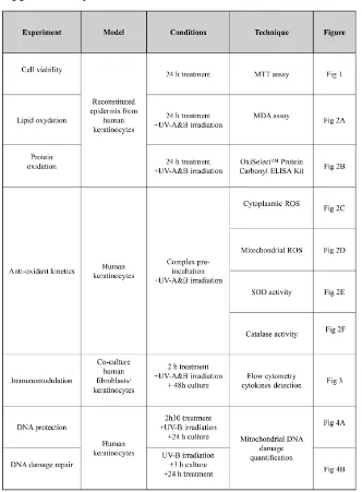
The cytotoxicity of each complex was assessed using the colorimetric MTT [3-(4,5-Dimethylthiazol-2-Yl)-2,5-Diphenyltetrazolium Bromide] assay (Abcam, Cambridge, USA). Reconstructed epidermis treated for 24 hours with the dif-ferent tested products were incubated for 3 hours at 37˚C with the MTT reagent (Figure S1). Dimethyl sulfoxide was then added to dissolve formazan crystals and the absorbance was measured at 570 nm.
2.5. Malondialdehyde (MDA) Extraction and Quantification
The lipid peroxidation was determined by measuring MDA content in recon-structed epidermis. After 24 hours of treatment with the tested products and UV-A (5 J/cm2)-UV-B irradiations (150 mJ/cm2) (Figure S1), epidermis
homo-genates were resuspended in a specific extraction buffer containing Tris-HCl 50 mM, NaCl 0.1 M, EDTA 20 mM, HCl 0.1 N and thiobarbituric acid (TBA) 0.67%. Cells were incubated for one hour at 50˚C in the dark, chilled in cold wa-ter and n-butanol was added. Samples were centrifuged at 10,000 g at 0˚C for 10 min and the upper phase was collected for MDA detection. The MDA-TBA complex was separated on a High Performance Liquid Chromatograph (HPLC) equipped with a Bischoff Model 2.200 pump (Bischoff Chromatography, Leon-berg, Germany), on a Ultrasep C18 (30 cm × 0.18 cm, 6 mm porosity) column. The MDA-TBA complex was eluted with methanol: H2O (40:60, v/v) and
moni-tored by fluorescence detection through a 821-F detector (Jasco, Easton, USA) with excitation at 515 nm and emission at 553 nm. A standard curve was used for the quantification.
2.6. Reactive Oxygen Species (ROS) Quantification
ROS were quantified after keratinocyte UV-irradiation [(UV-A (5 J/cm2) +
UV-B (100 mJ/cm2)] following a 0 min, 30 min and 3 hours kinetics using a
DOI: 10.4236/jcdsa.2019.92016 193 J. Cosmetics, Dermatological Sciences and Applications ROS (mROS) quantification, human keratinocytes were incubated with Mito-SOXTM Red reagent (Thermo Fisher Scientific) for 15 min at 37˚C, in the dark.
Cells were then washed twice with Phosphate Buffer Saline (PBS) and analysed by flow cytometry. In parallel, cells were incubated with the CM-H2DCFDA
probe (Thermo Fisher Scientific) for 15 min at 37˚C, in the dark, for the cytop-lasmic ROS detection. Cells were washed, as previously, twice in PBS 1X and 1 × 10⁴ cells were collected for flow cytometry analysis.
2.7. Superoxyde Dismutase (SOD) and Catalase Activities
Quantification
According to the same experimental procedure used for ROS quantification (Figure S1) Cells were sonicated twice in Tris-HCl (pH 7.5), for 30 sec, and cen-trifuge for 10 min at 12,000 g. Samples were kept at −80˚C and total protein concentration was measured by BCA kit (Thermo Fisher Scientific). SOD activi-ty was measured using a SOD-WST kit (Dojindo Molecular Technologies, Mu-nich, Germany) and catalase activity by the Amplex Red Assay Kit Catalase (Thermo Fisher Scientific). Calibration curves were prepared using purified en-zyme preparation. One unit of catalase will decompose 1 µmole of H2O2 per
minute in optimal enzymatic conditions (pH 7.0, 25 ˚C). One unit of SOD activ-ity corresponds to the enzyme quantactiv-ity required to inhibit 50% of WST-1 for-mazan per minute.
2.8. Cytokine Detection and Quantification
Fibroblasts/keratinocytes co-cultured cells were plated one day before the expe-riment in a serum-free growth medium. The different compounds were applied to the cells for 2 hours before cell activation by UV-A/UV-B exposure (Figure S1). Cell culture supernatant (25 µL) was collected after 48 hours and analysed with FlowCytomix for pro- and anti-inflammatory cytokines detection (Bender Med-Systems, Wien, Austria).
2.9. Oxidized Protein Quantification
Protein concentration was determined on reconstructed epidermis using Brad-ford protein quantification assay (Bio-Rad, Hercules, USA) and absorbance val-ues were measured at 595 nm using bovin serum albumin (BSA, 10 μg/mL) as standard solution.
Protein oxidation in BSA standard and in samples treated for 24 hours and irradiated by UV-A (5 J/cm2) and UV-B (100 mJ/cm2) (Figure S1) with the
complex were analysed using the OxiSelectTM Protein Carbonyl ELISA Kit
(Cli-nisciences, Nanterre, France) following manufacturer’s instructions.
2.10. Mitochondrial DNA (mDNA) Extraction, Amplification and
DNA Damage Quantification
DOI: 10.4236/jcdsa.2019.92016 194 J. Cosmetics, Dermatological Sciences and Applications the complex (5% BS + 2% IO for 150 min), UV-B irradiated (100 mJ/cm2) and
collected 24 hours later (Figure S1). In order to study the repairing effect, cells were first irradiated with UV-B, grown for 3 hours and the complex was then added to the culture medium for 24 hours (Figure S1).
DNA was extracted using Invisorb® Spin DNA Extraction kit (Eurobio, Les Ulis, France) following manufacturer’s instructions. Due to a low proportion in the total DNA extract, mDNA was amplified using REPLI-g Mitochondrial DNA (Qiagen, Courtaboeuf, France) and DNA damage [apurinic/apyrimidinic (AP) sites] were quantified in the samples using DNA Damage quantification kit (Biovision, Mountain View, USA) following manufacturer’s instructions. The optical density (OD) was analysed at 450 nm and sample DNA readings were applied to the calibration curve prepared with standard Aldehyde Reactive Probe-DNA (ARP/DNA) solutions (Dojindo Molecular Technologies).
2.11. Statistical Methods
The data were analyzed statistically using Microsoft Excel®. Error bars represent the standard deviation of three independent experiments. Differences were con-sidered statistically significant when p-value < 0.05 using Student’s t-test.
3. Results
3.1. Cell Viability Assay
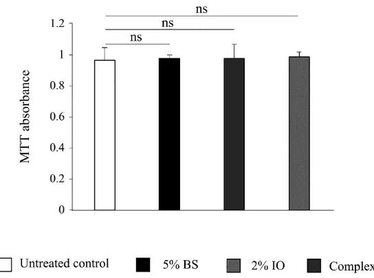
[image:7.595.238.510.469.671.2]Cells metabolic activity was measured by MTT assay after 24 hours of treatment on reconstituted epidermis. No significant effect was observed on cell prolifera-tion for the organic BS at 5%, IO extracts at 2% or with the combinaprolifera-tion of the two compounds (BS 5% + IO 2%) compared to the control (Figure 1).
Figure 1. Effect of birch sap, Inonotus obliquus extracts or the complex on cell viability.
DOI: 10.4236/jcdsa.2019.92016 195 J. Cosmetics, Dermatological Sciences and Applications
3.2. Antioxidant Effect
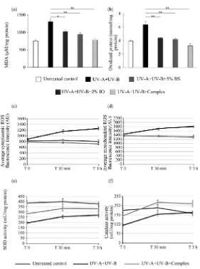
[image:8.595.235.516.245.623.2]The antioxidant effect was studied on UV-irradiated reconstituted epidermis af-ter 24 h treatment with the different compounds. Lipid peroxidation was meas-ured by MDA detection. Increased MDA detection was significantly reduced by epidermis treatment with the BS 5% (−22%, p < 0.05) or the IO 2% alone (−28%, p < 0.01) before oxidative stress induction by UV-irradiation. The complex of the two compounds showed higher reduction of lipid peroxidation revealing a cumulative effect (−41%, p < 0.01) (Figure 2(a)). Detection of protein oxidation was also measured in the same conditions and showed significant reduction of the oxidative stress in the BS 5%-, IO 2%- or complex-treated samples (−31%, −35%, −49%, respectively) with higher antioxidant effect of the complex.
Figure 2. Anti-oxidant activity analysis on reconstituted epidermis or cultured
keratinocytes. (a) (b) Bar graphs represent MDA (a) and oxidized protein (b) quantification after UV-A (5 J/cm2) + UV-B (100 mJ/cm2) irradiation or not
DOI: 10.4236/jcdsa.2019.92016 196 J. Cosmetics, Dermatological Sciences and Applications Cytoplasmic and mitochondrial ROS kinetics were followed 0 min, 30 min and 3 hours after UV-A + UV-B irradiation. The keratinocytes treatment with the complex significantly reduced the production of both cytoplasmic (Figure 2(c)) and mitochondrial ROS (Figure 2(d)) (−43%, p < 0.001 and −28%, p < 0.001 respectively in comparison with the irradiated batch) measured after 3 hours incubation, and showed a kinetic similar to the unirradiated cells (un-treated control). In parallel, the antioxidant enzyme activity of the SOD (Figure 2(e)) and the catalase (Figure 2(f)) were determined in the same conditions. Both enzymes activities were significantly increased by the BS 5% + IO 2% com-plex treatment prior keratinocytes irradiation (+22%, p < 0.001 and +29%, p < 0.001 respectively) revealing the protective property of the complex on SOD and catalase activity. Together these results showed the antioxidant properties of the BS 5% + IO 2% complex against UV-induced oxidative stress.
3.3. Immunomodulatory Effect on Human
Keratinocytes/Fibroblasts Co-Culture
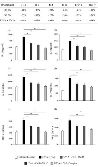
Skin UV irradiations induce keratinocytes activation with an inflammatory re-sponse and cytokines production that can further damage the cells. Immuno-modulatory effect was investigated on human keratinocytes/fibroblasts co-culture after 2 hours of treatment with the different compounds followed by keratinocytes activation in response to UV irradiation (UV-A + UV-B). Flow cytometry quantitative detection in the growth medium of various interleukins [Il-1β, IL-6, IL-8, IL-10, TNF-α and interferon-γ (IFN-γ)] showed a significant reduction of secretion in the BS or IO extracts keratinocytes treated conditions, with an even stronger decrease with the BS + IO complex treatment (Figures 3(a)-(f) and Table 1). These cytokines are involved in acute and chronic in-flammatory response after UV irradiation, revealing the anti-inin-flammatory ac-tivity and the modulation of immune response by the organic birch sap and chaga mushroom extract used separately or in a complex.
3.4. UV-B Protective and Repairing Effect
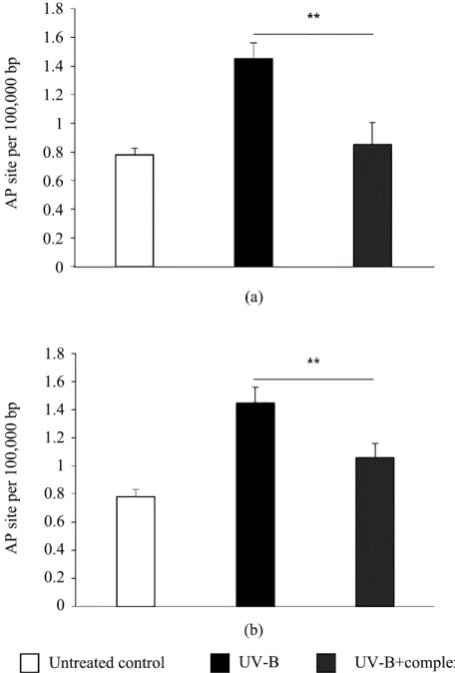
UV-B irradiation-induced DNA damage, measured by the number of apurin-ic/apyrimidinic sites, were significantly reduced (−41%, p < 0.01) in comparison with the control after 150 min treatment with the complex revealing the protec-tive effect on human keratinocytes (Figure 4(a)). Additionally, the BS 5% + IO 2% complex showed a regenerative effect with a significant lower (−27%, p < 0.01) DNA damage quantity when the complex was applied for 24 hours on the cells after UV-B-induced DNA damage (Figure 4(b)).
4. Discussion
DOI: 10.4236/jcdsa.2019.92016 197 J. Cosmetics, Dermatological Sciences and Applications
Table 1. Cytokines quantification on keratinocytes/fibroblasts co-culture after UV-A +
UV-B irradiation in the different conditions. Percent change from untreated UV-irradiated control. BS: Birch sap, IO: Inonotus obliquus.
Interleukins Il-1β Il-6 Il-8 Il-10 TNF-α IFN-γ
BS 5% −28% −26% −22% −24% −24% −22%
IO 2% −35% −32% −27% −30% −29% −24%
BS 5% + IO 2% −48% −49% −38% −40% −48% −38%
Figure 3. Complex immunomodulation effect on keratinocytes/fibroblasts co-culture
af-ter UV-A+UV-B irradiation. (a)-(f) Bar graphs represent inaf-terleukin IL-1β (a), inaf-terleu- interleu-kin IL-6 (b), interleuinterleu-kin IL-8 (c), interleuinterleu-kin IL-10 (d), TNF-α (e) and IFN-γ (f) quantifi-cation after UV-A (5 J/cm2) + UV-B (100 mJ/cm2) irradiation or not (untreated control).
DOI: 10.4236/jcdsa.2019.92016 198 J. Cosmetics, Dermatological Sciences and Applications
Figure 4. UV-B protective and regenerative effect on UV-B irradiated
hu-man keratinocytes. (a) (b) Complex (BS 5% + IO 2%) protective (a) and re-generative (b) effect against UV-B induced mDNA damages.
UV irradiations, including UV-A and UV-B, is a major component of skin exposome altering the physiology and integrity of epidermal cells at different le-vels.
One of the first effect of both UV-A and UV-B irradiations is the formation of free radicals, leading to the generation of a deleterious oxidative stress [24].
In particular, the abnormal production of ROS triggered by UV exposure may cause protein and lipid peroxidation inducing the formation of highly reactive carbonyl species including MDA and 4-hydroxy-2-nonenal [25].
In the present work we observed that birch sap, chaga mushroom extracts and the tested natural complex composed by 5% of organic birch sap and 2% of cha-ga mushroom extracts were able to decrease significantly the content of both cy-toplasmic and mitochondrial UV-induced ROS in cultured keratinocytes.
As expected, the tested products decreased strongly also the content of MDA and oxidized proteins following UV irradiations.
DOI: 10.4236/jcdsa.2019.92016 199 J. Cosmetics, Dermatological Sciences and Applications activity, which protect the skin against the oxidative stress induced by UV irrad-iations [26] by reducing free radical content.
In this context it should be noted that birch sap extract shows a relatively high content of different metallic elements, including copper and zinc [27], both met-als presenting an important anti-oxidant activity. In particular, it has been pre-viously shown that copper is able to induce SOD transcription and activity in cultured fibroblasts [28] [29]. The presence of copper and zinc in birch sap ex-tracts could explain the strong anti-oxidant activity of the BS 5% - IO 2% com-plex against UV irradiations.
In parallel chaga mushroom extract are rich in polyphenols [27], previously described as potent anti-oxidant [30]. As a consequence, the association of BS and IO extracts in the natural complex tested in the present work presents an important activity against the oxidative stress induced by UV-A and UV-B ir-radiation invitro.
As previously mentioned, the exposure of the skin to UV-A and UV-B can produce important damages to both genomic and mitochondrial DNA [2] [3] contributing profoundly to skin-age associated modifications [31]. Here we demonstrated that birch sap and chaga mushroom extracts exhibit a significant DNA protection and repair activity at both nuclear and mitochondrial levels.
Different mechanisms could be involved in the observed activities. Firstly, or-ganic BS extracts are rich in zinc. As previously described, zinc is able to protect against apoptosis and DNA damages induced by UV-A and UV-B irradiation in HaCat keratinocytes and fibroblasts invitro [32] [33]. As a result, the presence of zinc in the natural complex tested herein could explain in part the observed activity.
Secondly, polyphenols of chaga mushroom extract in addition to their an-ti-oxidant activity could act as a sunscreen due to their ability to absorb the whole UV-B spectrum and a part of UV-A spectrum [34]. In parallel, polyphe-nols are able to repair the UV-induced cyclobutane pyrimidine dimers via the stimulation of IL-12 expression [35] and by inducing the expression of genes encoding for proteins involved in nucleotide excision repair [36].
The role of UV exposure in the initiation of an important inflammatory process in the cutaneous tissue has also been documented [37]. The response of the skin to UV irradiation starts with the synthesis of IL-1, IFN-γ and TNF-α
which in turn induce the synthesis of other pro-inflammatory molecules in kera-tinocytes such as IL-6 and IL-8. In addition, UV irradiation can induce the syn-thesis of IL-10, a cytokine acting to attenuate inflammatory damages in the skin [38].
Moreover, the cytokine cascade induced upon UV irradiation triggers the ac-tivation of the transcriptional factors AP-1, which in turn regulates positively the expression of several MMPs, including MMP-1, MMP-3 and MMP-9 resulting in extracellular matrix degradation [39].
DOI: 10.4236/jcdsa.2019.92016 200 J. Cosmetics, Dermatological Sciences and Applications of different pro-inflammatory cytokines, including IL-1β, IL-6, IL-8, TNF-α and IFN-γ, following the exposure of human keratinocytes/ fibroblasts co-culture to UV-A and UV-B. Consequently, by reducing the inflammatory response of ke-ratinocytes to UV irradiations, lower levels of IL-10 were also detected.
On one hand, this activity could be explained by the presence in birch sap ex-tracts of botulin and betulinic acid, two molecules with a described an-ti-inflammatory activity [40].
On the other hand, Inonotusoubliquus extracts are rich in compounds pre-senting an anti-inflammatory activity including ergosterols, lanosterol, inotodiol and trametenolic acid [41] [42] which could be able to reduce the inflammatory cascade induced in skin cells after UV irradiations.
5. Conclusions
In the work described here, we demonstrated for the first time that birch sap and chaga mushroom extracts protect the skin against the UV-induced damages. In particular we have shown that these natural compounds present a strong DNA protection and repair activity as well as anti-oxidant and anti-inflammatory properties on UV-exposed keratinocytes and fibroblasts in vitro. Nevertheless, this study has been conducted on cellular models lacking the biological com-plexity of the skin tissue. In this sense, the photo-protection activity of birch sap and chaga mushroom extracts should be confirmed on 3D skin models, such as reconstructed epidermis or human skin explants exvivo. These biological mod-els are composed of different types of skin cells and their use could help to better characterize the protection activity of birch sap and chaga mushroom extracts against UV.
Moreover, the use of human skin explant exvivo as experimental model could elucidate the kinetics of penetration of both extracts across the stratum cor-neum.
The results obtained here, in particular the anti-oxidant and the an-ti-inflammatory properties, suggest that birch sap and chaga mushroom extracts complex might present additional activities of interest for the dermocosmetic field, including anti-pollution activity. To confirm this hypothesis, the protective role of birch sap and chaga mushroom complex against environmental pollu-tants should be demonstrated following the exposure of 3D skin model to exter-nal aggressions including heavy metals, volatile organic compounds and poly-cyclic aromatic hydrocarbons.
To conclude, taken together our findings suggest that Betulaalba and Inono-tusobliquus extracts represent valid natural ingredients for the development of cosmetic products with photo-protection activity.
Acknowledgements
DOI: 10.4236/jcdsa.2019.92016 201 J. Cosmetics, Dermatological Sciences and Applications
Conflicts of Interest
MS and BP are employees of Inderma laboratory.
References
[1] D’Orazio, J., Jarrett, S., Amaro-Ortiz, A. and Scott, T. (2103) UV Radiation and the Skin. International Journal of Molecular Sciences, 14, 12222-12248.
https://www.mdpi.com/1422-0067/14/6/12222
https://doi.org/10.3390/ijms140612222
[2] Shibutani, S., Takeshita, M. and Grollman, A.P. (1991) Insertion of Specific Base during DNA Synthesis Past the Oxidation Damaged Base 8-Oxodg. Nature, 349, 431-434. https://www.nature.com/articles/349431a0
https://doi.org/10.1038/349431a0
[3] Pfeifer, G.P. (1997) Formation and Processing of UV Photoproducts: Effects of DNA Sequence and Chromatin Environment. Photochemistry and Photobiology, 65, 270-283. https://doi.org/10.1111/j.1751-1097.1997.tb08560.x
https://onlinelibrary.wiley.com/doi/abs/10.1111/j.1751-1097.1997.tb08560.x
[4] Thiele, J.J., Traber, M.G. and Packer, L. (1998) Depletion of Human Stratum Cor-neum Vitamin E: An Early and Sensitive in Vivo Marker of UV Induced Pho-to-Oxidation. Journal of Investigative Dermatology, 110, 756-761.
https://www.jidonline.org/article/S0022-202X(15)40076-4/fulltext
https://doi.org/10.1046/j.1523-1747.1998.00169.x
[5] Ogura, R., Sugiyama, M., Nishi, J. and Haramaki, N. (1991) Mechanism of Lipid Radical Formation Following Exposure of Epidermal Homogenate to Ultraviolet Light. Journal of Investigative Dermatology, 97, 1044-1047.
https://doi.org/10.1111/1523-1747.ep12492553
[6] Wood, J.M. and Schallreuter, K.U. (2006) UVA-Irradiated Pheomelanin Alters the Structure of Catalase and Decreases Its Activity in Human Skin. Journal of Investig-ative Dermatology, 126, 13-14.https://doi.org/10.1038/sj.jid.5700051
[7] Meguro, S., Arai, Y., Masukawa, K., Uie, K. and Tokimitsu, I. (1999) Stratum Cor-neum Lipid. Abnormalities in UVB-Irradiated Skin. Photochemistry and Photobi-ology, 69, 317-321.https://doi.org/10.1111/j.1751-1097.1999.tb03292.x
[8] Biniek, K., Levi, K. and Dauskardt, R.H. (2006) Solar UV Radiation Reduces the Barrier Function of Human Skin. Proceedings of the National Academy of Sciences of the United States of America, 109, 17111-17116.
https://www.pnas.org/content/109/42/17111
https://doi.org/10.1073/pnas.1206851109
[9] Hasegawa, T., Kaneko, F. and Niwa, Y. (1992) Changes in Lipid Peroxide Levels and Activity of Reactive Oxygen Scavenging Enzymes in Skin, Serum and Liver Follow-ing UVB Irradiation in Mice. Life Sciences, 50, 1893-1903.
https://doi.org/10.1016/0024-3205(92)90550-9
[10] Zheng, Y., Xu, Q., Chen, H., Chen, Q., Gong, Z. and Lai, W. (2017) Transcriptome Analysis of Ultraviolet A-Induced Photoaging Cells with Deep Sequencing. The Journal of Dermatology, 45, 175-181. https://doi.org/10.1111/1346-8138.14157
https://onlinelibrary.wiley.com/doi/pdf/10.1111/1346-8138.14157
[11] Zheng, Y., Lai, W., Wan, M. and Maibach, H.I. (2011) Expression of Cathepsins in Human Skin Photoaging. Skin Pharmacology and Physiology, 24, 10-21.
https://doi.org/10.1159/000314725
DOI: 10.4236/jcdsa.2019.92016 202 J. Cosmetics, Dermatological Sciences and Applications
https://europepmc.org/abstract/med/15773537
[13] Li, D., Turi, T.G., Schuck, A., Freedberg, I.M., Khitrov, G. and Blumenberg, M. (2001) Rays and Arrays: The Transcriptional Program in the Response of Human Epidermal Keratinocytes to UVB Illumination. The FASEB Journal, 15, 2533-2550.
https://doi.org/10.1096/fj.01-0172fje
[14] Clydesdale, G.J., Dandie, G.W. and Muller, H.K. (2001) Ultraviolet Light Induced Injury: Immunological and Inflammatory Effects. Immunology & Cell Biology, 79, 547-568. https://doi.org/10.1046/j.1440-1711.2001.01047.x
[15] Cui, Y., Kim, D.S. and Park, K.C. (2005) Antioxidanteffect of Inonotus obliquus. Journal of Ethnopharmacology, 96, 79-85.https://doi.org/10.1016/j.jep.2004.08.037 [16] Shibnev, V.A., Mishin, D.V., Garaev, T.M., Finogenova, N.P., Botikov, A.G. and
Deryabin, P.G. (2011) Antiviral Activity of Inonotus obliquus Fungus Extract to-wards Infection Caused by Hepatitis C Virus in Cell Cultures. Bulletin of Experi-mental Biology and Medicine, 151, 612-614.
https://link.springer.com/article/10.1007%2Fs10517-011-1395-8?LI=true
https://doi.org/10.1007/s10517-011-1395-8
[17] Géry, A., Dubreule, C., André, V., Rioult, J.P., Bouchart, V., Heutte, N., Eldin de Pécoulas, P., Krivomaz, T. and Garon, D. (2018) Chaga (Inonotus obliquus), a Fu-ture Potential Medicinal Fungus in Oncology? A Chemical Study and a Comparison of the Cytotoxicity against Human Lung Adenocarcinoma Cells (a549) and Human Bronchial Epithelial Cells (BEAS-2B). Integrative Cancer Therapies, 17, 832-843. https://doi.org/10.1177/1534735418757912
[18] Yun, J.S., Pahk, J.W., Lee, J.S., Shin, W.C., Lee, S.Y. and Hong, E.K. (2011) Inonotus obliquus Protects against Oxidative Stress-Induced Apoptosis and Premature Se-nescence. Molecules and Cells, 31, 423-429.
https://doi.org/10.1007/s10059-011-0256-7
[19] Lee, E.J. and Cha, H.J. (2019) Inonotus obliquus Extract as an Inhibitor of α-MSH-Induced Melanogenesis in B16F10 Mouse Melanoma Cells. Cosmetics, 6, 9.
https://doi.org/10.3390/cosmetics6010009
[20] Rastogi, S., Pandey, M.M. and Kumar Singh Rawat, A. (2015) Medicinal Plants of the Genus Betula—Traditional Uses and a Phytochemical-Pharmacological Review. Journal of Ethnopharmacology, 159, 62-83.
https://doi.org/10.1016/j.jep.2014.11.010
[21] Svanberg, I., Sõukand, R., Łuczaj, Ł., Kalle, R., Zyryanova, O., Dénes, A., Papp, N., Nedelcheva, A., Šeškauskaitė, D., Kołodziejska-Degórska, I. and Kolosova, V. (2012) Uses of Tree Saps in Northern and Eastern Parts of Europe. Acta Societatis Botani-corum Poloniae, 81, 343-357.https://doi.org/10.5586/asbp.2012.036
[22] Saewan, N. and Jimtaisong, A. (2015) Natural Products as Photoprotection. Journal of Cosmetic Dermatology, 14, 47-63.https://doi.org/10.1111/jocd.12123
[23] Krutmann, J., Bouloc, A., Sore, G., Bernard, B.A. and Passeron, T. (2017) The Skin Aging Exposome. Journal of Dermatological Science, 85, 1527-1561.
https://doi.org/10.1016/j.jdermsci.2016.09.015
[24] de Jager, T.L., Cockrell, A.E. and Du Plessis, S.S. (2017) Ultraviolet Light Induced Generation of Reactive Oxygen Species. Advances in Experimental Medicine and Biology, 996, 15-23.https://doi.org/10.1007/978-3-319-56017-5_2
DOI: 10.4236/jcdsa.2019.92016 203 J. Cosmetics, Dermatological Sciences and Applications
https://www.ncbi.nlm.nih.gov/pmc/articles/PMC3973651
https://doi.org/10.1016/j.jphotobiol.2014.01.019
[26] Sander, C.S., Chang, H., Salzmann, S., Muller, C.S., Ekanayake-Mudiyanselage, S., Elsner, P. and Thiele, J.J. (2002) Photoaging Is Associated with Protein Oxidation in Human Skin in Vivo. Journal of Investigative Dermatology, 118, 618-625.
https://doi.org/10.1046/j.1523-1747.2002.01708.x
[27] Grabek-Lejko, D., Kasprzyk, I., Zaguła, G. and Puchalski, C. (2017) The Bioactive and Mineral Compounds in Birch Sap Collected in Different Types of Habitats. Bal-tic Forestry, 23, 230-239.
https://www.balticforestry.mi.lt/bf/PDF_Articles/2017-23[2]/Baltic%20Forestry%20
2017.2_394-401.pdf
[28] Powell, S.R. (2000) The Antioxidant Properties of Zinc. The Journal of Nutrition, 130, 1447S-1454S.https://doi.org/10.1093/jn/130.5.1447S
[29] Itoh, S., Ozumi, K., Kim, H.W., Nakagawa, O., McKinney, R.D., Folz, R.J., Zelko, I.N., Ushio-Fukai, M. and Fukai, T. (2009) Novel Mechanism for Regulation of Extracellular SOD Transcription and Activity by Copper: Role of Antioxidant-1. Free Radical Biology & Medicine, 46, 95-104.
https://www.sciencedirect.com/science/article/pii/S0891584908005789?via%3Dihub
https://doi.org/10.1016/j.freeradbiomed.2008.09.039
[30] Lee, I.K., Kim, Y.S., Jang, Y.W., Jung, J.Y. and Yun, B.S. (2007) New Antioxidant Polyphenols from the Medicinal Mushroom Inonotus obliquus. Bioorganic & Me-dicinal Chemistry Letters, 17, 6678-6681.
https://doi.org/10.1016/j.bmcl.2007.10.072
[31] Yarosh, D.B. (2016) DNA Damage and Repair in Skin Aging. In: Farage, M., Miller, K. and Maibach, H., Eds., Textbook of Aging Skin, Springer, Berlin, Heidelberg, 1-13. https://doi.org/10.1007/978-3-642-27814-3_31-3
[32] Parat, M.O., Richard, M.J., Pollet, S., Hadjur, C., Favier, A. and Béani, J.C. (1997) Zinc and DNA Fragmentation in Keratinocyte Apoptosis: Its Inhibitory Effect in UVB Irradiated Cells. Journal of Photochemistry and Photobiology B: Biology, 37, 101-106.https://doi.org/10.1016/S1011-1344(96)07334-4
[33] Leccia, M.T., Richard, M.J., Favier, A. and Béani, J.C. (1997) Zinc Protects against Ultraviolet A1-Induced DNA Damage and Apoptosis in Cultured Human Fibrob-lasts. Biological Trace Element Research, 69, 177-190.
https://link.springer.com/article/10.1007/BF02783870
https://doi.org/10.1007/BF02783870
[34] Nichols, J.A. and Katiyar, S.K. (2010) Skin Photoprotection by Natural Polyphenols: Anti-Inflammatory, Antioxidant and DNA Repair Mechanisms. Archives of Der-matological Research, 302, 71-83. https://doi.org/10.1007/s00403-009-1001-3
https://link.springer.com/article/10.1007%2Fs00403-009-1001-3
[35] Meeran, S.M., Mantena, S.K., Elmets, C.A. and Katiyar, S.K. (2006) (-)-Epigallocatechin-3-Gallate Prevents Photocarcinogenesis in Mice through In-terleukin-12-Dependent DNA Repair. Cancer Research, 66, 5512-5520.
http://cancerres.aacrjournals.org/content/66/10/5512.long
https://doi.org/10.1158/0008-5472.CAN-06-0218
[36] Katiyar, S.K., Vaid, M., van Steeg, H. and Meeran, S.M. (2010) Green Tea Polyphe-nols Prevent UV-Induced Immunosuppression by Rapid Repair of DNA Damage and Enhancement of Nucleotide Excision Repair Genes. Cancer Prevention Re-search, 3, 179-189. https://doi.org/10.1158/1940-6207.CAPR-09-0044
DOI: 10.4236/jcdsa.2019.92016 204 J. Cosmetics, Dermatological Sciences and Applications [37] Kondo, S. (1999) The Roles of Keratinocyte-Derived Cytokines in the Epidermis
and Their Possible Responses to UVA-Irradiation. Journal of Investigative Derma-tology Symposium Proceedings, 4, 177-183.
https://doi.org/10.1038/sj.jidsp.5640205
https://www.jidsponline.org/article/S1087-0024(15)30258-6/pdf
[38] Iyer, S.S. and Cheng, G. (2012) Role of Interleukin 10 Transcriptional Regulation in Inflammation and Autoimmune Disease. Critical Reviews in Immunology, 32, 23-63. https://doi.org/10.1615/CritRevImmunol.v32.i1.30
https://www.ncbi.nlm.nih.gov/pmc/articles/PMC3410706/pdf/nihms377104.pdf
[39] Rittié, L. and Fisher, G.J. (2002) UV-Light-Induced Signal Cascades and Skin Aging. Ageing Research Reviews, 1, 705-720.
https://www.sciencedirect.com/science/article/pii/S1568163702000247?via%3Dihub
https://doi.org/10.1016/S1568-1637(02)00024-7
[40] Flekhter, O.B., Medvedeva, N.I., Karachurina, L.T., Baltina, L.A., Galin, F.Z., Zaru-dii, F.S. and Tolstikov, G.A. (2005) Synthesis and Pharmacological Activity of Betu-lin, Betulinic Acid, and Allobetulin Esters. Pharmaceutical Chemistry Journal, 39, 401-404. https://link.springer.com/article/10.1007/s11094-005-0167-z
https://doi.org/10.1007/s11094-005-0167-z
[41] Park, Y.M., Won, J.H., Kim, Y.H., Choi, J.W., Park, H.J. and Lee, K.T. (2005) In Vivo and in Vitro Anti-Inflammatory and Anti-Nociceptive Effects of the Methanol Extract of Inonotus obliquus. Journal of Ethnopharmacology, 101, 120-128.
https://doi.org/10.1016/j.jep.2005.04.003
DOI: 10.4236/jcdsa.2019.92016 205 J. Cosmetics, Dermatological Sciences and Applications
