BONE MARROW ANGIOGENESIS AND MICROVESSEL DENSITY IN PATIENTS WITH
Richa Sharma, Sunita Singh, Sudhir Atri,
Department of Pathology and Clinical Hematology,
ARTICLE INFO ABSTRACT
Introduction:
anemia (AA) according to the severity as non severe (NsAA), severe (sAA) and very severe (VsAA) and to study microvessel density using immunohistochemical stain using CD 34 an
analysis of microvessel density by computer assisted quantitative analyser and to compare it in the three subcategories of aplastic anemia with control group.
prospective study was conducted in the Departmen
Thirty cases of AA patients and 10 cases of control were taken. AA patients were diagnosed on the basis of complete blood count, peripheral blood smear, bone marrow aspiration examination and bone marrow tre
VsAA using camitta et al criteria. IHC using CD 34 was performed to look for angiogenesis and to calculate microvessel density (MVD).
and 3rd decade of life. Females were more commonly affected with a male to female ratio (11/19) of 0.58:1. In Aplastic anemia patients (Group I), using Camitta et al criteria 30 cases were categorized into NsAA (10 cases), sAA (12 cases) and
is a good marker of angiogenesis. MVD showed a decreasing trend with increase in severity of the disease.
angiogenesis and mean MVD score shows a decreasing trend from anemia
Copyright©2019, Richa Sharma et al. This is an open distribution, and reproduction in any medium, provided
INTRODUCTION
Aplastic anemia (AA) is a life threatening disorder that is characterized by deficiency of pluripotent hematopoietic progenitor cells with consecutive bone marrow (BM) aplasia and peripheral pancytopenia1. Aplastic anemia is pancytopenia with hypocellular bone marrow in the absence of infiltration and fibrosis having cellularity <25% however us
cellularity is even <10% (Marsh et al., 2009).
AA shows geographical variability. The incidence of AA varied from 10- 52.7% among patients with pancyto
(Khunger et al., 2002). Factors that temporarily or permanently
injure the bone marrow which further affect the blood cell production are radiation and chemotherapy treatments, exposure to toxic chemicals, use of certain drugs, autoimmune disorders, viral infection, pregnancy and idiopathic ( 1989). Recently the fact that angiogenesis may have an important role in aplastic anemia has attracted considerable attention and this has lead to conducting of new studies regarding the role of angiogenesis in AA (Erdem
The induction of angiogenesis is a complex and dynamic processes mediated by many pro-angiogenic and anti angiogenic molecules. Disruption of the balance
ISSN: 0975-833X
Article History:
Received 24th May, 2019
Received in revised form
20th June, 2019
Accepted 28th July, 2019
Published online 31st August, 2019
Citation: Richa Sharma, Sunita Singh, Sudhir Atri, RenukaVerma, Manali satiza and Rajeev Sen
density in patients with Aplastic Anemia”, International Journal of Current Research
Key Words:
Aplastic Anemia, Angiogenesis, Morphometric analysis
*Corresponding author: Renuka Verma
RESEARCH ARTICLE
BONE MARROW ANGIOGENESIS AND MICROVESSEL DENSITY IN PATIENTS WITH
APLASTIC ANEMIA
, Sunita Singh, Sudhir Atri, *Renuka Verma, Manali satiza and
ology and Clinical Hematology, Pt. B.D. Sharma, PGIMS, UHS, Rohtak, Haryana, India
ABSTRACT
Introduction: The present study was conducted with the aim to diagnose and categorize aplastic anemia (AA) according to the severity as non severe (NsAA), severe (sAA) and very severe (VsAA) and to study microvessel density using immunohistochemical stain using CD 34 an
analysis of microvessel density by computer assisted quantitative analyser and to compare it in the three subcategories of aplastic anemia with control group. Material and Methods:
prospective study was conducted in the Department of Pathology at Pt.
Thirty cases of AA patients and 10 cases of control were taken. AA patients were diagnosed on the basis of complete blood count, peripheral blood smear, bone marrow aspiration examination and bone marrow trephine biopsy (using Jamshidi’s needle). AA patients were categorized as sAA, NsAA and VsAA using camitta et al criteria. IHC using CD 34 was performed to look for angiogenesis and to calculate microvessel density (MVD). Results: Majority of patients (56%) of AA were between 1st and 3rd decade of life. Females were more commonly affected with a male to female ratio (11/19) of 0.58:1. In Aplastic anemia patients (Group I), using Camitta et al criteria 30 cases were categorized NsAA (10 cases), sAA (12 cases) and VsAA (8 cases). MVD was calculated using CD 34 which is a good marker of angiogenesis. MVD showed a decreasing trend with increase in severity of the disease. Conclusion: The study shows that Aplastic anemia is associate
angiogenesis and mean MVD score shows a decreasing trend from anemia.
access article distributed under the Creative Commons Attribution the original work is properly cited.
anemia (AA) is a life threatening disorder that is characterized by deficiency of pluripotent hematopoietic progenitor cells with consecutive bone marrow (BM) aplasia and peripheral pancytopenia1. Aplastic anemia is pancytopenia row in the absence of infiltration and fibrosis having cellularity <25% however usually 2009). The incidence of AA shows geographical variability. The incidence of AA 52.7% among patients with pancytopenia Factors that temporarily or permanently injure the bone marrow which further affect the blood cell production are radiation and chemotherapy treatments, exposure to toxic chemicals, use of certain drugs, autoimmune ction, pregnancy and idiopathic (Nissen, Recently the fact that angiogenesis may have an important role in aplastic anemia has attracted considerable attention and this has lead to conducting of new studies
Erdem et al., 2006).
The induction of angiogenesis is a complex and dynamic angiogenic and
anti-balance between
pro-angiogenic and anti-angiogenic factors in favour
angiogenic factors leads to formation of new vascular structure. VEGF is one of the most relevant angiogenic factors as its expression shows close correlation with the vessel density in several human tumors and inhibition of VEGF by specific antibodies suppresses tumor growth
Blythe, 2008). Angiogenesis is quantified through the staining of blood vessels with various endothelial markers. Various
endothelial cell markers used for quantification of
angiogenesis include CD31, CD34, CD105
(Wang et al., 2008). Morphometry means measurement of
form, and is defined as the quantitative description of geometric features of structure with any dimension
1991). This is more restricted definition of morphometry than that of Weibel, who includes stereology in the definition of morphometry (Weibel, 1979).
advantages over conventional visual assessment; objectivity, reproducibility and the ability to detect changes not immediately apparent to the naked eye.
measurement is called morphometry. The sequence of steps involved in the analysis of microscopic image is ‘Microscopic image CCD video camera
processing Imagesegmentation
International Journal of Current Research Vol. 11, Issue, 08, pp.6192-6197, August, 2019
DOI: https://doi.org/10.24941/ijcr.36206.08.2019
Richa Sharma, Sunita Singh, Sudhir Atri, RenukaVerma, Manali satiza and Rajeev Sen, 2019. “Bone marrow angiogenesis and microvessel International Journal of Current Research, 11, (08), 6192-6197
BONE MARROW ANGIOGENESIS AND MICROVESSEL DENSITY IN PATIENTS WITH
satiza and Rajeev Sen
Sharma, PGIMS, UHS, Rohtak, Haryana, India
The present study was conducted with the aim to diagnose and categorize aplastic anemia (AA) according to the severity as non severe (NsAA), severe (sAA) and very severe (VsAA) and to study microvessel density using immunohistochemical stain using CD 34 and morphometric analysis of microvessel density by computer assisted quantitative analyser and to compare it in the Material and Methods: The present t of Pathology at Pt. B.D. Sharma PGIMS, Rohtak. Thirty cases of AA patients and 10 cases of control were taken. AA patients were diagnosed on the basis of complete blood count, peripheral blood smear, bone marrow aspiration examination and bone phine biopsy (using Jamshidi’s needle). AA patients were categorized as sAA, NsAA and VsAA using camitta et al criteria. IHC using CD 34 was performed to look for angiogenesis and to Majority of patients (56%) of AA were between 1st and 3rd decade of life. Females were more commonly affected with a male to female ratio (11/19) of 0.58:1. In Aplastic anemia patients (Group I), using Camitta et al criteria 30 cases were categorized VsAA (8 cases). MVD was calculated using CD 34 which is a good marker of angiogenesis. MVD showed a decreasing trend with increase in severity of the The study shows that Aplastic anemia is associated with reduced bone marrow angiogenesis and mean MVD score shows a decreasing trend from non severe to very severe Aplastic
License, which permits unrestricted use,
angiogenic factors in favour of pro-angiogenic factors leads to formation of new vascular
VEGF is one of the most relevant angiogenic factors as its expression shows close correlation with the vessel density in several human tumors and inhibition of VEGF by dies suppresses tumor growth (Jackson and Angiogenesis is quantified through the staining of blood vessels with various endothelial markers. Various
endothelial cell markers used for quantification of
angiogenesis include CD31, CD34, CD105 and factor VIII Morphometry means measurement of form, and is defined as the quantitative description of geometric features of structure with any dimension (Baak, This is more restricted definition of morphometry than ibel, who includes stereology in the definition of , 1979). Measurement has several advantages over conventional visual assessment; objectivity, reproducibility and the ability to detect changes not immediately apparent to the naked eye. This concept of measurement is called morphometry. The sequence of steps involved in the analysis of microscopic image is ‘Microscopic Video frame grabber Image Interactive measurement Data
INTERNATIONAL JOURNAL OF CURRENT RESEARCH
recording Data analysis. Morphometry enables the pathologist to obtain densitometric as well as morphometric parameters like the number of vessels with a certain dimension range, the vessel luminal area, vessel luminal perimeter and the number of immune-stained areas per microscopic field (Hamilton and Allen, 1995). The present study was conducted to diagnose and categorise aplastic anemia according to the severity and to study microvessel density using immunohistochemical stain CD 34 and to compare the microvessel density in three subcategories of aplastic anemia with control group.
MATERIALS AND METHODS
The present study was conducted in Pt. B.D. Sharma, PGIMS, UHS, Rohtak from year 2017 to 2019. The study comprised of patients diagnosed as AA on the basis of complete blood count, peripheral blood smear, bone marrow aspiration examination and bone marrow trephine biopsy (using Jamshidi’s needle). The bone marrow aspiration and biopsy was performed after an informed consent of the patient in the Department of Medicine and was processed routinely. Thirty cases of AA patients and 10 cases of control were taken. All the sections were stained for Haematoxylin and Eosin stain. All AA cases were categorised in NsAA, sAA, and VsAA
using Camitta et al. criterion (Camitta et al., 1982) (Table 1).
Bone Marrow cellularity was calculated according to Tuzuner and Bennet reference standards (Tuzuner and Bennett, 1994).
Control: Control patients constituted the bone marrow
biopsies of patients with iron deficiency anaemias and marrow for staging of Hodgkins lymphoma.
Inclusion Criteria: Only newly diagnosed cases and who
were not yet started on any treatment were included in the study.
Exclusion Criteria: Patients on immunosuppressive therapy
were excluded from the study.
Immunohistochemical analysis: IHC was performed on 3-5
μm in thick sections from 10% formalin fixed paraffin- embedded specimens, according to the streptoavidin-biotin immunoperoxidase technique (Dako-cytomation). Multiple slides were evaluated, and ideal section was used for IHC
staining Positive and negative controls were run
simultaneously. Strong brown nuclear immunoreactivity was considered as positive staining.
Control for IHC staining: Sections of normal bone marrow
biopsy were used as positive controls for anti CD34 antibodies. Negative controls were obtained by substituting the primary antibody with an antibody of irrelevant specificity.
Statistical Analysis
A retrospective study was carried out for all the variables included in the study. Chi square test was used to compare the categorical values. P value less than 0.05 was accepted as clinically significant using SPSS version 20.0. Morphometric Analysis: The quantitative morphometric studies were done by image analysis. Images provided by a charged device video camera coupled with Olympus BX51 microscope at a magnification 400 X were stored on a host computer based on Pentium 4 processor with operating system Microsoft
Windows Vista/ XP through a digital frame grabber and processing was done by image analysis software Image Pro Plus Version 6.3. Microvessel (MV) was defined as any highlighted endothelial cell or endothelial cell cluster, tumor cells and other connective tissue elements. Vessel lumen was not necessary for a structure to be defined as microvessel. Large vessels and vessels in the periosteum or bone were excluded because vascularisation is not representative of neo-angiogenesis in these areas. Areas of staining with no discrete breaks were counted as a single vessel. MVD was assessed by light microscopy in representative areas with highest number of capillaries and small venules (neovascular ‘‘hot spots’’) according to the method that was first described by Weidner et al.13 The sections were scanned first at low magnification (100X), and the most intense area of neovascularisation (hot spot) was identified. Microvessel (MV) counts were performed on 400 X. The MVD were determined by two independent observers in each case. Computer assisted image analysis was performed on all cells, tissues and vessels expressing antibody staining, avoiding confounding background and included all positive staining vessels.
RESULTS
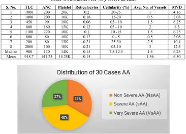
Out of total 40 cases taken in the study; 10 cases were taken as control and total of 30 cases of AA were categorized using Camitta et al criteria and it was observed that 10 cases were categorised as non-severe (NsAA), 12 as severe AA (sAA) and 8 cases as very severe AA (VsAA) (Figure 1). Maximum number of cases of AA were between the age group of 10 to 30 years constituting total of 17/30 patients suggesting 56% of total cases. (Figure 2) Cases from almost all age groups were taken to represent as controls. Out of total 30 cases of AA, 63% were females, suggesting mild female preponderance. In control group, 60% were males. Fifty three percent (16/30) of AA cases had TLC count ranging between 1000- 2000. Only 2 (6.5%) patients had count less than 500/cumm and only 4 patients (13%) had more than 2000/cumm. Twenty three percent of AA cases had ANC <200µL, 33% cases had ANC between 200-500µL and 43% cases had ANC >500 µL. All patients had thrombocytopenia with approximately 77% (23/30) patients had platelet count less than 30,000/cumm. Quantum of corrected reticulocyte count suggested that 60% of cases had reticulocyte count <0.5%, 23% cases had reticulocyte count between 0.5-1% and 16% cases had reticulocyte count more than 1%. All the cases showed reduction in bone marrow cellularity in AA cases with and only 3% had cellularity more than 30%. On IHC with CD34 staining, No. of vessels varied from 2 to 8 as per high power field when an average of 10 views were taken. MVD varied from 8.33 to 33.33/mm2. Mean microvessel density of 7.41+/- 3.9 was observed in AA cases with median of 6.25 and range from 2.08 to 14.5. Mean micro- vessel density of 23.74 +/- 7.9 with median of 25 and range from 8.3 to 33.3 was observed in controls.
NsAA patients showed: Median TLC count of 2050, median
ANC count of 800, reduced platelet count of 33.5K and reticulocyte count of 0.59 on PBF. Bone marrow biopsy showed cellularity with a median of 20-25% and mean average no. of vessels of 2 with mean MVD of 8.5 with median of 9.3 (Table 2).
sAA patients showed: Median 1500 TLC, <500 ANC, median
Table 1. Camitta et al. classification for Aplastic anemia
Severity Criteria
Severe (sAA)
Bone marrow cellularity < 25% (or if <30%, 25-50% residual hematopoietic cells) and atleast two of the following:
•Peripheral blood absolute neutrophil count (ANC) <500/µl •Peripheral blood platelet counts <20,000/µL
•Peripheral blood corrected reticulocytes <1%
Very Severe(vsAA) As severe, but peripheral blood absolute neutrophil count (ANC)
<200/µL
Non Severe (NsAA) Hypocellular bone marrow with peripheral blood cytopenias not fulfilling
criteria for severe or very severe aplastic anemia
Table 2. Parameters for non-severe aa cases
S. No. TLC ANC Platelet Reticulocytes Cellularity(%) Avg No. of Vessels MVD
1 1100 800 30K 0.41 25-30 3 12.5
2 3400 510 35K 1.3 25-30 0.5 2.08
3 3000 1050 35K 0.56 30-35 3 12.5
4 2000 1160 45K 1.2 25-30 1 4.16
5 2000 740 20K 1.4 15-20 1 4.16
6 2100 504 25K 0.22 20-25 1.5 6.25
7 2000 800 50K 1.2 20-25 2 8.3
8 2100 840 32K 0.17 10-15 2.5 10.41
9 3500 1200 25K 0.54 10-15 3 12.5
10 2000 540 55K 0.63 0-5 3 12.5
Median 2050 800 33.5K 0.59 20-25 2.25 9.35
Mean 2320 814 35.2K 0.76 2.05 8.53
Table 3. Parameters for severe aa cases
S.No. TLC ANC Platelet Reticulocytes Cellularity(%) Avg. No. of Vessels MVD
1 1000 400 22K 0.17 20-25 2 8.3
2 1200 360 12K 0.14 10-15 2.5 10.4
3 2000 500 15K 0.56 20-25 3.5 14.5
4 1000 300 10K 0.5 15-20 1.5 6.25
5 1200 480 15K 1.32 20-25 1 4.16
6 1500 330 12K 0.58 15-20 1 4.16
7 1500 600 10K 0.2 25-30 1 4.16
8 4000 920 8K 0.32 20-25 3.5 14.5
9 2000 600 15K 0.58 20-25 1.5 6.25
10 1100 495 32K 0.21 20-25 1 4.16
11 2000 500 20K 0.65 20-25 0.5 2.08
12 1500 450 20K 0.21 15-20 1.5 6.25
Median 1500 487.5 15K 0.41 20-25 1.5 6.2
[image:3.595.114.481.516.779.2]Mean 1666.7 494.6 15.9 0.45 1.7 7.1
Table 4. Parameters for very severe aa cases
S. No. TLC ANC Platelet Reticulocytes Cellularity (%) Avg. No. of Vessels MVD
1 1000 200 20K 0.2 20-25 1 4.16
2 1000 200 10K 0.18 15-20 0.5 2.08
3 450 90 18K 0.06 05 - 10 1.5 6.25
4 800 160 15K 0.12 05 - 10 2 8.3
5 1100 220 10K 0.1 10 -15 1.5 6.25
6 800 80 18K 0.12 0 - 5 0.5 2.08
7 200 80 13K 0.21 25-30 2.5 10.4
8 2000 100 10K 0.21 05-10 3 12.5
Median 900 130 14K 0.15 7.5-12.5 1.5 6.25
Mean 918.7 141.25 14.25K 0.15 1.56 6.50
Figure 2. Demography on the basis of age
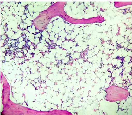
[image:4.595.305.560.51.280.2]Figure 3. Bone marrow sections from AA patient with increase in fat: cell ratio (H & E;200X)
[image:4.595.307.561.319.536.2]Figure 4. Increased fat: cell ratio in AA patient (H & E; 400X)
Figure 5. Reduced number of microvessels in severe Aplastic anemia (CD 34; 400X)
Fig. 6. Morphometric representation of reduced number of microvessels in severe Aplastic anemia (CD34; 400X)
Bone marrow biopsy showed cellularity with a median of 20-25% and mean no. of vessels of 1.7 and mean MVD of 7.1 were observed (Table 3).
VsAA patients showed: Median ANC count of 130, < 15K
[image:4.595.35.291.337.561.2] [image:4.595.35.291.596.789.2]DISCUSSION
AA is classified as non-severe (NsAA), severe (sAA) and very severe (VsAA) based on the degree of the peripheral blood cytopenia and classified on the basis of camitta et al criteria taking into account the bone marrow cellularity, peripheral blood, absolute neutrophil count, platelet count and corrected reticulocyte count. Total of 30 diagnosed cases of AA, on the basis of complete blood count, peripheral blood smear and bone marrow aspiration examination were further evaluated by bone marrow trephine biopsy, performed using Jamshidi’s needle and was processed routinely. Only newly diagnosed cases and who were not yet started on any treatment were
included in the study. Patients who were on
immunosuppressive therapy were excluded from this study. Ten cases as control were also analysed. All the sections were stained for H&E stain. The cases were categorised in NsAA, sAA, and VsAA on basis of reticulocyte count and cellularity. IHC staining for CD 34 was done on all cases included in the study and average no. of vessels and microvessel density were further analysed with Olympus BX 51 using objectives at 40x (being 0.24 for 100 eyepiece small divisions). In our study majority of (56%) patients of AA were between 1st and 3rd decade of life. The youngest patient was 9 years old and the oldest patient was 60 years old with mean age of 27.5 years. Our study showed slight female preponderance in AA cases with a male to female ratio (11/19) of 0.58:1. All 30 cases of AA in our study were subcategorised into NsAA, sAA and VsAA (10,12 and 08 respectively) on the basis of Camitta et al criteria which is mostly widely accepted standard for the diagnosis of Aplastic Anemia. Our study suggested that median values of TLC, ANC, Platelets, reticulocyte and bone marrow cellularity showed a decreasing trend in various subcategories of AA ( Table 2, 3 and 4) Same criteria was used
by the Fureder et al. (2006), Wu et al. (2015), Gupta et al.
(2017), Somasundaram et al. (2009) and Yoon et al. (2012)
for sub-classification of AA. Their studies also suggested reducing trend in median values of ANC, Platelets and corrected Reticulocytes, However Yoon et al18 emphasized that ANC should be an essential and not an optional criteria for diagnosis and classification of AA contrary to the study of
Camitta et al. (1982) which also relates well with our study.
Bacigalupo et al. (2017) also in their study emphasized the
importance of ANC for predicting the severity of AA and further assessment of prognosis during the treatment, thus we also conclude ANC being a good marker for the assessment of severity of AA. Our study also suggests reducing TLC in subcategories of AA. All the cases diagnosed as AA and the control group were subjected to IHC staining for CD 34 antibody and average number of blood vessels and mean vessel density was analysed. Controls which included cases with Iron deficiency anemia and marrow for staging with Hodgkin’s Lymphoma showed a mean vessel number of 5.7+/-1.9, MVD of 23.74+/-7.91. However in cases of AA the mean vessel of 1.78+/-0.95 and MVD of 7.4+/-3.9 was observed suggesting reduced angiogenesis. Our results were in concordance with
the studies of Fureder et al. (2006), Wu et al. (2015). They
observed a mean bone marrow MVD in AA significantly lower than that in control group (P<0.001). Further average number of vessels and mean vessel density was analysed in subcategories of AA. There was a decreasing number in the average no. of vessels per field as well as decreasing trend of MVD with the increasing severity of the disease.
Somasundaram et al. (2009), Gupta et al. (2017) and Ji et al.
(2006) also observed in their study that increasing severity of the disease is associated with decrease in the microvessel density hence reduced angiogenesis. In our study a statistically significant difference in MVD of NsAA vs sAA and NsAA vs VsAA whereas there was no statistically significant decrease in MVD of sAA vs. NsAA was observed. Our study is in
concordance with the study of Somasundaram et al. (2009) and
Ji et al. (2006) They also observed that the MVD of sAA vs NsAA patients and the MVD of VsAA vs. NsAA being significantly different. However there was no significant difference between sAA and VsAA. However the study of
Gupta et al. (2017) and Fureder et al. (2006) in their study
reported significant difference in MVD of controls and AA patients however no apparent difference in the MVD when patients in various subcategories of AA (NsAA vs. sAA vs. VsAA) were compared. These findings probably hint towards decreasing angiogenesis with increasing severity but many more studies with more number of cases will be required to solve the dilemma.
Conclusion
Our findings demonstrate that Camitta et al. classification of
AA on the basis of peripheral blood absolute neutrophil count, platelet count, corrected reticulocyte count and bone marrow cellularity is essential with ANC being the true indicator of classification and not an optional criterion, with further assessment of TLC also being a good tool for classification. The MVD is a good marker of angiogenesis in patients with AA and there is a significant decrease in angiogenesis in bone marrow as the severity of AA increases.
REFERENCES
Baak JPA. 1991. Manual of quantitative pathology in cancer diagnosis and prognosis. Berlin: Springer-Verlag, 7-18 Bacigalupo A. 2017. How I treat acquired aplastic anemia.
Blood, 129(11):1428-36.
Camitta BM, Storb R, Thomas ED. 1982. Aplastic anemia:
pathogenesis, diagnosis, treatment, and prognosis. N Engl J
Med., 306: 645-718
Erdem F, Giindogdu M, Kiziltunc A. 2006. Serum Vascular Endothelial Growth Factor Level in Patients with
Haematological Malignancies. Eur J Gen Med., 3:116-20.
Fureder W, Krauth MT, Sperr WR, Sonneck K, Klupp IS, Mullauer L et al. 2006. Evaluation of angiogenesis and vascular endothelial growth factor expression in the bone
marrow of patients with Aplastic Anemia. AJP,
168:123-29.
Gupta P, Khurana N, Singh T,Gupta D, Dhingra KK. 2009. Bone Marrow angiogenesis in Aplastic Anemia- a study of CD 34 and VEGF Expressions in Bone Marrow Biopsies.
Haematology, 14:16-21.
Hamilton PW, Allen DC. 1995. Morphometry in
histopathology. J Pathol., 175:369-79.
Jackson P, Blythe D. 2008. Immunohistochemical techniques. In: Bancroft JD, Gamble M, editors. Theory and practice of histological techniques. 6th ed. Philadelphia: Churchil Livingstone., P.433-72.
Ji JL, Liu H, Sun C, Jiang SH, Ding RS. 2006. Expression of Vascular Endothelial Growth Factor in Patients with Aplastic Anemia and its Significance. Zhongguo Shi Yan Xue Ye XueZaZhi,14: 285-8.
Khunger JM, Arylselvi S, Sharma U. et al. 2002. Pancytopenia
- a clinicohematological study of 200 cases. Indian J
Marsh JC, Ball SE, Cavenagh J. et al. 2009. Guidelines for the
diagnosis and management of aplastic anaemia. Br J
Haematol., 147: 43-70.
Nishida N, Yano H, Nishida T. et al. 2006. Angiogenesis in
cancer. Vas Health Risk Manag., 2:213- 9.
Nissen DC. 1989. Pathophysiology of Aplastic Anemia.
Bailliersclin haematol., 2:37-49.
Somasundaram V, Tevatia MS, Purohit A, Ahuja A,Mahapatra M, Tyagi S et al. 2017. Evaluation of Bone Marrow Microvessel Density in Patients with Aplastic Anemia.
Indian J Hematol blood transfuse, 33: 169-74.
Tuzuner N, Bennett JM. 1994. Reference standards for bone
marrow cellularity. Leuk Res., 18: 645–47.
Wang D, Stockard CR, Harkins L, Lott P, Salih C, Yuan K. et
al. 2008. Immunohistochemistry in the evaluation of
neovascularization in tumor xenografts. Biotech histochem,
83:179-89.
Weibel ER. Stereological methods. In: Weibel ER, editor. Practical methods for biological morphometry. Vol 1. London: Academic Press. 1979; 44-5.
Wu L, Mo W, Zhang W, Deng H, Li Y,Zhou R. et al. 2015.
Impairment of hematopoietic stem cell niches in patients
with aplastic anemia; Int J Hematol., 102:645–53.
Yoon HH, Huh SJ, Lee JH, Lee S, Kim SH, Kwon HC, Kim HJ. 2012. Should we still use Camitta's criteria for severe
aplastic anemia?, Korean J Hematol., 47(2):126-30.
Young, NS. and Maciejewski, J. 1997. The Pathophysiology of
Acquired Aplastic Anemia. N Engl J Med., 1997; 336:
1365-72.