With g text-figures Printed in Great Britain
FACTORS INFLUENCING SUBMERGENCE AND THE
HEART RATE IN THE FROG
BY D. R. JONES* AND G. SHELTON*
Department of Zoology, The University, Southampton
{Received 14 November 1963)
INTRODUCTION
In view of the intermediate position held by the amphibians between air-breathing and water-breathing vertebrates, surprisingly little is known of the external or internal factors regulating respiratory exchange and related circulatory phenomena in these animals. Spurway & Haldane (1953) have suggested that the breathing behaviour in the Amphibia represents the complete expression of a pattern which becomes truncated in the air-breathing descendants and regained in diving birds, reptiles and mammals. Whether this controversial view is accepted or not, there can be no doubt that a study of respiratory exchange in the Amphibia must give some insight into the development of an air-breathing from the earlier water-breathing system. Before examining the detailed neural pathways involved in respiratory regulation, however, it is important to know what environmental factors govern gas exchange in air and water and so make it possible for the animal to submerge or to breathe in air.
It has been known for some time that submersion produces respiratory and circu-latory changes of approximately the same type in those reptiles, birds and mam-mals which are habitual and well-adapted divers (Andersen, 1961, 1963; Irving, Scholander & Grinnell, 1941; Johansen, 1959; Scholander, 1940; Scholander, Irving & Grinnell, 1942) and to some extent in pearl divers who are not so well adapted (Scholander, Hammel, LeMessurier, Hemingsen & Garey, 1962). These changes consist of apnoea and a slowing of the heart (bradycardia) together with a curtailment of blood flow in which the muscles, periphery, and most of the visceral organs are depleted of blood. As far as has been established the circulatory changes are entirely reflex, whereas the apnoea may be reflex or may contain 'voluntary' components. Certainly many factors are known to affect the inhibition of the rhythmic respiratory centre and in man at least, as Spurway & Haldane (1953) point out, excitation always overcomes the inhibitory processes for the lungs of drowned men always contain water. Whether excitation tends to become dominant and so perhaps be the significant factor in causing the other diving vertebrates to break surface is not clear.
Although a certain amount is known about the mechanics of lung ventilation in Amphibia (Krogh, 1941; de Marneffe-Foulon, 1962) and about the relative importance of skin and lungs as the exchanging surface (Krogh, 1904; Dolk & Postma, 1927), there is very little information for these semi-aquatic animals which can be compared with that available for the less well-adapted diving vertebrates. During submersion the requirements of an amphibious vertebrate would be different in some respects from those of the diving but purely air-breathing types. Rhythmic activity in the respiratory
418 D. R. JONES AND G. SHELTON
centre must be inhibited in both cases but the demands placed on the circulatory system and on the general body metabolism ought to be dissimilar. Poczopko (i960) has shown that in the frog there is an increase during submersion in cutaneous circu-lation as measured by the number of open skin capillaries. He also suggests on the basis of a decrease in heart rate, as determined on the exposed heart, that there is a reduction in the flow of blood to other organs. The diving bradycardia has been con-firmed by Leivestad (i960) working on the toad, and by using a calorimetric method this author has found that at 200 C. the tissue metabolism is reduced to 20% of the resting level in air as also is the oxygen consumption. The animal does not appear to build up an oxygen debt, however. The situation in the amphibians so far examined seems therefore to be complicated in that it falls between that seen in diving birds and mammals where an oxygen debt must be incurred during submersion, and that in a truly amphibious animal where the same respiratory and metabolic equilibria could be maintained in both aquatic and terrestrial enviroments. Though the submerged amphibian increases the exchange capabilities of the skin it appears likely that the body surface is inadequate to meet the demands placed on it at temperatures approach-ing 20° C, so that some radical reorganization of the metabolism is necessary if oxygen debt is to be avoided.
Under these circumstances the aquatic environment must place a fairly severe restriction on the amphibian and it is significant to have some measure of the capacity of the animal for submerging and remaining submerged. In the present paper an attempt is made to determine the influence of environmental gas concentrations on the balance between submergence and air breathing in the frog and the effect the change from one type of respiratory exchange to the other has on breathing and heart rate.
METHODS
The experiments described have been carried out on some 150 frogs {Rana
tem-poraria L.), varying in weight from 18-40 g. The work was done in the winter, spring
Factors influencing submergence and heart rate in the frog 419
Oxygen and carbon dioxide content of the incoming water was varied by saturation with gas mixtures in which the constituent gases were accurately monitored by means of flow-meters. As a further safeguard samples of the water were taken regularly from the tank and the concentrations of the two respiratory gases were determined. Oxygen was measured by Winkler's method, the thiosulphate titration being usually replaced by measuring the optical density of the liberated iodine in an E.E.L. absorptiometer. Free carbon dioxide was determined from the pH and carbonate equilibrium, the former being measured on a direct reading meter and the latter by sulphuric acid titration to pH 4.
The electrocardiogram (e.c.g.) was obtained by means of steel or silver wire electrodes which were insulated with varnish except at the tip. When the animal was attached to the Perspex board the electrode could be fixed to the board and located under the thorax without the necessity of piercing the skin. At other times the electrode was pushed through the skin into the region of the pectoral girdle. When the animal was free to move about in the tank the electrodes had to be sewn into position with a loop of cotton and connected to the amplifier with very fine, varnished, copper wire. The indifferent electrode was located either in the water of the tank or in the limbs of the frog. The e.c.g. signal was amplified in a Tektronix 122 preamplifier and displayed on an A.E.I, pen recorder or on a Tektronix 502 oscilloscope.
The experiments in the 12 1. tank were carried out on unanaesthetized animals and in all of these at least 1 hr. was allowed to elapse after establishing the animal in the apparatus before any recordings were taken. For the experiments in which vagoto-mized frogs were used, anaesthetized animals were fixed to a wax block in a much smaller tank (1 1.). The anaesthetic was Sandoz MS 222 in solution in the water bathing the frog. For the vagotomy itself the animal was deeply anaesthetized in 300 mg. MS 222 per litre. The whole of the visceral branch of the vagus was sectioned bilaterally in the angle of the jaw just central to the point of origin of the cardiac branch. The approach to the nerve in this region involved a 1 cm. aperture in the skin and the minimum of damage to the animal. Post-mortem examinations were made to determine that the operations had been successful. The frog was allowed to recover from the anaesthetic after vagotomy and then re-anaesthetized at a light level (50-100 mg. MS 222 per litre) for the experiments on heart rate.
RESULTS
(a) The effect of gas tension changes on submergence
during this manoeuvre. As the oxygen content of the water was reduced below air saturation the animal spent more time at the surface breathing and, below 5 mg. oxygen per litre, air breathing was always seen in the experiments, even those of only 2 hr. duration.
J.10
m 5
V
E
-53 mln.
• 4 hr. experiment • 3 hr. experiment A 2 hr. experiment
J _
10 15
Oxygen concentration (mg./l.)
Fig. 1. The relationship between oxygen content of the water in which frogs are submerged and the time which they spend visiting the surface to breathe air. Temperatures 7-80 C.
When similar experiments were carried out in water containing higher concen-trations of carbon dioxide than the air-saturation value, it was found that the animals were not on the whole very sensitive to this gas. Though the behaviour of the frogs was rather variable the presence of increasing concentrations of carbon dioxide had little effect until levels around 100 mg./l. were reached (Fig. 2). Concentrations higher than this led to the animals spending a consistently longer time at the surface than they did at lower concentrations.
(b) The heart rate during submergence
the submergence transient increases in heart rate were observed, usually related to short bursts of activity, but complete release from the bradycardia did not occur until the animal was brought up into air. In general it was found that the heavier frogs showed a greater decline in rate than the lighter ones (Fig. 3). When the diving brady-cardia was fully developed the heart occasionally became arrhythmic; atrio-ventricular blockage was observed on only a few occasions. The PR interval increased as the heart rate decreased whereas the RT interval often shortened, though, not consistently so. These changes were reversed when the animal surfaced. The full extent of the decline in heart rate usually took some 15-30 min. to become apparent and in this respect the amphibian differs from the diving reptiles, birds and mammals in which a much more rapid response is seen. The recovery to the pre-submergence rate was, on the other hand, a very rapid process and was usually complete in a minute or less.
6 0 r
-W
=" 20
• 3 hr. experiment
A 2 hr. experiment
_L 10 100 Carbon dioxide concentration (mg./l.)
1000
Fig. 2. The relationship between carbon dioxide content of the water in which frogs are submerged and the time which they spend visiting the surface to breathe air. Temperatures 8-io° C.
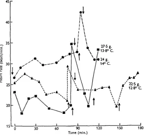
rate tended to be quite low and the occasional visit to the surface was invariably followed by an increase in rate (Fig. 4).
Several factors may be involved, together or separately, in the appearance of the bradycardia. Submergence may give rise to changes in the general metabolism due to shortage of oxygen and perhaps excess carbon dioxide, as well as to the more direct stimuli produced by the water surrounding the body or the cessation of rhythmic
50
£ 30 E
1?
2 e
s
20
10
10 20 30 •W SO
Time (mln.)
60 70 80 90
Fig. 3. The effect of submersion on the heart rate of five frogs. The downward-pointing arrow indicates submersion, the upward ones emersion.
Factors influencing submergence and heart rate in the frog
« r
423
90 Time (mln.)
[image:7.451.80.375.74.351.2]Fig. 4. The heart rate of three frogs moving freely in the experimental tank. Selected sections from recordings taken over longer periods of time. Arrows indicate submersion and emersion as in Fig. 3.
Table 1. The reduction of heart rate produced by total immersion, prevention of buccal
movements in air, and prevention of buccal movements toith the trunk in water
Temperature 13—15° C. The heart rate at the beginning of each experiment and the lowest rate achieved after treatment are given. The lowest rate is also expressed as a percentage of the initial rate in air.
Buccal cavity clamped ind Frog 1
Frog 2 Frog 3 Frog 4
Total immersion 42-25(60%) 40-25 (63 %) 45-24 (55 %) 30-25(64%)
Floor of buccal cavity clamped 41-35 (85 %) 42-36(86%) 43-35 (81 %) 43-35 (81 %)
body immersed 44-30(68%) 45-26(58%) 45-27(60%)
until the clamp was removed. Submersion is thus not an indispensable factor, though it may be a contributory one, in the production of bradycardia.
When the animal was submerged in water whose oxygen content had been lowered below the air-saturation level it was found that the bradycardia became more pro-nounced. The critical level for producing an effect greater than that seen in air-saturated water was about 5 mg. oxygen per litre and the greatest reduction in rate occurred when the water was devoid of oxygen (Fig. 5). A curious feature of these experiments was that after submergence in water containing oxygen below this critical amount there appeared to be some acclimation on the part of the frog. Subsequent immersions in water containing oxygen at air saturation did not cause the same reduc-tions in heart rate as were produced before the treatment in water depleted of oxygen (Fig. 5). Repeated immersion in water saturated with air never produced any evi-dence of this type of acclimation.
50
£ 40
s c e
d
2 20
104
r\
i
60 120 180
Time (mln.)
240 300 360
Fig. 5- Bradycardia produced by submersion of a frog (36 g.) in water of varying oxygen content. 170 C. Arrows indicate submersion and emersion as in Fig. 3. The oxygen content of the water is given in mg. per litre in the relevant sections of the diagram.
The effect of low concentrations of carbon dioxide on the heart rate was negligible, and not until gas mixtures containing more than 2-3 % carbon dioxide were used, and the concentration in water reached approximately 50 mg./l., was there any noticeable enhancement of the normal bradycardia (Fig. 6). Extremely high concentrations around 200 mg. carbon dioxide per litre had a very marked depressant effect on heart rate, and recovery from this treatment was slow because breathing movements were not immediately resumed when the animal was brought to the surface. When regular breathing eventually developed there was a pronounced increase in heart rate.
Factors influencing submergence and heart rate in the frog 425
as 0-3%. The oxygen content of the gas was continuously monitored by means of a polarographic oxygen electrode and was never found to exceed this level, which was assumed to be insignificant in this type of experiment.
50
I 2 nig. J I 136 mg.t I 2 mg. J I 84 mg. j I 2 mg. j I 39 mg.
40
30
20
10 4 60 120J _
Time (mln.)
180 240
Fig. 6. Bradycardia produced by submersion of a frog (29-5 g.) in water of varying carbon dioxide content. 150 C. Arrows indicate submersion and emersion as in Fig. 3. The carbon dioxide content of the water is given in mg. per litre in the relevant sections of the diagram.
usual alternation of pulmonary and buccal ventilation did not develop. Instead the movements were grouped into periods, 10-15 sec. in length, when the lungs were ventilated, sometimes alternating with periods of entirely buccal ventilation. Lung-filling movements continued throughout the period at the surface but the buccal respirations, when present, decreased in amplitude and stopped completely so that long respiratory pauses (up to 60 sec.) separated the groups of pulmonary movements (Fig. ye).
i—1 1—1—i »—1 •—*
A M M / \ A M M M A ( W W M M ^
_L
Time—5 sec. Intervals
Fig. 7. The effect of submersion and emersion into air and nitrogen on the heart rate and breathing movements of a frog. 180 C. Upper trace: e.c.g. Lower trace: breathing movements (down on trace = expansion of the buccal cavity), (a) In air: heart rate 62/min.; Breathing rate, total 102/min., lungs 45/min. (6) 15 min. after submersion: heart rate ao/min. (c) 5 min. after emersion into air: heart rate 56/min.; breathing rate, total 138/min. lungs 93/min. (d) 20 min. after submersion, showing emersion into nitrogen: heart rate 18/min. up to 37's/min. (e) After 15 min. in nitrogen: heart rate 24/min.; breathing rate, total 85/min. all lung, breathing bursts 50 sec. apart. (/) After 40 min. in air: heart rate 61/min.; breathing rate, total 144/min., lungs 73/min.
Water
ft
Watert
Nitrogen Watert
Air• « r
^ 3 0
20 _ L - i - J_ _ L
10 20 30 40 50 60 70 80_L J _ _L Time (mln.)
J _ _L _L J
[image:11.451.58.413.47.309.2]90 100 110 120 130
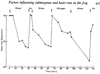
Fig. 8. The effect of emersion into air and nitrogen on the heart rate of a frog (25 g.). 130 C. Arrows indicate submersion and emersion as in Fig. 3.
I
60
50
30
1 1 I I L
10 20 30 40 50 60 0 10
Time (mln.)
I I I I L I 1 1 20 30 40 50 60 70 Fig. 9. Bradycardia produced by submersion of a frog before (a) and after (6) bilateral
vagotomy. 170 C. Arrows indicate submersion and emersion as in Fig, 3.
(c) The effect of vagotomy on diving bradycardia
[image:11.451.51.404.350.533.2]428 D. R. JONES AND G. SHELTON
anaesthetized frog, was often found to be at the same level or even lower than before vagotomy. The significance of this observation may be questionable since the anaes-thetic used is known to have an accelerating effect on fish hearts (Shelton & Randall, 1962) and will probably have some effect here. When the animal was submerged the decline in heart rate was seen as before (Fig. 96). A remarkable degree of agreement was obtained in four out of ten experiments between the final rate achieved after 30 min. submergence in both the intact and vagotomized animals. The speed at which the bradycardia was developed varied slightly and in most cases no consistent changes were seen which could be attributed with certainty to the nerve section. The recovery processes tended to occur more slowly than in the intact animal as Fig. 9 shows, though not always so clearly as seen in this figure. In one experiment evidence was obtained that vagal Inhibition was playing a part in the development of diving bradycardia. The heart rate fell rapidly on submergence, the frequency fluctuated considerably, and the beat became arrhythmic after 5 min. When the vagi were sectioned in this animal bradycardia was produced on submersion but over a much longer time course and the beat remained rhythmic for the full 30 min. period. Clearly some individual variation exists in the way in which the heart rate is decreased when the frog submerges. These results suggest that a considerable part of the decrease is independent of vagal inhibition but that this nerve may sometimes play a part in both the development and cessation of bradycardia.
DISCUSSION
Factors influencing submergence and heart rate in the frog 429
oxygen must be conserved from the outset and sent to organs such as the brain where oxygen shortage would be most damaging. In these animals a rapid curtailment of blood flow through the majority of organs is required and, in fact, the heart rate falls quickly as the transfer from one type of circulation to the other is reflexly produced. Oxygen concentration has been demonstrated to be a very important factor in determining both the ability of the animal to remain submerged and the extent to which the heart rate is reduced when submerged. At concentrations below 5 mg. of oxygen per litre the effect on both of these is very noticeable. Carbon dioxide has also been shown to have its effect both on bradycardia and on the ability to remain sub-merged, but at concentrations which are so high as to be unlikely in nature. Krogh (1904), and many other authors, have suggested that this gas is eliminated very rapidly through the skin in Amphibia and so accumulation in the body is unlikely unless the animal finds itself in environments containing very high levels.
Oxygen concentration is not the only important factor having an effect on heart rate, however. The experiments in which frogs were brought up into nitrogen show quite clearly that the breathing movements themselves are in some way able to influence heart rate. The release of rhythmic breathing can, without changing the animal's oxygen supply, briefly terminate a bradycardia produced by submersion. The converse appears to be true only to a slight extent. The sudden stoppage of rhythmic breathing has an immediate effect on heart rate but this is usually small compared with the slow, steady decline which follows.
An explanation of the results described in this paper can be given in terms of the controlling influence of oxygen concentration and the connexion between breathing movements and heart rate. Submersion slows the heart because the breathing move-ments cease and the oxygen exchange is curtailed. Total submersion is more effective in the production of bradycardia than is stopping the breathing movements with a clamp or by immersion of the head alone because it gives a more complete anoxia, diffusion of oxygen through water being slower than in air. Emersion of the frog releases the bradycardia immediately because the onset of breathing has a stimulating effect on the heart and there is also, under normal circumstances, a recovery from anoxia. Other factors concerned in control have clearly not been eliminated completely but for the present there is no need to postulate more than the two above.
fall in frequency. At the moment therefore it is difficult to decide how the response to oxygen lack is mediated. What does seem certain is that the influence of the breathing movements on heart rate must be an effect mediated by the nervous system. It is difficult to imagine a mechanism other than a nervous one which would be effective. The experiments on vagotomized animals show that, in some cases, sudden changes in heart rate seen before vagotomy cannot be produced after the operation. In addition, preliminary experiments using the adrenaline antagonist, yohimbine hydrochloride, indicate that the sympathetic nervous system may be involved in the quick recovery of heart rate when breathing begins after submersion.
SUMMARY
1. The ability of the frog to remain submerged declines as the oxygen concentra-tion in the water falls or the carbon dioxide content rises. The critical oxygen con-centration appears to be about 5 mg./l. and the critical carbon dioxide concon-centration 100 mg./l. at temperatures around io° C.
2. Submergence results in a decrease in heart rate which develops over a period of 15-30 min. but which disappears immediately the animal surfaces and breathes. The bradycardia is accentuated by oxygen lack or carbon dioxide excess.
3. During submergence the heart is influenced by two main factors, the shortage of oxygen and the cessation of breathing movements, both of which contribute to the decrease in rate. The former can still affect rate after vagotomy. The connexion between breathing and heart rate is dependent on the nervous system, though the detailed pathway is not worked out.
D. R. J. wishes to thank the Department of Scientific and Industrial Research for financial support during the period of this research.
REFERENCES
ANDERSEN, H. T . (1961). Physiological adjustments to prolonged diving in the American alligator.
Acta pkysiol. scand. 53, 23-45.
ANDERSEN, H. T. (1963). Factors determining the circulatory adjustments to diving, I and II. Acta pkysiol. scand. 58, 173—200.
CLARK, A. J., EGCLETON, M. G., EGGLETON, P., GADDIE, R. & STEWART, C. P. (1938). The Metabolism
of the Frog's Heart. Edinburgh: Oliver and Boyd.
DOLK.H. E. &POSTMA,N. (1927). Uber die Hautund die Lungenatmung von Rana temporaria. Z.vergl.
Pkysiol. 5,
417-44-IRVING, L., SCHOLANDER, P. F. & GRINNELL, S. W. (1941). Significance of the heart rate to the diving ability of seals. J. Cell. Comp. Pkysiol. 18, 283-97.
JOHANSHN, K. (1959). Heart activity during experimental diving of snakes. Amer. J. Pkysiol. 197, 604-6.
KROGH, A. (1904). On the cutaneous and pulmonary respiration of the frog. Skand. Arch. Pkysiol. 15, 328-419.
KROGH, A. (1941). Comparative Physiology of Respiratory Mechanisms. Philadelphia: University of Pennsylvania Press.
LEIVESTAD, H. (i960). The effect of prolonged submersion on the metabolism and the heart rate in the toad. Arbok Univ. Bergen. (Mat.-nat. serie), 5, 1—15.
DB MARNEFFE-FOULON, C. (1962). Contribution a l'etude du mecanisme et du controle des mouvements respiratoires chez Rana. Ann. Soc. zool. Belg. 93, 81-132.
POCZOPKO, P. (i960). Changes in blood circulation in Rana esculenta L. while diving. Zool. Polon. 10,
Factors influencing submergence and heart rate in the frog 431
SCHOLANDER, P. F . , IRVING, L . & GRINNELL, S. W. (1942). On the temperature and metabolism of the seal during diving. J. CM. Comp. Phytiol. 19, 67-78.
SCHOLANDER, P . F . , HAMMEL, H . T . , LEMESSURIKR, D . H . , HEMMINOSEN, E. & GAREY, W . (1962). Circulatory adjustments in pearl divers. J. Appl. Physiol. 17, 184-90.
SHELTON, G. & RANDALL, D . J. (1962). T h e relationship between heart beat and respiration in teleost fish. Comp. Biochem. Pkytiol. 7, 237-50.
SMYTH, D . M . (1939). T h e central and reflex control of respiration in the frog. J. Pkytiol. 95, 305—27. SPURWAY, H . & HALDANE, J. B. S. (1953). Breathing in newts with a general survey. Behaviour, 6,