ON THE STRUCTURE OF THE CELLS BEARING THE
VELAR CILIA OF THE NUDIBRANCH VELIGER
BY G. S. CARTER.
{Received istkjuly, 1928.)
(With Seven Text-figures.)
THE CELLS BEARING THE VELAR CILIA OF THE NUDIBRANCH VELIGER. THE object of the present paper is to extend the previous observations of the author (3) on the nervous control of the cilia of the Molluscan velum and to provide a description of the cytoplasmic structure of the cells which bear these cilia. It is hoped that a revision of this subject, based as far as possible on observations made on the living cell rather than on fixed material, may be of use in the study of the mechanism of ciliary activity from both the chemical and structural points of view. In order to increase the definiteness of the observations, they were restricted to one type of veliger. That of Aeolidia papillosa was chosen on account of the ease with which material of this species could be obtained. The veligers were studied at the stage at which they are still contained within the egg-capsule but can be seen actively moving within it. These movements may be observed for several days before the larvae escape, and during this period the structure of the velum alters very little, except by growth. By the time that the movements of the cilia are active, the cells of the velum are fully developed and the beat of the cilia has the form seen in the free-swimming veliger. The intermissions of the beat, described in the previous paper, already occur and the nerve-supply of the velum is complete. This stage, therefore, gives a convenient point at which the veligers can be readily obtained in bulk and the cells studied as fully effective organs.
Various methods were used in the investigation of these cells. Most of the internal structure can be made out in the living cell by the use of vital stains and several of these were used. The cells were studied both in their natural position on the velum and free in the water after the veliger had been crushed. Such cells may often be seen to move through the water, driven by the beat of their cilia, which continues for some time. The ciliary mechanism has therefore not been damaged by this treatment.
These observations were supported by study of the cells in fixed material, and for this purpose as many different methods as possible were used. For the study of the general structure of the cells, formalin, Flemming's chrom-osmic mixture with and without acetic acid and Carnoy's fluid were used as fixatives, and as stains haematoxylin and Mann's methyl blue-eosin mixture: for that of the chromatin,
G. S. CARTER
Feulgen's nucleoprotein stain (fuchsin-sulphuric acid)1: for that of the Golgi apparatus and the mitochondria, Golgi's rapid bichromate-silver nitrate method, da Fano's silver impregnation, and the Mann-Kopsch osmic acid method: and for that of the lipoids of the cell, the method of Marchi was used in addition. Finally for the demonstration of the nerve-endings, several of these methods and Lowit's gold-impregnation produced useful results. Iodine was used as a test for glycogen and starch in the fresh cell and in fixed material.
OBSERVATIONS.
In the general arrangement of the structure of the cell, in the presence on each cell of three cilia and in the resolution of the cilium into a series of triangular plates, the ciliated cells of these veligers agree precisely with those described in the previous papers (2,3). No further account of these points will be given here. In certain minor respects the cytoplasmic structure is different from that previously described and these will be mentioned.
(1) T H E CYTOPLASMIC CILIARY APPARATUS.
(a) Fresh Material.
The appearance of the unstained cell in side and end view is shown in Fig. 1. By far the clearest view of the ciliary apparatus within the cell is given by the use of iodine as a stain. On treatment with this substance the whole series of granules below the cilium appear black. The structure thus displayed is shown in Fig. 2. Diagrams of the appearance of the cell when it has been stained with various vital dyes are given in Fig. 3.
It will be seen (Figs. 2 and 3 d) that below each cilium there occurs a double • row of granules (s.g.), exactly on the surface of the cell. These lie distal to the true
basal granules (b.g.), which are about 1 JJL from the surface. These two sets of granules are connected by rod-shaped bodies (the "stabchen" of German authors), which are also stained black by iodine. The basal granules are not round but are extended somewhat towards the interior of the cell. The granules on the surface of the cell are stained by none of the vital dyes except thionin but the basal granules are stained by several of these. A structure of this type has frequently been des-cribed in ciliated cells from fixed material (e.g. Frenzel(6), in Molluscs).
Below the basal granules is an area of modified protoplasm which can be seen in cells stained with iodine and several of the vital dyes (Figs. 2, 3 a, d,f) to be striated in a direction parallel to the axis of the cilium. Being clearly a part of the ciliary apparatus, this area will be called the "ciliary protoplasm" (c.p.). It is in this part of the cell that the so-called internal fib res of the cilium have often been described from fixed material. These are said to be fibres passing from the basal granules into the cytoplasm of the cell. Appearances of fibres in this position are
1
common in fixed material of these ciliated cells as of others, especially in material fixed by Flemming's fluid or by the Mann-Kopsch method (Fig. 5), but the im-pression given by the living cell is that the striations represent divisions between columnar elements of the protoplasm rather than anything of the nature of true fibres. The appearance of the striations in the living cell is certainly not that of fibres. It is not difficult to imagine either that columnar elements of the kind sug-gested might shrink on fixation and appear as fibres in fixed material or that the protoplasm between these elements might stain more darkly than the elements themselves and equally give the appearance of fibres. Since the internal fibres are said to end upon the basal granules the former is the more probable explanation
Fig. 1. The ciliated cell in side and end view, c.p., ciliary protoplasm;
f.v., reserve food vacuoles; n.r.v.,
[image:3.641.336.582.424.579.2]"neutral red" vacuoles; n.,position of nucleus.
Fig. 2. Ciliary apparatus stained with iodine. The cell is killed by the treatment but the pro-toplasm not coagulated, b.g., basal granules;
c.p., ciliary protoplasm; m.b.g., methylene blue
granules; s.g., surface granules; St., rods between surface and basal granules ("stabchen"); str., striations of the ciliated protoplasm.
of their origin. For, although owing to the small scale of the structures it is not possible to determine definitely whether the striations seen in the living cell end directly upon the basal granules or between them, it appeared more probable that the latter was true (Fig. 2, where they are shown thus). That the "internal fibres" are artefacts is the opinion of many recent workers on the ciliated cell (e.g. Kolacevdz), Saguchi(i6>).
In the living cell there is no indication of any cone-shaped arrangement of the cytoplasmic elements in this part of the cell, an arrangement which has often been shown in figures derived from the study of fixed material of ciliated cells. In some cases the striations appear to become closer together towards the interior of the cell (Figs. 2, 3 6, tf) but this is never so pronounced as to make a cone-shaped arrangement possible within the cell. On the other hand when fibre-like structures are seen in fixed material of these cells, they sometimes appear to form a complete
100 G. S. CARTER
cone, exactly in the manner that has been described in other ciliated cells. There can be little doubt therefore that at least in these cells both the "internal fibres" and the cone-shaped arrangement are artefacts due to the fixation of the cell.
When stained with certain of the vital stains (thionin and dahlia) (Fig. 3 d,f), this part of the protoplasm appears to be granular. This appearance is not seen in fixed material.
a, Methylene Blue, "b. Neutral Red.
°~°c
(i!«•
•is • if3
-X
^ _ ^
UUI
c. Sudan III. Rile Bluer
d. Thionin.
UUi
8cf
o .
8s
°
Mr
ii
e. Janus Green. f. D a h l i a .
IPS
S
««
Fig. 3. The ciliated cells in side and end view, stained with various vital stains.
(b) Fixed Material.
In material fixed in Carnoy's fluid or in formalin, both the basal granules and the methylene blue granules are preserved but it is very difficult to differentiate the surface granules from the cuticle of the cell and the "stabchen" are very rarely visible. Both the inner layers of granules stain with haematoxylin and with Mann's stain. When stained with the latter they take on the same colouration as the cyto-plasm but in a more intensified form. None of these structures stain in material prepared by Feulgen's method. There can be no doubt therefore that none of them contain nucleoproteins.
When stained with osmic acid the two inner layers of granules always give a lipoid reaction. Thus both layers are blackened in Flemming's fluid and both appear very black in Mann-Kopsch preparations. It is noticeable that the colour which they take up in the latter preparations is very resistant to the action of turpentine and much more so than that of the other lipoids of the cell. When treated with osmic acid after bichromate (Marchi's method) none of these granules are stained. The lipoids in them must therefore be other than true fats. This is confirmed by the fact that they do not stain with Sudan III or Nile blue. When the cell was examined with the polariscope, no double refraction could be seen in them but this may have been due to their small size. Neither the surface granules or the "stabchen" are to be seen in these preparations.
The basal granules and the methylene blue granules are readily impregnated with silver. In preparations made by the Golgi method they may be seen, but faintly, and they are easily visible as black objects in material prepared by da Fano's method, being the only structures in the cytoplasm of the cell which are blackened in these preparations apart from the Golgi apparatus.
The iodine reaction in the basal and methylene blue granules may be obtained in material fixed in Carnoy's fluid. Neither in fixed or fresh material does the black colour given by the iodine disappear when the tissue is heated. Nor did treatment of the tissue with diastase have any effect upon the reaction. It is therefore clear that the colour does not indicate the presence of starch in the granules but it may not improbably be due to the presence of some related carbohydrate substance.
From these observations it must be concluded that these two layers of granules of the ciliary apparatus are similar in general chemical constitution but the dif-ferences in their reactions to some of the vital stains show that they are not identical: that they contain lipoid material but not true fats: and that possibly carbohydrates are also present in them. It is not so easy to form any definite conclusion of the constitution of the surface granules and the "stabchen" but their reaction with iodine in the living cell indicates that they may also contain carbohydrate.
io2 G. S. CARTER
(2) THE VACUOLES OF THE CYTOPLASM.
It is shown in Figs. 1 and 3 that two types of vacuole occur in the cell. Those of the smaller type are only found in the distal part of the cell and on the side of the ciliary apparatus towards the centre of the disc of the velum, i.e. on the side away from that towards which the effective beat is directed. These vacuoles are very evident when the cell is stained with neutral red, taking on a bright red colour. They are here called the "neutral red vacuoles" for this reason. Their contents are shown to be acid by this reaction. They are also stained by Sudan III and Nile blue and must therefore be considered to contain true fats. Their colour when stained with Nile blue is distinctly blue and the fat must therefore be in the form of fatty acid.
Throughout the rest of the cytoplasm, with the exception of a small area in the distal part of the cell and on the opposite side of the ciliary apparatus to that of the neutral red vacuoles, are large vacuoles which stain only lightly or not at all in the vital stains. A few of these near the position of the neutral red vacuoles form an exception to this statement in that they sometimes take on a red colour when the cell is stained with neutral red.
Both types of these vacuoles give lipoid reactions in fixed material. They are blackened in Mann-Kopsch preparations and lose their stain readily in turpentine. They are also black in material fixed with Flemming's fluid either with or without acetic acid. They are blackened with osmic acid after the action of bichromate. No double refraction could be observed in them with the polariscope.
It must therefore be concluded that the neutral red vacuoles contain fatly acids and that the other vacuoles contain lipoids similar to these but not identical with them, since they do not stain in Sudan III and Nile blue. In all probability they also contain many other constituents.
(3) MITOCHONDRIA.
When the cell is stained with Janus green or dahlia, small curved rod-shaped bodies may be seen lying close to the large lipoid vacuoles towards the base of the cell (Fig. 3 e,f). These bodies are stained black in Mann-Kopsch preparations and can easily be recognised when the colour has been extracted from the vacuoles with turpentine. They are also stained black in formalin-iron haematoxylin preparations. They are not blackened in da Fano preparations. It is therefore concluded that they are the mitochondria of the cell.
mito-chondria with the vacuoles is of interest in view of the many theories which have been put forward that the mitochondria are in some way connected with the chemical activity of the cell.
(4) GOLGI APPARATUS.
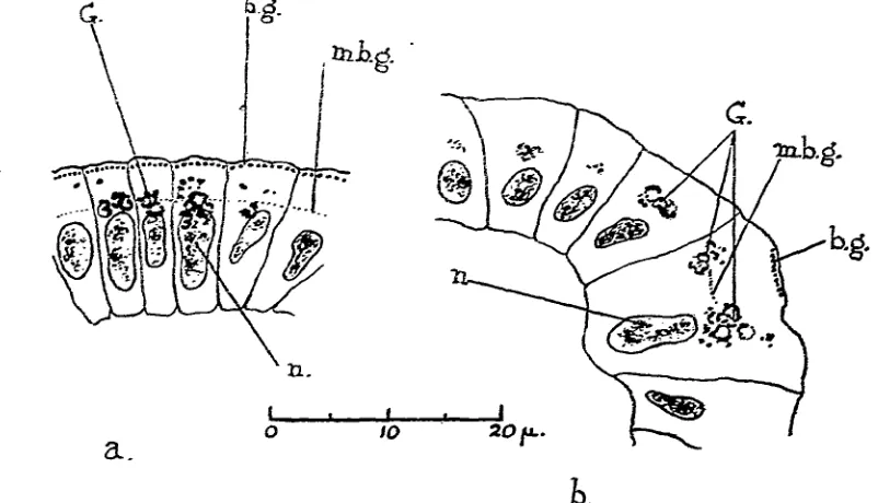
In da Fano preparations the Golgi apparatus may be seen lying just distal to the nucleus at the level of the base of the ciliary protoplasm (Fig. 4 a, b). It is more usually confined to the side of the cell towards the centre of the disc of the velum, where the space of the cytoplasm is larger than on the other side of the cell. Some-times it is to be seen on both sides of the base of the ciliary protoplasm but it never extends into the area of the ciliary apparatus. It presents the same appearance in Mann-Kopsch preparations and in them has the typical reactions of the apparatus
a.
20 f. [image:7.641.112.513.378.608.2]h.
Fig. 4. Da Fano preparations showing sections of the ciliated cells. 3/x.
(a) In a plane perpendicular to that of the beat of the cilium and
tangential to the disc of the velum, (b) In a plane parallel to that of the beat of the tilium: the centre of the velar disc towards the lower side of the figure. b.g., basal granules; m.b.g., methyleneblue granules;
G., Golgi apparatus; n., nucleus.
in being more resistant to the action of turpentine than the mitochondria or the lipoid vacuoles. It is, however, no more resistant than the granules of the ciliary apparatus. The position of the Golgi apparatus agrees with that observed by Rau and Ludfordd4) in the ciliated cells of vertebrates and by Hirschler(n) in the flagellated epithelium of sponges.
(5) CHROMATIN AND NUCLEUS.
104 G. S. CARTER
(6) NERVE-SUPPLY.
Nerve-fibrils passing from the cerebral ganglia to the ciliated cells and ending between them were described in the previous paper (3). Further investigation has shown that intracellular fibrils arise from these where they lie in the spaces between the ciliated cells, and pass into the interior of the ciliated cells themselves. These intracellular fibrils end in the neighbourhood of the basal granules (Figs. 5,6, i.n.t.). They may be seen somewhat faintly blackened in Mann-Kopsch (Fig. 4) and da Fano preparations, and still less clearly in preparations made by the Golgi method. They are displayed most clearly by Lowit's gold chloride impregnation (Fig. 6). They may be seen in fresh cells stained with thionin.
Fig. 5. Section of the ciliated cells in a plane tangential to the velum, (as in Fig. 3<z). Mann-Kopsch method. 6/x. b.g., basal granules; c.p., ciliary protoplasm; G., Golgi apparatus; m.b.g., methylene blue granules; n., nucleus; n.f., nerve fibrils below the cells; n.t.y intracellular nerve terminations.
Fig. 6. Section fof the ciliated cell in a plane parallel to the beat of the cilium (as in Fig. 36). Lowit's method. 6/x.. £.w.£.,Eintracellular nerve ter-minations; n., nucleus; n'., nuclei of neighbouring ceils; n.f'., intercellular nerve fibrils.
In the end view of the cell, these fibrils lie over the fibre-like structures which are often to be seen in the ciliary protoplasm in fixed preparations ("internal fibres ") and may easily be confused with them. Their level in the sections is, how-ever, different from that of the ciliary protoplasm, to which they are completely external, and they can therefore be clearly distinguished from the structures within it. The arrangement of all the structures in this part of the cell will be clear from a comparison of Figs. 5 and 6 or by reference to the diagrams of Fig. 7.
In one respect the nerve supply of these cells differs from that described in the previous paper. There thickened areas on the intercellular fibrils on the level of the outer part of the nucleus were described (Aeolis drummondi—Facelina curta). These would apparently occupy the points at which the intracellular fibrils branch from the fibrils between the cells. They were not observed in the veligers of Aeolidia
papillosa.
DISCUSSION.
(i) THE CHEMICAL MECHANISM OF CILIARY ACTIVITY.
A theory of the chemical reactions from which the energy which is used in the movement of the cilium is derived has been put forward by Gray (8). His
observa-o 10
c.
Fig. 7. Diagrams of the structure of the ciliated cells in side and end views. a.G., area of the Golgi apparatus; b.g., basal granules; c, cilium; c.p., ciliary protoplasm; f.v., reserve food vacuoles; i.n.t., intracellular nerve terminations; m., mitochondria; m.h.g., methylene blue granules; «./., nerve fibrils below the cells; n.f'., nerve fibrils between the cells; n.r.v., neutral red vacuoles; o.n., outline of the nucleus; s.g., surface granules; t.p., triangular plates of the cilium.
carbo-io6 G. S. CARTER
hydrate. On other evidence he suggested that this protein was probably of the nature of a glycoprotein. He analysed the chemical changes involved in the upkeep of the activity of the cilium into three parts, thus:
(1) A nonoxidative reaction, interference with which alters the rate of the beat. From this reaction an acid active substance is probably derived.
(2) A nonoxidative reaction, in which this latter substance is concerned.
(3) An oxidative reaction, necessary for prolonged activity but not immediately essential for the beat of the cilium.
The parallel between this series of reactions and the reactions which are known to occur in muscle is clear, and was pointed out by Gray.
The observations recorded in this paper are not opposed to such a theory. It is especially upon the latter part of this series of changes that they seem to have some bearing. The breakdown of a glycoprotein and the utilisation of its protein con-stituent for the production of energy would naturally set free the carbohydrate portion in the neighbourhood of the ciliary apparatus. Attempts to demonstrate the presence of carbohydrates in this part of the ciliated cell have been made but have not been successful. Boyland, however (recorded by Gray (9), p. 108), has found that the gills of Pecten contain glycogen in measurable but small quantities. The affinity of the granules of the ciliary apparatus for iodine suggests the possibility that they contain some carbohydrate substance, although, as has been stated above, this substance is certainly not starch.
It is at first sight astonishing, if the theory gives a true account of the changes going on within the ciliated cell, that it should not be possible to demonstrate the presence of carbohydrate in other parts of the cell. On the other hand fats and lipoids have been shown to be very plentiful in these cells, being present both in the granules of the ciliary apparatus and in all the vacuoles of the cytoplasm. That fat is a form in which the reserve food-materials of the animal cell are frequently stored is well known and that it may be transformed into carbohydrate in the body is also true (e.g. Smedley(i7)). It seems probable that in these lipoids we see the material which is used in the form of the carbohydrate portion of the glycoprotein in the ciliary apparatus: that the fats of the vacuoles at the base of the cell are trans-formed into carbohydrate, combined with the protein which must also be present among the reserve food materials of the cell and used in the form of a glycoprotein to give the energy needed for ciliary activity. That these changes probably take place in the ciliary apparatus itself is indicated by the fact that no carbohydrate is to be found elsewhere in the cell. Gray (8) was unable to demonstrate the presence of a glycoprotein in the ciliated cells of the gills of Mytilns except in the immediate neighbourhood of the ciliary apparatus. In the cells described in this paper none could be found in any part of the cell by the use of thionin. These observations do not disprove the momentary formation of such a substance in the cell but they make it improbable that it is present as a permanent reserve food material.
of the cell in this form; and it seems probable that the vacuoles which have been called the "neutral red vacuoles" in this paper play an important part in this process. These vacuoles were shown to contain a fatty acid. Pyruvic acid is known to be formed in the transformation of carbohydrate into fat (Smedleyd7)) and this or a similar acid may be the source of their staining properties. That they play some such part in the metabolism of the cell is supported by the observation that they can be seen to circulate slowly around this portion of the cell. This can be most easily observed in an actively beating cell of which a side view can be obtained, preferably one still attached to the velum but slightly displaced. The vacuoles could not be seen to pass over into the area of the larger vacuoles at the base of the cell but it is clear that a circulation such as that which was observed would make the migration of substances about the cell more easy.
This view of their function is also supported by the fact, mentioned above, that a few of the larger vacuoles of the second type, lying near them, may sometimes be seen to be acid when stained with neutral red. In the rest of the cell the large vacuoles are never so. That some of the vacuoles in this position should be acid is to be expected if material is passed on to them from the strongly acid "neutral red vacuoles."
The transformation of carbohydrate to fat is an oxidative reaction and it is therefore suggested that it may be the third of the series demanded by Gray's theory, a reaction necessary for the continued activity of the cilium but not im-mediately essential for its beat, a reaction, in fact, which plays the same part in the activity of the cilium that the removal of lactic acid does in that of muscle.
(2) T H E FUNCTIONS OF THE VARIOUS STRUCTURES OF THE CILIARY APPARATUS IN THE ACTIVITY OF THE CILIUM.
The arrangement of the ciliary organs which has been described above supports very strongly the conclusion of the previous papers that in these cells the apparatus is complex and that the unit of ciliary activity is not the whole cilium but one of the simpler units of which the cilium is built up. This simpler unit consists of an ex-ternal fibre with the cytoplasmic organs associated with it, namely, a columnar element of the ciliary protoplasm and one of each of the different types of granules. In considering the mechanism of ciliary activity attention must therefore be directed to this unit and not to the whole cilium.
108 G . S. C A R T E R
supporting function; and the chemical differentiation which has been shown to be present in the granules suggests that they have at any rate other functions besides this. It is not clear that more support than is provided by the cuticle of the cell is needed by the beat of a fibre of such small dimensions.
The chemical structure of all the granules of the ciliary apparatus has been shown to be very similar, so far as the observations recorded above determine it. It has been suggested that they are concerned with the latter part of the series of changes underlying ciliary activity. It is very probable that they are also the position where other chemical changes take place, but their arrangement at intervals along the columnar elements of the ciliary protoplasm is of a type which might be ex-pected to occur if they were chiefly concerned in collecting and removing the waste products of ciliary activity. It would seem probable that the ciliary protoplasm itself is the seat of the changes which provide the chemical energy for the cilium.
(3) THE GOLGI APPARATUS AND THE CILIARY STRUCTURES.
The parallel between the staining reactions of the Golgi apparatus and of the granules associated with the ciliary structures, so far as their lipoid constituents are concerned, has been noted and is striking. It clearly indicates some similarity between the lipoids in these various parts of the cell. The known association of the Golgi apparatus with active chemical changes in the ceil (e.g. in secretory cells) is parallel to the connection of these granules with the activity of the cilium.
The position of the Golgi apparatus, surrounding the inner part of the ciliary protoplasm and tying between it and the vacuoles, which clearly contain the reserve food materials of the cell and are therefore the source from which the energy of the ciliary movement is ultimately derived, suggests that this modified area of the cell also has some part to play in the mechanism lying behind the beat of the cilium and probably in the supply of chemical energy to the ciliary apparatus. Whether this is the case and, if so, what that part is, it is at present impossible to say. Such a function would be in line with the commonly accredited functions of the Golgi apparatus in other cells.
(4) THE INTRACELLULAR NERVE-FIBRILS.
The best-known cases in which intracellular nerve-fibrils in ciliated cells have previously been described are those of the epithelium of the alimentary canal of
Anodonta, figured by Apathy d), and of the tentacles of Sipuncdus (Metalnikoffd3)).
Only the latter described connections between these fibrils and the nervous system of the animal.
their ends lay alternate to the basal granules and not directly upon them. If the true structure of the ciliary protoplasm is that which has been described above, parallel columnar elements of protoplasm, it would seem very probable that the deposition of the metal might occur on the surfaces between these elements and give the appearance of fibrils in fixed preparations. The conical arrangement of the fibrils described by Apathy, in view of the conical arrangement which is often seen in the "internal fibres," supports this view. It may also be remarked that the ciliated cells studied by Apathy, those of the epithelium of the alimentary canal of Anodonta, are not of a type in which nervous control might be expected to exist. Whether or not this interpretation of his results is the true one, it is not important for the purposes of this paper to decide. From the description which has been given of the fibrils in the ciliated cells of these veiigers, there can be little doubt that they are not structures of the kind figured by him. That they are in fact nervous is maintained partly on account of their properties in staining by methods which are recognised as specific for nerve-fibrils and partly on the evidence presented in the previous paper.
SUMMARY.
(1) The cytoplasmic structure of the cells bearing the velar cilia of the veliger of Aeolidia papillosa is described, partly from the observation of living cells stained with vital dyes and partly from the study of fixed material.
(2) Intracellular nerve-fibrils passing into these cells and ending in the neighbourhood of the basal granules are described. These lie in the cytoplasm outside the ciliary apparatus and are distinct in nature from those described in ciliated cells by previous authors.
(3) The bearings of these results on the theory of ciliary activity are discussed.
LITERATURE LIST.
(1) APATHY, S. (1897). Mitt. Zool. Stat. Neapel, 12, 495. (2) CARTER, G. S. (1924). Proc. Roy. Soc. London, B. 96, 115. (3) (1926). Brit. Journ. Exp. Biol. 4, 1.
(4) EISMOND, J. (1899). Anat. Anz. 18, Erganzh. 125. (5) ERHARD, H. (1910). Arch. Zellforsch. 4, 309.
(6) FRENZEL, J. (1886). Arch. Mikr. Anat. 28, 53.
(7) GATENBY, J. B. (1921). Eighth edition of The Microtomist's Vade-Mecum. By A. Bolles Lee, Churchill, London.
(8) GRAY, J. (1924). Proc. Roy. Soc. London, B. 96, 95. (9) (1928). Ciliary Movement. Camb. Univ. Press.
(10) GRINFELTT, E. and EUZIERE, J. (1913). C.R. Ass. Anat. 15, 101. (11) HIRSCHLER, J. (1914). Anat. Anz. 47, 289.
(12) KOLACEV, A. (1910). Arch. Mikr. Anat. 76. (13) METALNIKOFF, S. (1900). Z. zviss. Zool. 68, 261.
(14) RAU, A. S. and LUDFORD, R. J. (1925). Quart. Journ. Micr. Sri. 69, 509. (15) ROBERTSON, M. (1927). Parasitology, 19, 375.
(16) SAGUCHI, S. (1917). Journ. Morph. 29, 217.