ORIGINAL RESEARCH
Restored Activation of Primary Motor Area from
Motor Reorganization and Improved Motor
Function after Brain Tumor Resection
N. Shinoura Y. Suzuki R. Yamada T. Kodama M. Takahashi K. Yagi
BACKGROUND AND PURPOSE: Reorganization of brain function may result in preservation of motor function in patients with brain tumors. The goal of the present study was to investigate whether function of the primary motor area (M1) was restored and whether motor function improved after brain tumor resection.
METHODS: Five patients with metastatic brain tumors located within or near M1 underwent awake surgery with intraoperative cortical mapping and continuous task monitoring. Preoperative and post-operative functional MR imaging (fMRI) was performed during hand clenching, and diffusion tensor imaging (DTI) was performed in 1 case to further characterize the area activated in fMRI.
RESULTS: Preoperative fMRI performed during hand clenching demonstrated reorganization of motor function. In patients with severe paresis (cases 3, 4, and 5), clenching of the affected hand induced a large blood oxygen level– dependent response in the right hemisphere, mainly in the anterior temporal lobe, despite the location site of the tumor. Postoperative fMRI during hand clenching demonstrated activation of the contralateral M1. Furthermore, in case 5, DTI detected tracts, possibly the inferior longitudinal fasciculus, arising from anterior temporal activated area as well as tracts connecting the premotor and M1 activated area. This patient demonstrated mirror movement of the hand during the course of motor function recovery.
CONCLUSIONS:Tumor resection resulted in restoration of M1 function and improved motor function in patients with preoperative reorganization of M1 function. Furthermore, the preoperative reorgani-zation of motor function in cases with severe paresis may be related to changes in the right hemisphere, including the temporal lobe.
F
unctional MR imaging (fMRI) studies and intraoperative cortical mapping have demonstrated reorganization of the motor area in response to gradual compression of the primary motor cortex (M1) by a tumor.1-4This reorganization allows for variable preservation of motor function and can occur in one of several patterns, including perilesional extension, shifts from primary to secondary motor areas, or shifts to homolo-gous areas of the unaffected hemisphere.5-11However, the sta-tus of this reorganization after tumor resection is not known. Thus, the goal of the present study was to investigate whether function of the M1 is restored and whether motor function improves after brain tumor resection.Materials and Methods
Patient
The degree of paresis before and after surgery is indicated by using an objective scale as follows: 5⫽normal, contraction against powerful resistance, 5⫺ ⫽between 5 and 4, 4⫽good, contraction against gravity and some resistance, 3⫽fair, contraction against gravity only, 2⫽poor, movement only with gravity eliminated, 1⫽trace, flicker of contraction, and 0⫽complete paralysis.
Case 1.A 57-year-old woman with breast cancer experienced on-set of right hand paresis (scale 5⫺) after developing epilepsy 1 month before surgery. MR imaging showed a heterogeneously enhancing tumor within a left frontal lesion (Fig 1A-1).
Case 2.A 71-year-old man with lung cancer experienced onset of left-sided hemiparesis (scale 5⫺) 1 month before surgery. MR imag-ing demonstrated a heterogeneously enhancimag-ing tumor within a right parietal lesion (Fig 2A-1).
Case 3.A 49-year-old man with colon cancer experienced onset of right-sided hemiparesis (upper extremity [scale 4] affected more than lower extremity [scale 5⫺]) and aphasia 1 month before surgery. MR imaging demonstrated a heterogeneously enhancing tumor within a left frontal lesion (Fig 3A-1).
Case 4.A 71-year-old woman with colon cancer experienced on-set of left-sided hemiparesis (scale 4) 1 month before surgery. MR imaging demonstrated a heterogeneously enhancing tumor within a right frontal lesion (Fig 4A-1).
Case 5.A 76-year-old man with renal cancer experienced onset of left-sided hemiparesis 1 month before surgery (scale 4). Weakness of the left arm progressed after several episodes of partial seizures in-volving the left arm. MR imaging showed a heterogeneously enhanc-ing mass within a right frontal lesion (Fig 5A-1).
Preoperative paresis was relatively severe in cases 3, 4, and 5 com-pared with cases 1 and 2. Informed consent to perform fMRI, diffu-sion tensor imaging (DTI), and awake surgery was obtained from each patient.
fMRI and Image Analysis
The fMRI and image analysis was performed as described previous-ly.12In brief, the fMRI was performed with a 1.5T General Electric
Signa Horizon LX imager (GE Yokokawa Medical System, Tokyo, Japan) with an acquisition sequence of: gradient-echo– echo-planar imaging (EPI) echo time [TE]/repetition time [TR]⫽82.5/3000 ms, 128⫻128 matrix, with a 24⫻24-cm field of view. The reconstructed voxel size was 1.88⫻1.88⫻2.3 mm3. Five-millimeter thickness
sections were obtained with 70 repetitions, and a block design of Received July 18, 2005; accepted after revision October 17.
From the Department of Neurosurgery, Komagome Metropolitan Hospital, Tokyo, Japan (N.S., R.Y., M.T.); and Department of Radiologic Technology, Tokyo Metropolitan University of Health Sciences, Tokyo, Japan (Y.S., T.K., K.Y.).
Address correspondence and reprint requests to: Nobusada Shinoura, Department of Neurosurgery, Komagome Metropolitan Hospital, 3-18-22 Hon-Komagome, Bunkyo-ku, Tokyo 113-8677, Japan; e-mail: shinoura@cick.jp
FUNCTIONAL
ORIGINAL
30-second interval was used. fMRI was per-formed 1 week before surgery and 1 month after surgery, and the patient was asked to perform repetitive closing of the hand to a fist (hand clenching), because functional hand motor recovery is usually complete at this point in patients who had surgery for brain tumor. The fMRI acquisition time was approximately 3.5 minutes. Anatomic im-ages were acquired with the use of a 3D fast-spoiled gradient echo sequence with TE of 2.4 ms, TR of 26.0 ms, flip angle of 30°, bandwidth of 31.25 kHz, image matrix of 256⫻256, and section thickness of 2.3 mm. After reconstruction, the EPI images were aligned to correct for head motion and were Fig 1.Case 1. A 57-year-old woman with metastatic brain tumor located inside the left-sided M1.
A-1, Preoperative MR imaging displays heterogeneously enhancing tumor in the M1.
A-2, Postoperative MR imaging shows ring enhancing lesion, but there was no residual tumor in the M1 accord-ing to the intraoperative observation for tumor cavity.
B, fMRI images activated by right hand clenching (B,
orange) and right elbow flexion (B,blue) before surgery.
C, fMRI images activated by right hand clenching (orange) and right elbow flexion (blue) after surgery. A relatively small area of the contralateral M1 was activated by right hand clenching before surgery, whereas a large area of the contralateral M1 was activated by right hand clenching after surgery.
Fig 2.Case 2. A 71-year-old man with metastatic brain tumor located inside the right-sided S1.
A-1, Preoperative MR imaging displays heterogeneously enhancing tumor in the S1.
A-2, Postoperative MR imaging shows no residual tumor in the S1.
B, fMRI images activated by left hand clenching (B,orange) and right hand clenching (B,blue) before surgery.
C, fMRI images activated by left hand clenching (orange) and right hand clenching (blue) after surgery. Left hand clenching (orange areainB) activated the contralateral M1 and S1 areas separately before surgery and (orange areain
[image:2.585.53.374.38.732.2]co-registered with anatomic images. EPI images were smoothed by using isotropic Gaussian kernels of 4 mm, and statistical analysis was performed by using the SPM program (London University, London, England).13The significance of the activations (Pvalue) was less than
0.05, depending on the degree of paresis of patients. When the degree of paresis was severe, we needed to increasePvalue to detect the activated area. The limitations of fMRI, especially when evaluating individual patients who might be uncomfortable to perform specific tasks because of their handicaps, should be noted.
DTI Image Analysis and Coregistration of fMRI and DTI Images
DTI and image analysis was performed as described previously.12In
brief, standard imaging gradients were used with a maximum strength of 22 mT/m and a slew rate of 77 mT/m/ms. All data were acquired by using a birdcage head coil. The DTI acquisition sequence
was a single-shot, spin-echo EPI, with TE of 98.6 ms, 128⫻128 acquisition matrix, and a 24⫻24-cm field of view.14Acquisitions of
contiguous 5-mm sections were obtained, covering the whole brain, with a b value of 1000 mm2/s in 42 noncollinear directions. The
re-constructed voxel size was 1.88⫻1.88⫻4.00 mm3. The DTI
acqui-sition time for 61 images was approximately 10 minutes. The diffu-sion tensor eigenvalues (1,2,3) and eigenvectors (1,2,3) were
calculated from DTI data, and fractional anisotropy (FA) maps were generated according to the Tensorlines algorithm,15,16which was a
combination of the tensor deflection algorithm at low FA and Stream-lines tracking at high FA,17-19with the use of DTI Analyzer (IDL v. 5.6;
Research System, Boulder, Colo). Stopping criteria (threshold 0.1) were used for analysis.
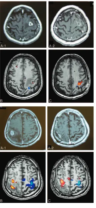
Using the highest significantly activated fMRI voxel in each task, coregistration of fMRI and DTI images were used to select the seed points on the FA maps within the white matter adjacent to the Fig 3.Case 3. A 49-year-old man with metastatic brain tumor located inside the left-sided premotor area.
A-1, Preoperative MR imaging displays heterogeneously enhancing tumor in the premotor area.
A-2, Postoperative MR imaging shows no residual tumor in the premotor area.
B, fMRI images activated by right hand clenching (B-1,B-3;orange) and left hand clenching (B-2,B-4; blue) before surgery,
[image:3.585.56.532.39.459.2]activated cortex during fMRI. For the tractography of pathways, the following (regions of interest) ROIs were selected in case 5: the right premotor and M1, and the right anterior temporal lobe.
Mapping and Awake Tumor Resection
Mapping and awake tumor resection was performed as described pre-viously.20In brief, the patient was positioned in the supine position
and given rigid head fixation (Sugita Headrest; Mizuho Medical Co, Tokyo, Japan) after administration of a local anesthetic agent at the pin sites and regional field block sites. Under general anesthesia with fentanyl and propofol by laryngeal mask airway, the skin was infil-trated with the same local anesthetic agent and incised, and a crani-otomy and incision of the dura was performed.
The laryngeal airway was then removed and oxygen was adminis-tered via a nasal cannula. Cortical mapping was performed by stimu-lating the cortex with the modified Ojemann stimulator.21,22To avoid
an intraoperative seizure, a low-stimulus setting (3–5 mA, 60-Hz bi-phasic square wave pulse of 1 ms/phase for 4 seconds’ duration) was used. Electrocorticography was performed to monitor for after-dis-charges during stimulations. The patients were observed by the
neu-rosurgeon, and movements of hand clenching were continuously monitored and reported to the operator.
Corticotomy was performed, avoiding the sites identified as elo-quent cortex by cortical mapping, and the tumor was removed in the usual fashion. Continuous adequate task judging from the fMRI and DTI images was performed during the removal of tumor near the eloquent motor area responsible for the movement of hand. The pa-tient was performing adequately tasks such as clenching the affected hand, bending the affected elbow, raising the affected arm, or bending the affected knee during the removal of the tumor that was located near the eloquent hand motor area as determined by fMRI and DTI. The fMRI and DTI data combined with a neuronavigation system was available to the neurosurgeon in the operating room, as described before.20 We have attempted to correlate preoperative fMRI and
brain mapping data by using a neuronavigation system. To prevent deterioration of motor function, continuous task under awake anes-thesia seems more useful in comparison with electrophysiologic monitoring of the motor strip during general anesthesia, because we could detect subtle deterioration of individual motor function by continuous task.20After completion of the tumor resection, the pa-Fig 4.Case 4. A 71-year-old woman with metastatic brain tumor located inside the right-sided premotor area.
A-1, Preoperative MR imaging displays heterogeneously enhancing tumor in the premotor area.
A-2,Postoperative MR imaging shows no residual tumor in the premotor area.
B, fMRI images activated by right hand clenching (blue) and left hand clenching (red) before surgery,
[image:4.585.54.535.41.382.2]tient was reintubated with a laryngeal airway, and general anesthesia with fentanyl and propofol was administered. After closure of dura, the bone flap was replaced, and the skin was closed in the usual manner.
Results
Cortical Mapping during Surgery and Postoperative Course
Case 1.Cortical stimulation mapping detected the hand
area of the M1 just beside the tumor. Corticotomy was per-formed in the caudal M1 area just beside the tumor. There was hemorrhage in the region of the tumor, and the right hand motor function worsened during tumor resection but im-proved by the end of awake surgery. It completely imim-proved a week after surgery (scale 5). Postoperative MR imaging dem-onstrated complete removal of the tumor (Fig 1A-2).
Case 2.Cortical stimulation mapping detected M1 rostral
to the tumor. Corticotomy was performed in the caudal pri-mary sensory area (S1) just beside the tumor. The patient did not develop new motor deficits at the end of awake surgery, and left-sided motor function improved by 1 week after sur-gery (scale 5). Postoperative MR imaging demonstrated com-plete removal of the tumor (Fig 2A-2).
Case 3.Cortical stimulation mapping detected M1 caudal
to the tumor. Corticotomy was performed in the rostral premo-tor area just beside the tumor. This patient did not develop new motor deficits at the end of awake surgery, and the right hemipa-resis and aphasia that was present preoperatively improved within a week after surgery (scale 5). Postoperative MR imaging demonstrated complete removal of the tumor (Fig 3A-2).
Case 4.Cortical stimulation mapping detected the M1
cau-dal to the tumor. Corticotomy was performed in the rostral Fig 5.Case 5. 76-year-old man with metastatic brain tumor located inside the right-sided M1.
A-1, Preoperative MR imaging displays heterogeneously enhancing tumor in the M1.
A-2, Postoperative MR imaging shows no residual tumor in the M1.
B, fMRI images activated by left hand clenching (orange) before surgery. fMRI by right hand clenching was not performed before surgery.
[image:5.585.55.536.39.439.2]premotor area just beside the tumor. The patient did not de-velop new motor deficits at the end of awake surgery. The left hemiparesis that was present preoperatively improved within a week after surgery (scale 5). Postoperative MR imaging dem-onstrated complete removal of the tumor (Fig 4A-2).
Case 5.Cortical stimulation mapping identified the hand
and arm area in the cortex just rostral to the M1, which was identified by fMRI and by intraoperative and postoperative neurologic observation, because the manipulation of the tu-mor located in M1 easily and frequently induced deterioration of paresis, which was observed by intraoperative and postop-erative neurologic examination. Corticotomy was performed in the motor area facing the central sulcus just beside the tu-mor. The patient experienced frequent epileptiform phenom-ena during tumor removal and experienced agnosia of the right arm just after opening the dura. The patient demon-strated deficits in hand and arm muscle strength postopera-tively, but these deficits resolved by postoperative day 14 (scale 5⫺). Postoperative MR imaging demonstrated complete re-moval of the tumor (Fig 5A-2). Postoperatively, the patient evinced mirror movement when trying to perform unilateral hand clenching on either side.
Preoperative and Postoperative fMRI
Case 1.Left hand clenching (Fig 1B,orange area) activated
a relatively small area of the contralateral M1 before surgery but activated a large area of the contralateral M1 after surgery (Fig 1C,orange area).
Case 2.Right hand clenching (Fig 2B,orange area)
acti-vated the contralateral M1 and S1 areas separately before sur-gery but activated a large portion of the contralateral M1 and S1 and the contralateral supplementary motor area after sur-gery (Fig 2C,orange area).
Case 3.Right hand clenching (Fig 3B-3,orange area)
acti-vated areas in both the ipsilateral and contralateral M1 and in the ipsilateral anterior temporal lobe (Fig 3B-1,orange area) before surgery. Further, left hand clenching (Fig 3B-4,orange area) only activated the contralateral M1 before surgery. After surgery, right hand clenching (Fig 3C-2,orange area) or left hand clenching (Fig 3C-2,blue area) produced activation only in the contralateral M1. The activated area in the anterior tem-poral lobe by right hand clenching before surgery was not activated by right hand clenching after surgery (Fig 3C-1).
Case 4.Both left and right hand clenching (Fig 4B,redand
blue areas, respectively) activated the right temporal and fron-tal lobes before surgery. After surgery, left and right hand clenching only activated the anterior temporal lobe (Fig 4 C-1,-2,-3), and left hand clenching also activated a relatively small area of contralateral M1 (Fig 4C-6). There was an over-lapping between the preoperative fMRI and the cortical map-ping sites except case 4. In case 4, M1 site was determined by the postoperative fMRI and neurologic examination.
Case 5.Left hand clenching activated relatively small areas
in the ipsilateral and contralateral M1 (Fig 5B-2,-3, orange area) and relatively large areas in the ipsilateral anterior tem-poral lobe (Fig 5B-1,orange area) before surgery. After sur-gery, left hand clenching activated areas in the contralateral premotor and M1 (Fig 5C-4,orange area) and in the ipsilateral medial M1 close to the cortical rim (Fig 5C-3). Interestingly,
left (Fig 5C-1,orange area) or right (Fig 5C-1,blue area) hand clenching activated the area in the right anterior temporal lobe.
In summary, the increase of the blood oxygen level– depen-dent (BOLD) response in the secondary and ipsilateral motor areas occurred in cases 3, 4, and 5, and it was completely re-versible in case 3. Interestingly, paresis was relatively severe in cases 3, 4, and 5.
DTI of Activated Area in Case 5
To evaluate the connection between the activated area and surrounding cortex after surgery, tractography was performed with a focus on the highly activated area. In case 5, right pre-motor, M1, and anterior temporal areas were highly activated, and a tract connection was observed between premotor and M1 (Fig 6). The right anterior temporal areas were activated in response to right hand clenching but not left hand clenching before surgery and were activated by either right or left hand clenching after surgery (Fig 7). The tracts, possibly the inferior longitudinal fascicules (ILF), originated from the activated an-terior temporal lobes (Fig 7).
Discussion
The present study demonstrated that tumor resection resulted in restoration of M1 function and improved motor function in patients with preoperative reorganization of M1 function. Fig 6.Case 5. 3D reconstruction of fMRI and DTI images constructed during left hand (affected side) clenching after surgery. Fibers (blue) connecting the premotor area (yellow) and the M1 (white) were detected.
[image:6.585.301.534.42.193.2] [image:6.585.301.534.237.390.2]However, there are limitations to this study, such as small number of patients, lack of an objective correlation of fMRI with brain mapping, and intrinsic limitations to localize neu-ronal activity of the BOLD response. Previous studies using fMRI have demonstrated that the perilesional cortex, the ipsi-lateral primary motor area, and secondary motor areas (eg, supplementary motor area, premotor area, superior parietal cortex) that are not directly affected by tumor show activation in proportion to the size of lesion or degree of paresis.3,4,10,11,23 The signal intensity loss in lesions near the tumor may be related to tumor-induced hemodynamic change or to a loss of active neurons, resulting in a hemodynamic change by motor activation.4These secondary motor areas represent the basic functional units of the motor system because of the attenuated connections between the different secondary motor areas and a convergence of connections on M1.24,25Further, the shift of activation to secondary motor areas may mediate the compen-satory responses of the brain and result in variable preserva-tion of motor funcpreserva-tion.4The reversible increase of the BOLD response in the secondary motor area including right temporal lobe or ipsilateral M1 can be explained by the compensatory responses of brain in the patients with relatively severe paresis of scale 4 (cases 3, 4, and 5; Table 1). On the other hand, the area activated by hand clenching increased after surgery in patients with relatively mild paresis of scale 5⫺, possibly be-cause the degree of paresis was relatively mild and thus the compensatory mechanism did not work.
In the present study, the ipsilateral and contralateral M1 were activated in cases 3 and 5. Ipsilateral pathways may act as a functional reserve after damage to contralateral routes.6,11 Interestingly, the amount of crossing corticospinal fibers var-ies considerably, ranging between total crossing and no cross-ing at all.26,27Because the ipsilateral activated primary motor area in case 3 was much larger than that in case 5, the amount of uncrossed fibers may differ in a comparison of the 2 pa-tients, though the amount of uncrossed fibers should be de-termined in future study.
Previous studies have demonstrated an increase in the ac-tivation of ipsilateral motor pathways, secondary motor areas, or cingulate cortex in patients recovering from stroke-induced hemiparesis.9,28,29 Short- and long-term reorganization of motor areas after surgery of brain tumor have also been re-ported by Duffau and colleagues,1,2who used intraoperative brain mapping to demonstrated additional sites in M1 after removal of glioma.1In patients with incomplete glioma resec-tion who underwent a second operaresec-tion, peritumorous func-tional reorganization was detected by cortical mapping.2By contrast, the present study demonstrated that activation of M1 was restored after tumor resection in patients with
preopera-tive reorganization of motor area in response to compression by the tumor. This observation underscores the reversible na-ture of motor function reorganization and suggests that these changes may occur secondary to changes in hemodynamics rather than loss or regeneration of neurons. In addition, both the M1 and premotor areas were activated after surgery in case 5, and fiber connections were demonstrated between both ar-eas (Fig 6). This suggests that both the M1 and the premotor area were co-activated during hand clenching in the course of recovery as a result of the fiber connection of both areas, which is also demonstrated in healthy subjects. It is noteworthy that resection of tumor within or near primary motor area was highly challenging and difficult without inducing more severe paresis. However, this technical limitation seems to be over-come by using a combination of intraoperative cortical map-ping, continuous motor task in awake surgery, and preopera-tive fMRI with tractography, which was co-registered with the patient head position and displayed in the operating room.20
In cases 3, 4, and 5, the paresis of affected hand was rela-tively severe, and the right hemisphere, especially the temporal lobe, was activated by the clenching of affected hand. Because processing of spatial information is preferentially handled by the right hemisphere,30-32the right hemisphere, especially the temporal lobe, may play a role in the recovery of severe paresis by providing spatial information to the affected hand. Inter-estingly, mirror movements were observed after surgery in case 5. In acute stroke, mirror movement enhances ipsilateral cortical activity,33and patients with persistent mirror move-ments show increased activity in the medial region of the ipsi-lateral M1 close to the cortical rim.34Indeed, this patient also showed activation in the ipsilateral medial M1 close to the cortical rim (Fig 4B-3). The temporal lobe is associated with visual memory maintenance, and the ILF, which originated from this area, transfers signals from visual area to memory areas.35,36Thus, mirror movement may be related to this tem-poral area, and patients may use the visual memory of hand movement for the recovery of paresis. Moreover, use of these visual images by looking at a mirror can result in improvement in motor function in patients recovering from paresis.37,38 Further investigation to elucidate the mechanism of mirror movement during recovery from paresis would be of benefit. In conclusion, tumor resection resulted in restoration of M1 function in patients and improved motor function in patients with preoperative reorganization of M1 function.
Acknowledgments
We thank H. Shinoura for assistance with manuscript preparation.
Tumor location, main activated area in pre- and postoperative fMRI, and brain mapping
Patient No.
Tumor Location
Main Activated Area of Preoperative fMRI
Main Activated Area of Brain Mapping
Main Activated Area of Postoperative fMRI
1 L M1 L M1 (small) L M1 L M1 (large)
2 R S1 R M1 (small), R S1 R M1 R M1 (large), R S1
3 L PMA R and L M1, R T L M1 L M1
4 R PMA R F and T (large) R M1 R F and T (small), R M1
5 R M1 R T (large), R M1 (small) R PMA R T (small), R PMA and M1 (large)
References
1. Duffau H.Acute functional reorganization of the human motor cortex during resection of central lesions: a study using intraoperative brain mapping.
J Neurol Neurosurg Psychiatry2001;70:506 –13
2. Duffau H, Denvil D, Capelle L.Long term reshaping of language, sensory, and motor maps after glioma resection: a new parameter to integrate in the surgi-cal strategy.J Neurol Neurosurg Psychiatry2002;72:511–16
3. Fandino J, Kollias SS, Wieser HG, et al.Intraoperative validation of functional magnetic resonance imaging and cortical reorganization pattern in patients with brain tumors involving the primary motor cortex.J Neurosurg1999;91: 238 –50
4. Krings T, Topper R, Willmes K, et al.Activation in primary and secondary motor areas in patients with CNS neoplasms and weakness.Neurology2002; 58:381–90
5. Alkadhi H, Kollias SS, Crelier GR, et al.Plasticity of the human motor cortex in patients with arteriovenous malformations: a functional MR imaging study.
AJNR Am J Neuroradiol2000;21:1423–33
6. Caramia MD, Telera S, Palmieri MG, et al.Ipsilateral motor activation in pa-tients with cerebral gliomas.Neurology1998;51:196 –202
7. Cao Y, D’Olhaberriague L, Vikingstad EM, et al.Pilot study of functional MRI to assess cerebral activation of motor function after poststroke hemiparesis.
Stroke1998;29:112–22
8. Seitz RJ, Hoflich P, Binkofski F, et al.Role of the premotor cortex in recovery from middle cerebral artery infarction.Arch Neurol1998;55:1081– 88 9. Weiller C, Chollet F, Friston KJ, et al.Functional reorganization of the brain in
recovery from striatocapsular infarction in man.Ann Neurol1992;31:463–72 10. Wunderlich G, Knorr U, Herzog H, et al.Precentral glioma location deter-mines the displacement of cortical hand representation.Neurosurgery1998;2: 8 –26
11. Yoshiura T, Hasuo K, Mihara F, et al.Increased activity of the ipsilateral motor cortex during a hand motor task in patients with brain tumor and paresis.
AJNR Am J Neuroradiol1997;18:865– 69
12. Shinoura N, Suzuki Y, Yamada R, et al.Fibers connecting the primary motor and sensory area play a role in grasp stability of the hand.Neuroimage2005a; 25:936 – 41
13. Friston KJ, Frith CD, Liddle PF, et al.Comparing functional (PET) images: the assessment significant change.J Cereb Blood Flow Metab1991;11:690 –99 14. Reese TG, Heid O, Weisskoff RM, et al.Reduction of eddy-current-induced
distortion in diffusion MRI using a twice-refocused spin echo.Magn Reson Med2003;49:177– 82
15. Basser PJ, Mattiello J, LeBihan D.MR diffusion tensor spectroscopy and imag-ing.Biophys J1994;66:259 – 67
16. Pierpaoli C, Jezzard P, Basser PJ, et al.Diffusion tensor MR imaging of the human brain.Radiology1996;201:637– 48
17. Lazar M, Weinstein DM, Tsuruda JS, et al.White matter tractography using diffusion tensor deflection.Hum Brain Mapp2003;18:306 –21
18. Conturo TE, Lori NF, Cull TS, et al.Tracking neuronal fiber pathways in the living human brain.Proc Natl Acad Sci U S A1999;96:10422–27
19. Xue R, van Zijl PC, Crain BJ, et al.In vivo three-dimensional reconstruction of rat brain axonal projections by diffusion tensor imaging.Magn Reson Med
1999;42:1123–27
20. Shinoura N, Yamada R, Kodama T, et al.Preoperative fMRI, tractography and continuous task during awake surgery for maintenance of motor function following surgical resection of metastatic tumor spread to the primary motor area.Minim Invasive Neurosurg2005;48:85–90
21. Berger MS, Kincaid J, Ojemann GA, et al.Brain mapping techniques to maxi-mize resection, safety, and seizure control in children with brain tumors. Neu-rosurgery1989;25:786 –92
22. Ojemann G, Ojemann J, Lettich E, et al.Cortical language localization in left, dominant hemisphere. An electrical stimulation mapping investigation in 117 patients.J Neurosurg1989;71:316 –26
23. Seitz RJ, Huang Y, Knorr U, et al.Large-scale plasticity of the human motor cortex.Neuroreport1995;6:742– 44
24. Fink GR, Frackowiak RS, Pietrzyk U, et al.Multiple nonprimary motor areas in the human cortex.J Neurophysiol1997;77:2164 –74
25. Rizzolatti G, Luppino G, Matelli M.The organization of the cortical motor system: new concepts.Electorencephalogr Clin Neurophysiol1998;106:283–96 26. Cuatico W.The phenomenon of ipsilateral innervation. One case report.
J Neurosurg Sci1979;23:81– 86
27. Hosokawa S, Tsuji S, Uozumi T, et al.Ipsilateral hemiplegia caused by right internal capsule and thalamic hemorrhage: demonstration of predominant ipsilateral innervation of motor and sensory systems by MRI, MEP, and SEP.
Neurology1996;46:1146 – 49
28. Chottet F, DiPiero V, Wise RJ, et al.The functional anatomy of motor recovery after stroke in humans: a study with positron emission tomography.Ann Neu-rol1991;29:63–71
29. Cramer SC, Nelles G, Schaechter JD, et al.A functional MRI study of three motor tasks in the evaluation of stroke recovery.Neurorehabil Neural Repair
2001;15:1– 8
30. Dieterich M, Bense S, Lutz S, et al.Dominance for vestibular cortical function in the non-dominant hemisphere.Cereb Cortex2003;13:994 –1007 31. Nebes RD.Hemispheric specialization in commissurotomized man.Psychol
Bull1974;81:1–14
32. Stephan KE, Marshall JC, Friston KJ, et al.Lateralized cognitive processes and lateralized task control in the human brain.Science2003;301:384 – 86 33. Staines WR, McIlroy WE, Graham SJ, et al.Bilateral movement enhances
ipsi-lateral cortical activity in acute stroke: a pilot functional MRI study.Neurology
2001;56:401– 04
34. Leinsinger GL, Heiss DT, Jassoy AG, et al.Persistent mirror movements: func-tional MR imaging of the hand motor cortex.Radiology1997;203:545–52 35. Catani M, Jones DK, Donato R, et al.Occipito-temporal connections in the
human brain.Brain2003;126:1–15
36. Ranganath C, Cohen MX, Dam C, et al.Inferior temporal, prefrontal, and hippocampal contributions to visual working memory maintenance and as-sociative memory retrieval.J Neurosci2004;24:3917–25
37. Sathian K, Greenspan AI, Wolf SL.Doing it with mirrors: a case study of a novel approach to neurorehabilitation.Neurorehabil Neural Repair2000;14:73–76 38. Stevens JA, Stoykov MEP.Using motor imagery in the rehabilitation of