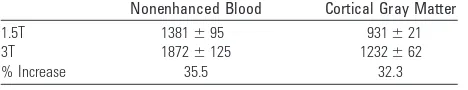
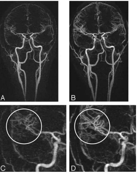
Intracranial Time Resolved Contrast Enhanced MR Angiography at 3T
Full text
Figure


Related documents
It is argued that Ratty and Mole’s experience passes the test of exemplifying all seven characteristics: ineffability, noesis, transiency, passivity, consciousness of the oneness
These included increasing their dose of medication, stockpiling medicines, using medications prescribed for other reasons or to other people to help them sleep, and
Draft genome assembly, annotation, and compar- ative analysis has identified variable content between strains with regards to the virulence factors, including toxin gene carriage,
Further, this study examined sources of variability in parental literacy support for 15 year olds attributable to factors related to parental learning support at the start of
Buoyant density in CsCI and guanine plus cytosine base content of the deoxyribonucleic acids of VE strains. Group VE-1
A well known result of Falconer showed that under mild assumptions the Hausdorff dimension of typical self-affine sets is equal to its Singularity dimension.. Heuter and
These studies 119,120,127 – 130 are described narratively below and a detailed summary is given in Table 28 [note that two studies, Cho et al.. laser plus injectable
Balls adsorbed at each rubella virus protein concentration were incubated with dilutions of a ru- bella antibody-positive serum and tested for binding of rubella-specific IgG
