Review Article
Molecular mechanisms of lymphatic metastasis in solid
tumors of the gastrointestinal tract
Melanie C Langheinrich1, Vera Schellerer1, Aristotelis Perrakis1, Clemens Lohmüller1, Claus Schildberg1,
Elisabeth Naschberger2, Michael Stürzl2, Werner Hohenberger1, Roland S Croner1
1Department of Surgery, University Hospital Erlangen; 2Department of Surgery, University Hospital Erlangen,Division of Molecular and Experimental Surgery, Germany
Received June 22, 2012; Accepted July 22, 2012; Epub September 5, 2012; Published September 15, 2012
Abstract: Tumor cell dissemination from the primary tumor site to distant organs is one of the characteristic proper-ties of malignant tumors and represents a crucial step in the progression of disease. Although the pattern of spread may vary in different types of carcinomas, dissemination via the lymphatic system represents a common event in metastasis. The extent of lymph node metastasis is one of the major determinants for the prognosis of patients
with gastrointestinal carcinomas and guides the therapeutically management. During the last decades, significant
attention has been given to the molecular mechanisms that control lymphatic metastasis. The process of lymphan-giogenesis has come into the focus. Lymphanlymphan-giogenesis, the formation of newly lymphatics, comprises a series of complex cellular events and is controlled by a balance between pro- and anti-lymphangiogenic signals. This article
will briefly describe the lymphatic system and then provide an overview of the molecular players involved in tumor
lymphangiogenesis.
Keywords: Lymphangiogenesis, lymphatic metastasis, molecular mechanisms, gastrointestinal tumors, lymphan-giogenic factors
Introduction
Solid tumors of the gastrointestinal tract such as esophageal, gastric, pancreatic colorectal cancer (CRC) are among the 10 most common malignancies worldwide. In Germany 2009 a total of ~215.000 people died suffering from
cancer. Looking at the specific cancer sites
colorectal cancer is the second leading cause of death, pancreatic cancer the fourth and
gas-tric cancer the fifth in men and sixth in women
(Cancer in Germany 2007/2008 8th edition 2012). Despite the existence of screening pro-grams, prevention strategies and multimodality appro-aches in the treatment of malignant gas-trointestinal tumors, they remain a major public health problem. Although the pattern of spread may vary in these different types of carcino-mas, dissemination from the primary tumor site to distant organs via the lymphatic system represents a common step in metastasis [1]. The presence of regional lymph node metasta-sis and the number of metastatic regional
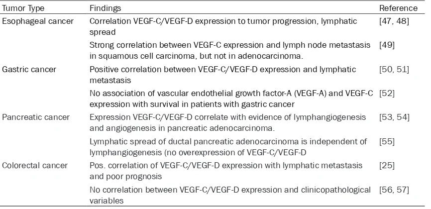
lymph nodes are of crucial importance for prog-nosis of patients with gastrointestinal carcino-mas. When lymphatic metastasis occurs patients have less favorable outcome (Table 1). Considering this and the high rate of incidence and mortality of gastrointestinal tumors it is critical to determine the molecular mechanisms of tumor dissemination. While the role of angio-genesis in cancer progression is well estab-lished, the role of the lymphatic system and the relation between lymphangiogenesis and tumor
metastasis is still incomplete clarified. This arti -cle provides a brief review of current knowledge in this area.
Structure and function of the lymphatic sys-tem
The identification of lymph specific markers
these lymphatic markers are not totally specific
to the lymph endothelium they play a crucial role in the development of the LS. The lymphat-ic vascular system is formed by lymphatlymphat-ic endo-thelial cells (LEC’s). LEC’s of initial capillaries typically express Prox-1, VEGFR-3, podoplanin, LYVE-1 and can secrete chemokines. After arte-riovenous differentiation, controlled by Notch/ COUP-TFII, Sox18 activates Prox-1 and induces the lymphatic differentiation program in the anterior cardinal vein [2].
Prox-1, the prospero-related homedomain
tran-scription factor is required for LEC specifica -tion. Downstream signaling of Prox-1 results in upregulation of LYVE-1 and VEGFR-3. The VEGF-C/VEGFR-3 is necessary for migration and survival of newly formed LEC’s.
LYVE-1, a homologue of the hyaluronic acid receptor CD44, is a member of the Link protein family and functions as a receptor for hyaluro-nan. Hyaluronan is a key mediator of cell migra-tion. LYVE-1 is used as a marker for distinguish-ing lymphatic vessels (LV) from blood vessels in
normal and tumor tissue. During embryogene-sis LYVE-1 is expressed in cardinal vein endo-thelium, almost simultaneous with Prox-1 expression. In contrast to Prox-1, LYVE-1 is not necessary for normal development or function of LV [3, 4].
Podoplanin is another specific marker for the
[image:2.612.90.522.93.342.2]lymphatic endothelium, due to the fact that it is expressed by developing and mature LEC’s but not by blood vessels. It is a transmembrane mucin like protein, which is expressed in nor-mal human tissue predominantly in lymph endothelial cells but also e.g. by podocytes, osteoblasts, alveolar Type I cells. Under normal conditions podoplanin is involved in LV forma-tion. Podoplanin knockout mice have lymphatic defects associated with dilated, malfunctioning lymphatic vessels and lymphedema and die at birth of respiratory failure [5, 6]. The expression of podoplanin is regulated by Prox-1. Since podoplanin is differentially expressed in a num-ber of neoplasms such as vascular tumors, germ cells tumors, esophageal squamous cell Table 1. The 5-year survival rate drops significantly from early tumors stage UICC I to advanced tumors
stage UICC III, when lymphatic metastasis occurs
Tumor type UICC I UICC III
Esophageal cancer > 50% < 15%
Gastric cancer > 80% < 30%
[image:2.612.88.521.96.155.2]Colorectal cancer >90% 30-60%
Figure 1. Scheme of differentially expressed genes between the lymphatic endothelium and the blood endothelium. Abbreviations: signal transducer and activator of transcription 6 (Stat6), monocyte chemotactic protein-1 (MCP-1), interleukin 6/8 (IL-6/8), intracellular adhesion molecule (ICAM), angiopoietin-1 (Ang-1), vascular endothelial growth factor/receptor (VEGF/R), cluster of Differentiation 44 (CD-44), insulin like growth factor 1/2 (IGF-1/2), fibroblast
carcinomas and gastric carcinomas, a role for podoplanin in tumor invasion and metastasis
has been suggested [7]. Recent findings indi -cate that podoplanin might favor invasion via its ability to remodel the cytoskeleton and thus increasing tumor cell motility [8].
The LS is a hierarchical network comprising cir-culating lymphocytes, blind ended capillaries, collector vessels, lymph nodes (LN) and lym-phoid organs and it serves key physiological
functions. It maintains fluid homeostasis by absorbing and draining e. g. interstitial fluid,
plasma proteins, extravasated cells and return-ing them back into the blood circulation, and the LS is an essential part of the body´s immu-nological surveillance system. Gene expression
profiles of LEC’s and blood endothelial cells
(BEC’s) have been analyzed and compared (Figure 1). About 300 genes are differentially expressed and the most obvious differences
were detected in genes coding for proinflamma
-tory cytokines/chemokines, cytoskeletal and cell matrix organization [9-12]. The difference in gene coding for cytoskeletal and cell matrix organization is consistent with the fact that
BEC’s are exposed to high blood flow/pressure
and therefore need stronger adhesion.
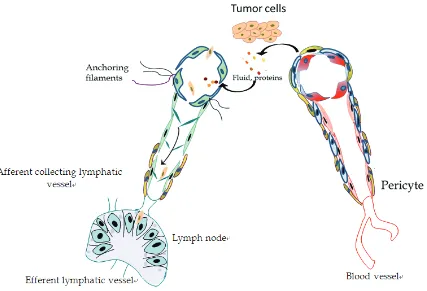
The function (fluid absorption and lymph trans
-port) is reflected in the structure of LV and dif -fers fundamentally from blood vessels (BV) (Figure 2). Initial lymphatic capillaries are about < 100 µm in diameter and characterized by loose intercellular junctions. In contrast to blood capillaries they have no or an incomplete basement membrane and the wall of LEC´s is jointed to the extracellular matrix by anchoring
filaments. Anchoring filaments, which contain elastic fibers, help the vessels to function. They
[image:3.612.96.523.70.361.2]prevent vessel collapse in conditions of high interstitial pressure via opening overlapping cell junctions and thereby widening the capil-lary lumen. Lymphatic capillaries merge into Figure 2. Structure of lymphatic vessels compared to blood vessel. Blood vessels are characterized by a complete basement membrane and are surrounded by pericytes and smooth muscle cells. Initial lymphatic vessels in con-trast have no or an incomplete basement membrane and are characterized by lose intercellular junctions and
collector lymphatic vessels. These collector LV consists of pericytes and have valves to help
propel a unidirectional flow to LN. Tumor cells
can take advantage of the structural LV design to promote their dissemination. The lack of a complete basement membrane or the loose intercellular junctions provides an easily acces-sible route for tumor cells. To facilitate tumor cell entry into the lymphatic vasculature altera-tion of the funcaltera-tional properties of the lymphat-ic endothelium could lead to adhesion and intravasation of tumor cells. Furthermore, che-mokines secreted by LEC’s, can mediate detachment, migration or invasion of tumor cells into LV.
Lymphangiogenesis
Tumor lymphangiogenesis
Carcinogenesis is a complex multistep process and dissemination from the primary tumor site to target organs via the lymphatic system repre-sents a common step in tumor cell spread. Gastric cancer frequently spreads to regional lymph nodes and gastrectomy with D1 and D2 lymphadenectomy has become a standard treatment procedure. Lymphangiogenesis, the process of formation of new lymphatic vessels, takes place in a variety of physiological and
pathophysiological processes such as
embry-onic development, regeneration, inflammation,
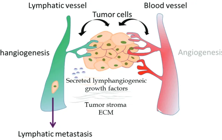
wound healing and lymph vascular malforma-tion [13]. There is an ongoing considerable debate in the literature whether lymphangio-genesis can be induced by tumor cells, whether lymphatic invasion requires the formation of new lymphatic vessels or uses preexisting lym-phatic vessels. Lymlym-phatic metastasis of tumor was classically viewed as a passive process, where tumor cells spread by utilizing preexist-ing lymphatic vessels e.g. via open junctions or by tumor eroding and not through the process of active new lymphatic formation (lymphangio-genesis). Until now there is mounting evidence that lymphangiogenesis does occur in tumors and that it promotes tumor progression. By analogy with angiogenesis, a shift in the bal-ance between pro lymphangiogenic and anti lymphangiogenic signaling, might lead to lym-phangiogenesis (Figure 3). The most obvious molecular regulators are represented in the following.
Molecular regulators of tumor lymphangiogen-esis
Vascular endothelial growth factors (VEGF):
[image:4.612.125.513.72.317.2]includes five members VEGF-A, VEGF-B,
VEGF-C, VEGF-D, PIGF, three tyrosine kinase receptors VEGFR-1, VEGFR-2, VEGFR-3 and two non protein kinase co-receptors (neutropilin -1 and neutropilin-2). VEGF-C and VEGF-D are the most important lymphangiogenic factors and exert this function via binding to VEGFR-2 and VEGFR-3 [14]. The rationale for this approach was derived from transgenic mouse models
[15]. In VEGF-C deficient mice lymphatic ves -sels fail to develop, while transgenic induction of VEGF-C leads to hyperplasia of lymphatic vessels [16-18]. The VEGF-C/VEGF-D/VEGFR-3 axis in adult human tissues is mainly expressed by lymphatic endothelial cells but also by a vari-ety of human carcinomas. Binding of growth factors leads to receptor dimerization, auto-phosphorylation of tyrosine residues, initiating of signal pathways and causes an increase in vascular permeability, proliferation, migration and survival of LEC’s in vitro and in vivo [19, 20]. In solid human tumors the
clinicopathologi-cal significance of VEGF-C/VEGF-D status cur -rently remains controversial (Table 2). For CRC
a significantly higher expression level of
VEGF-C/VEGFR-3 compared to normal tissue is described [21]. Figure 4 shows a histopatho-logical result of VEGF-C expression.
The majority of studies (such as esophageal, gastric, pancreatic or colorectal) have found a positive correlation between overexpression of
VEGF-C/VEGF-D on the one hand and vascular invasion, lymph vessel and lymph node involve-ment, distant metastasis and poor clinical out-come on the other hand [22-27]. However, in other studies such correlation could not be
con-firmed. The VEGF-C/VEGF-D/VEGFR-3 axis
exerts different biological effects on cancer cells to cause tumor progression. Some authors observed a lymphangiogenic switch in tumors, which means the ability of tumor cells to secrete their own growth factors leading to autocrine stimulation and via paracrine mechanisms tumor cells could induce the stimulation of other cells and thereby generating a (neo) vas-cularization of the tumor microenvironment. It is also of importance that the expression of VEGF-C/VEGF-D differs within the tumors. Some investigators showed that the expression
of VEGF-C in the invasion front is significantly
[image:5.612.91.524.96.306.2]higher than in tumor center, suggesting that the invasive edge may play a more important role in inducing tumor associated lymphangiogenesis compared to the rest of the tumor [2, 25, 28]. In addition, these studies have reported that lym-phatic vessels in the tumor margin were enlarged and dilated. Suggesting that tumor cells can easily use these gateways and intratu-moral LV might be poor in function [29]. The debate about the dominant role of intratumoral vs. peritumoral LV is still controversial. Taken together, several studies have provided evi-dence of an active involvement of the VEGF-C/ Table 2. Korrelation of VEGF-C/VEGF-D/VEGFR-3 expression in gastrointestinal tumors and clinical outcome.
Tumor Type Findings Reference
Esophageal cancer Correlation VEGF-C/VEGF-D expression to tumor progression, lymphatic
spread [47, 48]
Strong correlation between VEGF-C expression and lymph node metastasis
in squamous cell carcinoma, but not in adenocarcinoma. [49]
Gastric cancer Positive correlation between VEGF-C/VEGF-D expression and lymphatic
metastasis [50, 51]
No association of vascular endothelial growth factor-A (VEGF-A) and VEGF-C expression with survival in patients with gastric cancer [52]
Pancreatic cancer Expression VEGF-C/VEGF-D correlate with evidence of lymphangiogenesis
and angiogenesis in pancreatic adenocarcinoma. [53, 54] Lymphatic spread of ductal pancreatic adenocarcinoma is independent of
lymphangiogenesis (no overexpression of VEGF-C/VEGF-D [55] Colorectal cancer Pos. correlation of VEGF-C/VEGF-D expression with lymphatic metastasis
and poor prognosis [25]
No correlation between VEGF-C/VEGF-D expression and clinicopathological
VEGF-D/VEGFR-3 axis in lymphatic metastasis in gastrointestinal carcinomas.
Angiopoietins (Angs): Among the four distant Angs, Angiopoietin-1 (Ang-1) and Angiopoietin-2 (Ang-2) are the most intensive characterized members of the Ang family. Ang-1 acts via the receptor tyrosine kinase Tie2 as a constitutive Tie2 agonist, whereas Ang-2 is capable of act-ing as an agonist and antagonist in the interact-ing with Tie2. Tie2 is expressed on blood and lymph endothelial cells, but can also be found on cancers cells, monocytes and tumor associ-ated macrophages [30]. While Ang-1 is widely expressed in adult tissues, where it promotes vessel maturation and stabilization, Ang-2 expression occurs during vascular remodeling and angiogenesis. About the role of Angs in lymphangiogenesis little information is avail-able. Ang-1 is involved in LEC proliferation and
lymphatic vessel sprouting and Ang-2 deficient
mice exhibited severe lymphatic dysfunctions [31]. Overexpression of Angs and their receptor Tie2 in gastrointestinal tumor is associated with advanced disease and poor prognosis [32] [33, 34]. Ang-2 e.g. drives lymphatic metasta-sis of pancreatic ductal adenocarcinoma via lymphatic vascularization in the tumor stroma and through enhancing the capacity of tumor cells for adherence to endothelial cells. The
detailed function of Angs in lymphangiogenesis is still unclear.
Chemokines: Chemokine- s are a super family of ch- emoattractant cytokines and they are involved in a variety of immune res-ponses. Chemokines bi- nd to G-protein coupled receptors and they are grouped, according to the position of the cysteine residue, into four subfam-ilies: CXC, CXCR3, CC and C. Like mentioned before, LEC’s can secrete lymph-angiocrine cytokines su- ch as CCL21/CCL19, wh-ich act via CCR7 and CXCL12, which binds to CXCR4. They mediate homing of lymphocytes and migration of den-dritic cells into lymphatic vessels. To date, sev-eral sets of chemokines and their receptors have been suggested to play crucial roles in LN metastasis. High levels of expression of che-mokine receptors such CCR7 in gastric cancer, CCR7/CXCR4 in esophageal cancer, CX- CR3/CXCR4 and CCR7 in colorectal and CXCR4
in pancreatic cancer incre-ase the efficiency of
[image:6.612.89.398.72.302.2]tumor cells homing to metastatic target sites [13, 35, 36]. Patients with CXCR3 positive colon
cancer showed a significant shorter survival
compared to those without CXCR3 expres- sion.
Fibroblast growth factor (FGF): The fibroblast
growth factor family consists of structurally related ligands and four receptors (FGFR-1, FGFR-2, FGFR-3, FGFR-4). FGFR-1 and FGFR-2 are expressed in endothelial cells. The role of FGF-2 in angiogenesis has been well character-ized, but FGF-2 is also supposed to promote lymphangiogenesis. It might act directly via its receptor FGFR, which is upregulated by Prox-1, or through the protein kinase B (Akt)/mamma-lian target of rapamycin (mTOR)/p70 ribosomal S6 protein kinase (p70S6K) signaling pathway [37, 38]. Upregulation of FGFR-2 has been reported in pancreatic, gastric and colorectal cancer [39, 40].
Insulin like growth factor (IGF): The IGF system consists of the ligands insulin like growth factor 1 (IGF-1) and insulin like growth factor 2 (IGF-2) and their receptors IGF-1R, IGF-2R and 6 IGF binding proteins (IGFBP´s). IGF-1 and IGF-2 are polypeptide hormones. The IGF axis exerts diverse biological functions such as cell growth, differentiation and survival. Receptor signaling leads to activation of PI3K, Akt and various
[image:7.612.93.529.86.469.2]downstream networks. Recent findings indi -cate, that IGF-1/IGF-2 promote lymphangiogen-esis in a dependent and independent manner of the VEGF-C/VEGF-D/VEGFR-3 axis [41]. The role of the IGF system in tumorigenesis, cancer proliferation and survival is well documented [42, 43]. IGF-1R is frequently over expressed in several types of carcinomas (pancreatic, breast, colorectal, primary liver, sarcomas) and associated with invasion and metastasis.
Table 3. Lymphatic vascular factors and receptors. Adapted from [46] Lymphatic vascular
factor Biological activity Localization/Expression pattern VEGF-C/VEGF-D/
VEGFR-3 - growth factor/receptor- LEC: sprouting, migration proliferation, survial
- secreted dimeric glycoprotein growth factors - proteolytic procession of VEGF-C/-D precur-sors dictate angiogenic/lymphangiogenic potential
- VEGFR-3 is predominantly expressed in LECs that line inner surface of lymphatic vessels
LYVE-1 - endocytotic receptor for hyaluronan (major component of ECM and involved in cell migration, differentiation)
- type I integral membrane glycoprotein, lumi-nal/abluminal surface of LECs
Podoplanin - glomerular podocyte mucoprotein - expressed on lymphatic but not on blood vessel endothelium, osteoblasts, renal podocytes and lung alveolar
cells→cell motility
- single transmembrane protein
Prox-1 - homeobox gene for embryologic lym-phatic development and differentiation - expression: f.e. lens, heart, liver, pancreas
- subcellular localization controlled by compe-tition between nuclear localization signal and nuclear export signal
IGF - growth factor - produced in many tissue, function via auto-crine/paracrine mechanisms, circulating in the plasma in association with IGFBPs FGF - LEC migration and proliferation - FGF-1/FGF-2 lack signal sequences for
export out of the producer cell, most of the other members of the FGF family are secreted
HGF - growth factor - growth promoting activity of HGF requires proteolytic cleavage by extracellular serine proteinases
Ang1/Ang2/Tie2 - growth factor - Tie2 expressed on surface of endothelial cells
Hepatocyte growth factor (HGF): HGF belongs to the plasminogen prothrombin gene super-family and its activities are mediated by binding to the tyrosine kinase receptor c-Met. HGF is supposed to be involved in proliferation, migra-tion and tube formamigra-tion of LEC’s via down-stream ERK 1/2 and PI3K signaling. Several studies have shown a differentially expression of c-Met in solid tumors (colorectal cancer) and have indicated a positive correlation between c-Met expression metastatic spread and poor outcome [44, 45].
Conclusion
forms the basis for rational treatment options (neoadjuvant/adjuvant therapy). A Pubmed search for the term “lymphangiogenesis”
iden-tifies 1783 results versus 58778 results for the
term “angiogenesis”. This clearly shows that lymphangiogenesis is fairly behind in research compared to blood vessel formation. In the last
decades, since the identification of LEC specific
markers, our knowledge of mechanisms con-trolling lymphatic metastasis has increased
sig-nificantly (although each of these specific mark -ers has limitations) and lymphangiogenesis seems to play an important role in tumor spread. Tumor lymphangiogenesis is a complex multi step process, which is characterized by a
chain of events in which organ specific factors (f.e. lymph flow/lymph node density) and lym -phangiogenic factors are implicated, such as VEGF-C, VEGF-D, IGFs, HGF, angiopoietins and the list will undoubtedly continue to grow (Table 3).
A thorough understanding of these
mecha-nisms will define the concepts for future thera -peutic targeting of gastrointestinal tumors and will hopefully improve patient outcomes. Acknowledgements
This study was supported by the ELAN-Fond University of Erlangen-Nuremberg and the German Research Foundation (DFG CR 136/2-1).
Address correspondence to: Dr. Roland S Croner, Department of Surgery, University Hospital Erlangen, Krankenhausstrasse 12, 91054 Erlangen, Germany. Phone: +49 (0) 9131-8533296; E-mail: roland.cron-er@uk-erlangen.de.
References
[1] Leong SP, Cady B, Jablons DM, Garcia-Aguilar J, Reintgen D, Jakub J, Pendas S, Duhaime L, Cassell R, Gardner M, Giuliano R, Archie V, Cal-vin D, Mensha L, Shivers S, Cox C, Werner JA, Kitagawa Y, Kitajima M. Clinical patterns of metastasis. Cancer Metastasis Rev 2006; 25: 221.
[2] Alitalo K, Tammela T, Petrova TV. Lymphangio-genesis in development and human disease. Nature 2005; 438: 946.
[3] Van der Auwera I, Cao Y, Tille JC, Pepper MS, Jackson DG, Fox SB, Harris AL, Dirix LY, Ver-meulen PB. First international consensus on the methodology of lymphangiogenesis
quanti-fication in solid human tumours. Br J Cancer
2006; 95: 1611.
[4] Kato S, Shimoda H, Ji RC, Miura M.
Lymphan-giogenesis and expression of specific mole -cules as lymphatic endothelial cell markers. Anat Sci Int 2006; 81: 71.
[5] Yamanashi T, Nakanishi Y, Fujii G, Akishima-Fukasawa Y, Moriya Y, Kanai Y, Watanabe M,
Hirohashi S. Podoplanin expression identified in stromal fibroblasts as a favorable prognostic
marker in patients with colorectal carcinoma. Oncology 2009; 77: 53.
[6] Raica M, Cimpean AM, Ribatti D. The role of podoplanin in tumor progression and metasta-sis. Anticancer Res 2008; 28: 2997.
[7] Raica M, Ribatti D. Targeting tumor lymphan-giogenesis: an update. Curr Med Chem 2010; 17: 698.
[8] Cueni LN, Hegyi I, Shin JW, Albinger-Hegyi A, Gruber S, Kunstfeld R, Moch H, Detmar M. Tu-mor lymphangiogenesis and metastasis to lymph nodes induced by cancer cell expres-sion of podoplanin. Am J Pathol 2010; 177: 1004.
[9] Tammela T, Petrova TV, Alitalo K. Molecular lymphangiogenesis: new players. Trends Cell Biol 2005; 15: 434.
[10] Makinen T, Norrmen C, Petrova TV. Molecular mechanisms of lymphatic vascular develop-ment. Cell Mol Life Sci 2007; 64: 1915. [11] Saharinen P, Tammela T, Karkkainen MJ,
Ali-talo K. Lymphatic vasculature: development, molecular regulation and role in tumor
metas-tasis and inflammation. Trends Immunol 2004;
25: 387.
[12] Hirakawa S, Hong YK, Harvey N, Schacht V,
Matsuda K, Libermann T, Detmar M. Identifica
-tion of vascular lineage-specific genes by tran
-scriptional profiling of isolated blood vascular
and lymphatic endothelial cells. Am J Pathol 2003; 162: 575.
[13] Kawada K, Taketo MM. Significance and mech -anism of lymph node metastasis in cancer pro-gression. Cancer Res 2011; 71: 1214. [14] Lohela M, Bry M, Tammela T, Alitalo K. VEGFs
and receptors involved in angiogenesis versus lymphangiogenesis. Curr Opin Cell Biol 2009; 21: 154.
[15] Ribatti D. Transgenic mouse models of angio-genesis and lymphangioangio-genesis. Int Rev Cell Mol Biol 2008; 266: 1.
[16] Jeltsch M, Kaipainen A, Joukov V, Meng X, Lak-so M, Rauvala H, Swartz M, Fukumura D, Jain RK, Alitalo K. Hyperplasia of lymphatic vessels in VEGF-C transgenic mice. Science 1997; 276: 1423.
mouse embryos but leads to persistent lym-phatic hyperplasia in adult tissues. Am J Pathol 2008; 173: 1891.
[18] Karkkainen MJ, Haiko P, Sainio K, Partanen J, Taipale J, Petrova TV, Jeltsch M, Jackson DG, Talikka M, Rauvala H, Betsholtz C, Alitalo K. Vascular endothelial growth factor C is
re-quired for sprouting of the first lymphatic ves -sels from embryonic veins. Nat Immunol 2004; 5: 74.
[19] Roskoski R Jr. VEGF receptor protein-tyrosine kinases: structure and regulation. Biochem Biophys Res Commun 2008; 375: 287. [20] Birk DM, Barbato J, Mureebe L, Chaer RA.
Cur-rent insights on the biology and clinical as-pects of VEGF regulation. Vasc Endovascular Surg 2008; 42: 517.
[21] Omachi T, Kawai Y, Mizuno R, Nomiyama T, Mi-yagawa S, Ohhashi T, Nakayama J. Immunohis-tochemical demonstration of proliferating lym-phatic vessels in colorectal carcinoma and its
clinicopathological significance. Cancer Lett
2007; 246: 167.
[22] Tai XH, Ji WY, Sun XH. [Relationship among lymphangiogenesis, vascular endothelial gro wth factor-C mRNA expression and cervical lymphatic metastasis in laryngeal carcinoma]. Zhonghua Er Bi Yan Hou Tou Jing Wai Ke Za Zhi 2006; 41: 622.
[23] Gou HF, Chen XC, Zhu J, Jiang M, Yang Y, Cao D, Hou M. Expressions of COX-2 and VEGF-C in gastric cancer: correlations with lymphangio-genesis and prognostic implications. J Exp Clin Cancer Res 2011 ; 30: 14.
[24] Wang TB, Chen ZG, Wei XQ, Wei B, Dong WG. Serum vascular endothelial growth factor-C and lymphoangiogenesis are associated with the lymph node metastasis and prognosis of patients with colorectal cancer. ANZ J Surg 2011; 81: 694.
[25] Lin M, Lin HZ, Ma SP, Ji P, Xie D, Yu JX. Vascular endothelial growth factor-A and -C: expression and correlations with lymphatic metastasis and prognosis in colorectal cancer. Med Oncol 2011; 28: 151.
[26] Zhu C, Qi X, Chen Y, Sun B, Dai Y, Gu Y. PI3K/ Akt and MAPK/ERK1/2 signaling pathways are involved in IGF-1-induced VEGF-C upregulation in breast cancer. J Cancer Res Clin Oncol 2011; 137: 1587.
[27] Werynska B, Dziegiel P, Jankowska R. Role of lymphangiogenesis in lung cancer. Folia Histo-chem Cytobiol 2009; 47: 333.
[28] Acs G, Paragh G, Rakosy Z, Laronga C, Zhang PJ. The extent of retraction clefts correlates with lymphatic vessel density and VEGF-C ex-pression and predicts nodal metastasis and poor prognosis in early-stage breast carcino-ma. Mod Pathol 2012; 25: 163.
[29] Karnezis T, Shayan R, Caesar C, Roufail S, Har-ris NC, Ardipradja K, Zhang YF, Williams SP, Farnsworth RH, Chai MG, Rupasinghe TW, Tull DL, Baldwin ME, Sloan EK, Fox SB, Achen MG, Stacker SA. VEGF-D promotes tumor metasta-sis by regulating prostaglandins produced by the collecting lymphatic endothelium. Cancer Cell 2012; 21: 181.
[30] Huang H, Bhat A, Woodnutt G, Lappe R. Target-ing the ANGPT-TIE2 pathway in malignancy. Nat Rev Cancer 2010; 10: 575.
[31] Karpanen T, Alitalo K. Molecular biology and pathology of lymphangiogenesis. Annu Rev Pathol 2008; 3: 367.
[32] Schulz P, Fischer C, Detjen KM, Rieke S, Hilfen-haus G, von Marschall Z, Bohmig M, Koch I, Kehrberger J, Hauff P, Thierauch KH, Alves F, Wiedenmann B, Scholz A. Angiopoietin-2 drives lymphatic metastasis of pancreatic can-cer. FASEB J 2011; 25: 3325.
[33] Wang J, Wu K, Zhang D, Tang H, Xie H, Hong L, Pan Y, Lan M, Hu S, Ning X, Fan D. Expressions
and clinical significances of angiopoietin-1, -2
and Tie2 in human gastric cancer. Biochem Biophys Res Commun 2005; 337: 386. [34] Jo MJ, Lee JH, Nam BH, Kook MC, Ryu KW,
Choi IJ, Kim YW, Bae JM. Preoperative serum angiopoietin-2 levels correlate with lymph node status in patients with early gastric can-cer. Ann Surg Oncol 2009; 16: 2052.
[35] Singh R, Lillard JW, Jr., Singh S. Chemokines: key players in cancer progression and metas-tasis. Front Biosci (Schol Ed) 2011; 3: 1569. [36] Cui Y, Madeddu P. The role of chemokines,
cy-tokines and adhesion molecules in stem cell
trafficking and homing. Curr Pharm Des 2011;
17: 3271.
[37] Matsuo M, Yamada S, Koizumi K, Sakurai H,
Saiki I. Tumour-derived fibroblast growth fac -tor-2 exerts lymphangiogenic effects through Akt/mTOR/p70S6kinase pathway in rat lym-phatic endothelial cells. Eur J Cancer 2007; 43: 1748.
[38] Cueni LN, Detmar M. New insights into the mo-lecular control of the lymphatic vascular sys-tem and its role in disease. J Invest Dermatol 2006; 126: 2167.
[39] Sundlisaeter E, Rosland GV, Hertel JK, Sakari-assen PO, Almas B, Dicko A, Sondenaa K. In-creased lymphatic vascular density is seen before colorectal cancers reach stage II and growth factor FGF-2 is downregulated in tumor tissue compared with normal mucosa. APMIS 2009; 117: 212.
[40] Yamanaka Y, Friess H, Buchler M, Beger HG, Uchida E, Onda M, Kobrin MS, Korc M.
Overex-pression of acidic and basic fibroblast growth
with advanced tumor stage. Cancer Res 1993; 53: 5289.
[41] Da MX, Wu Z, Tian HW. Tumor lymphangiogen-esis and lymphangiogenic growth factors. Arch Med Res 2008; 39: 365.
[42] Weroha SJ, Haluska P. The insulin-like growth factor system in cancer. Endocrinol Metab Clin North Am 2012; 41: 335.
[43] Gao QG, Xie JX, Wong MS, Chen WF. IGF-I re-ceptor signaling pathway is involved in the neu-roprotective effect of genistein in the neuro-blastoma SK-N-SH cells. Eur J Pharmacol 2012; 677: 39.
[44] Kammula US, Kuntz EJ, Francone TD, Zeng Z, Shia J, Landmann RG, Paty PB, Weiser MR. Mo-lecular co-expression of the c-Met oncogene and hepatocyte growth factor in primary colon cancer predicts tumor stage and clinical out-come. Cancer Lett 2007; 248: 219.
[45] Organ SL, Tsao MS. An overview of the c-MET signaling pathway. Ther Adv Med Oncol 2011; 3: S7.
[46] Langheinrich M, Schellerer V, Oeckl K, Stürzl M, Naschberger E, Croner R. Molecular Mecha-nisms of Lymphatic metastasis. Colorectal Cancer Book 1 2011; 285: 298
[47] Noguchi T, Takeno S, Shibata T, Uchida Y, Yo-koyama S, Muller W. VEGF-C expression corre-lates with histological differentiation and me-tastasis in squamous cell carcinoma of the esophagus. Oncol Rep 2002; 9: 995.
[48] Kimura Y, Watanabe M, Ohga T, Saeki H, Ka-keji Y, Baba H, Maehara Y. Vascular endothelial growth factor C expression correlates with lym-phatic involvement and poor prognosis in pa-tients with esophageal squamous cell carcino-ma. Oncol Rep 2003; 10: 1747.
[49] Mobius C, Freire J, Becker I, Feith M, Brucher BL, Hennig M, Siewert JR, Stein HJ. VEGF-C ex-pression in squamous cell carcinoma and ad-enocarcinoma of the esophagus. World J Surg 2007; 31: 1768.
[50] Amioka T, Kitadai Y, Tanaka S, Haruma K, Yo-shihara M, Yasui W, Chayama K. Vascular en-dothelial growth factor-C expression predicts lymph node metastasis of human gastric carci-nomas invading the submucosa. Eur J Cancer 2002; 38: 1413.
[51] Wang W, Wu R, Sun G, Li X, Yuan W, Tang L. [Expression of VEGF-C and VEGF-D in gastric carcinoma and its relationship with lymph node metastases]. Zhong Nan Da Xue Xue Bao Yi Xue Ban 2010; 35: 335.
[52] Lee SJ, Kim JG, Sohn SK, Chae YS, Moon JH, Kim SN, Bae HI, Chung HY, Yu W. No associa-tion of vascular endothelial growth factor-A (VEGF-A) and VEGF-C expression with survival in patients with gastric cancer. Cancer Res Treat 2009; 41: 218.
[53] Schneider M, Buchler P, Giese N, Giese T, Wilt-ing J, Buchler MW, Friess H. Role of lymphan-giogenesis and lymphangiogenic factors dur-ing pancreatic cancer progression and lymphatic spread. Int J Oncol 2006; 28: 883. [54] Zhang B, Zhao WH, Zhou WY, Yu WS, Yu JM, Li
S. Expression of vascular endothelial growth factors-C and -D correlate with evidence of lym-phangiogenesis and angiogenesis in pan creatic adenocarcinoma. Cancer Detect Prev 2007; 31: 436.
[55] Sipos B, Kojima M, Tiemann K, Klapper W, Kruse ML, Kalthoff H, Schniewind B, Tepel J, Weich H, Kerjaschki D, Kloppel G. Lym-phatic spread of ductal pancreatic adenocarci-noma is independent of lymphangiogenesis. J Pathol 2005; 207: 301.
[56] George ML, Tutton MG, Janssen F, Arnaout A,
Abulafi AM, Eccles SA, Swift RI. A,
VEGF-C, and VEGF-D in colorectal cancer progres-sion. Neoplasia 2001; 3: 420.


![Table 3. Lymphatic vascular factors and receptors. Adapted from [46]](https://thumb-us.123doks.com/thumbv2/123dok_us/881433.600523/7.612.93.529.86.469/table-lymphatic-vascular-factors-receptors-adapted.webp)
