www.impactjournals.com/oncotarget/
Oncotarget, Vol. 7, No. 13
Reprogramming of energy metabolism as a driver of aging
Zhaoyang Feng
1, Richard W. Hanson
2,3,*, Nathan A. Berger
2,3,4and Alexander
Trubitsyn
51 Department of Pharmacology, School of Medicine, Case Western Reserve University, Cleveland, OH, USA 2 Department of Biochemistry, School of Medicine, Case Western Reserve University, Cleveland, OH, USA 3 Case Comprehensive Cancer Center, School of Medicine, Case Western Reserve University, Cleveland, OH, USA 4 Department of Medicine, School of Medicine, Case Western Reserve University, Cleveland, OH, USA
5 Institute of Biology and Soil Sciences of Far Eastern Brach of Russian Academy of Science, Vladivostok, Russia * Deceased
Correspondence to: Zhaoyang Feng, email: [email protected]
Keywords: aging, energy metabolism, mitochondria, glycolysis, physical activity, Gerotarget
Received: January 22, 2016 Accepted: February 11, 2016 Published: February 23, 2016
ABSTRACT
Aging is characterized by progressive loss of cellular function and integrity. It has been thought to be driven by stochastic molecular damage. However, genetic and environmental maneuvers enhancing mitochondrial function or inhibiting glycolysis extend lifespan and promote healthy aging in many species. In post-fertile
Caenorhabditis elegans, a progressive decline in phosphoenolpyruvate carboxykinase with age, and a reciprocal increase in pyruvate kinase shunt energy metabolism
from oxidative metabolism to anaerobic glycolysis. This reduces the efficiency and
total of energy generation. As a result, energy-dependent physical activity and other cellular functions decrease due to unmatched energy demand and supply. In return, decrease in physical activity accelerates this metabolic shift, forming a vicious cycle. This metabolic event is a determinant of aging, and is retarded by caloric restriction to counteract aging. In this review, we summarize these and other evidence supporting the idea that metabolic reprogramming is a driver of aging. We also suggest strategies to test this hypothesis
INTRODUCTION
Aging is hallmarked by the progressive loss of
cellular function and integrity that eventually leads to
vulnerability and death of organisms [1]. It lowers the
quality of life, and is a potent risk factor for cancer,
diabetes, and other prevalent diseases [2]. It has been
long thought that age-dependent accumulation of
stochastic damage of molecules drives aging [3]. Current
evidence demonstrate that a programmed event of energy
metabolism is a determinant of aging that can be modified
to modulate aging [4].
AGING INVOLVES REPROGRAMMING
OF ENERGY METABOLISM
Intermediary metabolism generates ATP from
nutrients, providing energy for cellular function and
maintenance. Alterations in energy metabolism are
linked to the aging process and aging-associated diseases
[5]. Substantial evidence has demonstrated that energy
production progressively decreases with age in all
organisms, mainly due to the decline in the function of
mitochondria [6]. Aged organisms also exhibit disrupted
homeostasis of carbohydrates, amino acids, and fatty
acids [5], major biological fuels [7]. The exact alterations
in energy metabolism that are associated with aging, their
physiological impact, and their contribution to aging are
unclear, impeding the understanding of aging mechanisms
and the development of mechanism-based strategies to
modulate aging.
is inconclusive whether oxidative damage is the cause
of aging-associated decline in mitochondrial function
or aging [12, 13]. Recently, we have reported that the
aging of
C. elegans
, a genetic model of aging that lives
about three weeks, is highlighted by a progressive
decline in cytosolic phosphoenolpyruvate carboxykinase
(PEPCK-C) after the reproductive peak, and a reciprocal
increase in pyruvate kinase (PK) [4]. While PK is an
enzyme of glycolysis, PEPCK-C is a metabolic enzyme
associated with longevity [4, 14, 15]. A key consequence
of this metabolic event is the shunt of energy metabolism
from oxidative metabolism to anaerobic glycolysis. In
all aerobic species, ATP can be generated both in the
presence and absence of oxygen. But 30-36 ATP can be
generated from one glucose molecule through oxidative
metabolism, while only 2 ATP are produced by anaerobic
glycolysis [7]. As a result, reciprocal changes in PEPCK-C
and PK with age reduce the efficiency of and total energy
production [4].
How do changes in PEPCK-C and PK, two cytosolic
enzymes, lead to decline in mitochondrial function?
The core of energy metabolism is the tricarboxylic acid
(TCA) cycle [7], a series of chemical reactions that
oxidize carbohydrates, fats and proteins into carbon
dioxide, generating NADH that is used by ETC to
produce ATP. Moreover, the TCA cycle intermediates
can be withdrawn from mitochondria to the cytosol, a
process called cataplerosis. This supplies carbons for
the synthesis of glyceride-glycerol, serine, and glucose,
as well as metabolites derived from these chemicals
[16]. Traditionally viewed as a rate-limiting enzyme of
gluconeogenesis and glyceroneogenesis, PEPCK-C is a
major cataplerotic enzyme that links the TCA cycle with
the metabolism of carbohydrates, fatty acids, amino acids,
and other metabolites [16]. At the biochemical level, the
activity of PEPCK-C is correlated tightly with the flux
of the TCA cycle but not gluconeogenesis [17]. At the
organismal level, PEPCK-C is required for the integration
of energy metabolism [18], and is critical for the
homeostasis of glucose, fatty acids, amino acids and other
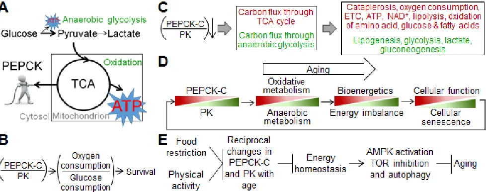
related metabolites (see ref [16] for review). In
C. elegans
,
PEPCK-C accelerates TCA cycle flux and promotes
oxidative metabolism [4], likely via increased cataplerosis
(Figure 1A). PEPCK-C also increases mitochondrial
respiration and counteracts its decline with age. On the
other hand, PK is a key glycolytic enzyme that greatly
favors the conversion of phosphoenolpyruvate to pyruvate,
promoting glycolysis [7]. During energy production,
pyruvate either enters mitochondria for oxidation, or is
converted to lactate by lactate dehydrogenase. PEPCK-C
shunts glucose metabolism toward oxidation, reducing
lactate production [4].
[image:2.612.63.552.441.634.2]The reciprocal changes in PEPCK-C and PK
with age, and their impact on oxidative metabolism
and anaerobic glycolysis are likely conserved from
C.
elegans
to humans. First, skeletal muscle of aging humans
from 10s to 70s exhibits an aging-associated decrease
in PEPCK-C activity, and increases in PK and lactate
dehydrogenase that cannot be explained by alterations in
numerical ratio of type I and type II muscle fibers with
age [19]. In aged skeletal muscle and liver of mammalian
animals,
PEPCK-C
mRNA is decreased and
PK
mRNA
is increased [20, 21]. Second, aging mammals including
humans display decreased mitochondrial function and
increased glycolysis in many tissues such as liver, skeletal
muscle and brain [22-25], as well as elevated lactate in
both tissues and serum [26]. Moreover, platelets of aged
humans exhibit reduced ATP production by mitochondria,
and increased ATP production by anaerobic glycolysis
[27], which likely reflects aging-associated changes in
energy metabolism of the whole body [27, 28].
RECIPROCAL CHANGES IN PEPCK-C
AND PK PROFOUNDLY IMPACT AGING
ORGANISMS
What are the physiological effects of reciprocal
changes of PEPCK-C and PK with age? The decline in
mitochondrial bioenergetics may subject aging organisms
to a relative energy deficiency, although the PK-driven
increase in glycolysis likely compensates for some
of the reduced energy production. A deficit in energy
supply reduces the function and integrity of many cells
and tissues, hence the survival of organisms, due to
unmatched energy demand and supply (Figure 1B). In
support of this view, PEPCK-C promotes physical activity,
fertility, autophagy, defense against osmotic and oxidative
stresses, and many other energy consuming processes
in various animal species [4, 14, 15, 29-34]. During the
aging of
C. elegans
, decline in PEPCK-C is coupled with
loss of physical activity, a major energy consumer [35],
and genetically enhanced PEPCK-C preserves physical
activity and extends lifespan in a dose-dependent manner
[4]. Of note, PEPCK-C promotes physical activity to
increase ATP turnover, AMP/ATP ratio (a key indicator of
cellular energy demand), the activation of 5’ AMP kinase
(AMPK, a major mediator of energy homeostasis activated
by higher AMP:ATP ratio [36]), fuel oxidation, ATP
content, and food intake, both acutely and chronically.
Many of these effects of PEPCK-C on energy demand and
supply, and cellular function and maintenance require the
activation of AMPK [4].
In addition to decline in energy production, the
following alterations in energy metabolism are predicted,
based on the metabolic roles of PEPCK-C, PK, and the
TCA cycle: 1) increase in synthesis and deposition of fats;
2) disrupted homeostasis of glucose and amino acids;
3) reduction in NAD
+; and 4) reduction in biosynthesis
associated with cataplerosis (Figure 1C).
Unlike glucose, fats can only be oxidized to produce
ATP [7]. The reduction in oxidative metabolism with age
would lead to decreased utilization of fats. On the other
hand, increased PK activity and glycolytic flux in aged
organisms should produce more pyruvate. In addition to
being oxidized in mitochondria or converted to lactate,
pyruvate can serve as a precursor of lipogenesis or
gluconeogenesis. Because mitochondrial function is
reduced, more pyruvate would be shunt to lipogenesis
or gluconeogenesis. Indeed, aging involves a shift
of fatty metabolism toward lipogenesis in mice [37]
and accumulation of fats in all organisms including
humans [38]. In mice, decline in PEPCK-C with age
underlies aging-associated reduction in lipolysis and the
coordinative down regulation of mitochondrial enzymes
[39]. Indeed, aged mice over-expressing PEPCK-C
exhibited less subcutaneous, visceral, and pericardial
fat deposit than even younger control mice fed the same
regular chow diet [31]
The shift of energy metabolism from oxidative
metabolism to anaerobic glycolysis suggests that
aged organisms demand more glucose as energy
source. Consistent with this view, gluconeogenesis is
elevated in aged yeast [40]. In aged mammals, basal
gluconeogenic capacity and blood glucose produced
through gluconeogenesis are increased [41], while hepatic
incorporation of glucose to glycogen is decreased [42].
In contrast to gluconeogenesis, glucose uptake into
skeletal muscle, brain and other energy consuming tissues
decreases with age due to reduced insulin signaling [43],
decreased insulin sensitivity [44], and reduced glucose
transporters [45]. An increased demand and supply
of glucose, while a reduction in usable glucose may
contribute to the disrupted homeostasis of glucose in
aged organisms [46]. Sarcopenia is the progressive loss
of muscle in aging humans and animals [47]. The lost
muscle mass, primarily proteins, results in increased
alanine and glutamine in circulation [48]. A fate of these
amino acids is to be oxidized in mitochondria. The shift
of energy metabolism away from oxidative metabolism
would reduce the disposal of these amino acids, and
contribute to the disrupted homeostasis of amino acids in
aged organisms [49].
Redox homeostasis is critical for cellular function
and integrity [50]. Significantly, aging is accompanied
by a progressive decline in intracellular NAD
+in species
including humans [51, 52]. NAD
+is an essential cofactor
for sirtuin, a deacetylase that promotes longevity and
healthy aging [53]. It is also a necessary substrate for poly
(ADP-ribose) polymerase [54], a critical enzyme in the
DNA repair process [54-56]. Decreased NAD
+increases
the vulnerability of cells to accumulation of DNA damage
with age [57-59]. The breakdown of glucose to pyruvate
consumes NAD
+[7]. NAD
+is regenerated through
fermentation that converts pyruvate to lactate. Increased
glycolytic flux in aged organisms would lead to increased
consumption of NAD
+. Insufficient regeneration of NAD
+could result in reduction in NAD
+.
acids relieves acidosis. Likely, decline in PEPCK-C with
age reduces cataplerosis and its related biosynthesis,
contributing to reduced proliferation and increased
vulnerability associated with aging.
Most cancer cells exhibit the Warburg effect [63],
higher glycolysis followed by lactate production, instead
of lower glycolysis followed by oxidation. Type II
diabetes is associated with metabolic changes paralleling
the Warburg effect, including expression of genes involved
in anaerobic glycolysis [64] and a decline in mitochondrial
function [65]. Thus, the shift of energy metabolism from
oxidative metabolism to glycolysis driven by reciprocal
changes in PEPCK-C and PK with age may promote
tumorigenesis and the prevalence of diabetes [66-68]. For
example, metformin, the most commonly prescribed drug
treating type II diabetes, may shunt energy metabolism
toward oxidative metabolism and away from glycolysis to
counteract cancers [68].
Collectively, the cataplerotic role of PEPCK-C acts
as a major adaptor of energy metabolism, and a carbon
valve for various biosynthetic pathways. Reciprocal
changes in PEPCK-C and PK after the reproductive
peak are a lead metabolic event associated with aging.
This event re-patterns metabolism of aging organisms,
decreases their cellular function and integrity, and
promotes the onset of aging-associated diseases.
RECIPROCAL CHANGES IN PEPCK-C
AND PK WITH AGE DETERMINE AGING
Evidence from mice and
C. elegans
demonstrate
that reciprocal changes in PEPCK-C and PK with age
determine aging. First, PEPCK-C counteracts loss of
cellular function and integrity with age. Specifically,
it extends fertility, retards aging-associated decrease
in physical activity, a negative indicator of health span
and lifespan [69], and enhances autophagic activity [4,
14]. Autophagy is a cell repair mechanism that removes
molecular wastes, and counteracts aging and aging-related
diseases [70-73]. Second, PEPCK-C retards cellular
senescence [4], assessed by the accumulation of molecular
wastes such as lipofuscin and β-galactosidase [74, 75],
and the expression of the proliferation restrictive marker
cyclin kinase inhibitor [76]. Cellular senescence may
contribute to aging [77]. Moreover, reciprocal changes in
PEPCK-C and PK with age are necessary and sufficient to
limit lifespan and fertility [4]. Last, PEPCK-C activity is
correlated with lifespan, and its enzyme level predicts life
expectancy [4].
Many effects of PEPCK-C on aging including
lifespan extension require the activation of AMPK
signaling and/or the inhibition of Target of Rapamycin
(TOR) signaling [4]. AMPK and TOR signaling are two
major molecular signals that control aging in species
including mammals [78-80]. The beneficial impact of
activation of AMPK and inhibition of TOR on lifespan
necessitates autophagy [81-83]. Consistently, PEPCK-C
enhances the activity of autophagy in aged
C. elegans
,
and requires the expression of autophagic genes to
promote longevity [4]. Interestingly, AMPK primarily
drives catabolism that produces energy and promotes
mitochondrial oxidation, and is a negative regulator of
glycolysis [84]. On the other hand, TOR signaling is a
cellular energy sensor of excessive nutrient and energy
that upregulates PK and glycolysis [85]. The anti-aging
effects mediated by TOR inhibition involve increased
energy metabolism and oxidative phosphorylation
complex [86-90].
In summary, reciprocal changes in PEPCK-C and
PK with age are a determinant of aging. The mechanisms
include disrupted energy homeostasis, as well as altered
AMPK, TOR signaling, and autophagy. Consistently,
mitochondrial bioenergetics and autophagic activity are
preserved in fibroblasts of centenarians [91]. Intriguingly,
declines in PEPCK-C and physical activity with age
promote each other to limit reproductive life and lifespan
[4] via a feedback mechanism [4, 14, 92, 93]. These
observations indicate that a vicious cycle of reciprocal
changes in PEPCK-C and PK, and decline in cellular
function and integrity drives aging (Figure 1D). The
causality between changes in metabolism and in cellular
function and integrity is unknown, and is likely a “chicken
and egg” dilemma.
RECIPROCAL CHANGES IN
PEPCK-C AND PK ARE A COMMON
DENOMINATOR OF AGING
Molecular signals, pharmacological reagents,
appropriate environmental stresses, and calorie restriction
(CR) extend lifespan and may also improve health in
various species. Strikingly, most of these maneuvers either
increase oxidative metabolism and energy production,
inhibit glycolysis, or both. In addition to AMPK and
TOR, insulin/IGF signaling (IIS) [94] and sirtuin [53] are
molecular signals that affect lifespan. Reduced IIS [95-99]
and sirtuin [51, 100-103] increase or stabilize PEPCK-C,
promote oxidative metabolism, and inhibit PK and/or
glycolysis.
and other lower organisms [16, 30, 110-112].
CR is the most robust intervention that extends
lifespan and improves health in species ranging from yeast
to non-human primates, via AMPK-TOR-autophagy axis
[80-83]. CR increases PEPCK-C activity and oxidative
metabolism while inhibiting PK activity and glycolysis
in animals [15, 113-116]. In humans, CR increases
mitochondrial biogenesis [117]. A plausible biological
reason underling this metabolic shift is to promote efficient
energy production and cataplerosis, in order to meet
the energy [118] and biosynthetic [119-121] need under
limited resources. Significantly, CR counteracts reciprocal
changes in PEPCK-C and PK with age to elicit anti-aging
effects including longevity in
C. elegans
[4]. On the other
hand, physical activity, which extends life expectancy in
humans [122], increases energy expenditure, PEPCK-C
and mitochondrial function. Notably, both mice and
C.
elegans
over-expressing PEPCK-C exhibited increased
physical activity, ate more, weighed less, had extended
fertility, and lived longer [4, 14]. Thus, energy balance,
achieved by reduced “energy in” from CR, enhanced
“energy out” from enhanced physical activity, or their
combination, counteracts reciprocal changes in PEPCK-C
and PK with age to retard aging.
In summary, reciprocal changes in PEPCK-C and
PK activity with age, and the consequent shift of energy
metabolism are a common denominator of aging. These
alterations can be retarded by CR, CR mimetics, and other
genetic and environmental factors to counteract aging via
AMPK and TOR pathways (Figure 1E).
CONCLUSIONS AND FUTURE
PERSPECTIVE
Reciprocal changes in PEPCK-C and PK with
age are likely part of a bigger reprogramming of energy
metabolism that profoundly affects the physiology of
aging organisms, thereby impacting the aging process. It
is important to obtain a complete picture of changes in
metabolism with age, and their influence on decline in
cellular function, cellular senescence, lifespan, and other
aging traits. Such investigation should focus on metabolic
pathways moving carbons into and out of the TCA cycle,
and those affecting the homeostasis of glucose, fats and
amino acids. This is because both CR and an optimized
ratio of macronutrients (carbohydrates, proteins and fats)
without reduction in total calorie intake extend lifespan
in mice [123-125], suggesting that alterations in these
metabolic pathways have significant but complex impact
on aging.
Evidence outlined here support the bioenergetics
theory of aging [126, 127], which proposes that the
decline in bioenergetics with age is the driver of aging.
Specifically, the decline in bioenergetics with age is a
pacemaker in the aging process, whereas other
aging-associated phenomena, such as the accumulation of
reactive species and the decline in repair mechanisms,
are secondary to the decline in bioenergetics. The sum of
all these changes leads to loss of physiological function
and eventually vulnerability and death of organisms.
The bioenergetics theory of aging also hypothesizes that
an unidentified primary genetic program (aging clock)
controls the decline in bioenergetics. However, the
presence of a vicious cycle between reciprocal changes
in PEPCK-C and PK, and decline in cellular function and
integrity indicates that a genetically programmed aging
clock may [128] or may not [129-131] be needed for the
decline in bioenergetics with age. For example, a
quasi-programmed hyperfunction, which has been proposed to
be associated with development and have harmful impact
on organisms, may start this vicious cycle without a
genetically programmed aging clock; while the pace of
this vicious cycle may be mediated by a genetic program.
In either case, it is critical to understand the mechanisms
underlying aging-associated changes in PEPCK-C and
PK, which are currently unknown. Remarkably, PEPCK-C
is acetylated in yeast [132] and human cells [133], and
PEPCK-C acetylation leads to its degradation [100].
Sirtuin, which promotes longevity in many species,
deacetylates and stabilizes PEPCK-C [100]. Deacetylation
mediated by sirtuin shunts energy metabolism away from
glycolysis and toward oxidative metabolism [134-137],
promoting energy production [138]. Sirtuin expression
and activity decrease with age [139, 140], and CR [141]
and exercise [142] increase sirtuin to decrease protein
acetylation. It will be interesting to examine if PEPCK-C
acetylation is involved in its decline with age, if sirtuin
slows this decline, and if sirtuin retards aging via altered
PEPCK-C stability.
Last, enhancing PEPCK-C is sufficient to delay
many key aging-associated metabolic and physiological
changes and increase lifespan [4, 14, 15]. Thus
interventions that sustain PEPCK-C represent a novel
strategy to counteract aging.
ACKNOWLEDGMENTS
We thank Transdisciplinary Research on Energetics
& Cancer directed by Dr. Nathan A. Berger, Nutrition
Obesity Research Center, Case Western Reserve
University (U54-CA-116867), Whitehall Foundation
(2013-08-82), department fund (ZF), and Ellison
Foundation (AG-SS-2420-10)(RWH and NAB) for
funding, and Drs. Thomas Sundermeier, Leslie Webster,
Hung-Ying Kao, and Johannes Von Lintig for discussion.
REFERENCES
1. Lopez-Otin C, Blasco MA, Partridge L, Serrano M and Kroemer G. The hallmarks of aging. Cell. 2013; 153:1194-1217.
2. Niccoli T and Partridge L. Ageing as a risk factor for disease. Curr Biol. 2012; 22:R741-752.
3. Miquel J, Economos AC, Fleming J and Johnson JE, Jr. Mitochondrial role in cell aging. Exp Gerontol. 1980; 15:575-591.
4. Yuan Y, Hakimi P, Kao C, Kao A, Liu R, Janocha A, Boyd-Tressler A, Hang X, Alhoraibi H, Slater E, Xia K, Cao P, Shue Q, Ching TT, Hsu AL, Erzurum SC, et al. Reciprocal changes in phosphoenolpyruvate carboxykinase and pyruvate kinase with age are a determinant of aging in C. elegans. J Biol Chem. 2015.
5. Barzilai N, Huffman DM, Muzumdar RH and Bartke A. The critical role of metabolic pathways in aging. Diabetes. 2012; 61:1315-1322.
6. Bratic I and Trifunovic A. Mitochondrial energy metabolism and ageing. Biochim Biophys Acta. 2010; 1797:961-967. 7. Voet D and Voet J. Biochemistry. (New York: John Wiley
& Sons, Inc).1995.
8. Sohal RS and Weindruch R. Oxidative stress, caloric restriction, and aging. Science. 1996; 273:59-63.
9. Hamilton ML, Van Remmen H, Drake JA, Yang H, Guo ZM, Kewitt K, Walter CA and Richardson A. Does oxidative damage to DNA increase with age? Proc Natl Acad Sci U S A. 2001; 98:10469-10474.
10. Xanthoudakis S, Miao G, Wang F, Pan YC and Curran T. Redox activation of Fos-Jun DNA binding activity is mediated by a DNA repair enzyme. EMBO J. 1992; 11:3323-3335.
11. Morel Y and Barouki R. Repression of gene expression by oxidative stress. Biochem J. 1999; 342 Pt 3:481-496. 12. Perez VI, Bokov A, Van Remmen H, Mele J, Ran Q, Ikeno
Y and Richardson A. Is the oxidative stress theory of aging dead? Biochim Biophys Acta. 2009; 1790:1005-1014. 13. Shokolenko IN, Wilson GL and Alexeyev MF. Aging: A
mitochondrial DNA perspective, critical analysis and an update. World J Exp Med. 2014; 4:46-57.
14. Hakimi P, Yang J, Casadesus G, Massillon D, Tolentino-Silva F, Nye CK, Cabrera ME, Hagen DR, Utter CB, Baghdy Y, Johnson DH, Wilson DL, Kirwan JP, Kalhan SC and Hanson RW. Overexpression of the cytosolic form of phosphoenolpyruvate carboxykinase (GTP) in skeletal muscle repatterns energy metabolism in the mouse. J Biol Chem. 2007; 282:32844-32855.
15. Yuan Y, Kadiyala CS, Ching TT, Hakimi P, Saha S, Xu H, Yuan C, Mullangi V, Wang L, Fivenson E, Hanson RW, Ewing R, Hsu AL, Miyagi M and Feng Z. Enhanced energy metabolism contributes to the extended life span of calorie-restricted Caenorhabditis elegans. J Biol Chem. 2012; 287:31414-31426.
16. Yang J, Kalhan SC and Hanson RW. What is the metabolic role of phosphoenolpyruvate carboxykinase? J Biol Chem. 2009; 284:27025-27029.
17. Burgess SC, He T, Yan Z, Lindner J, Sherry AD, Malloy CR, Browning JD and Magnuson MA. Cytosolic phosphoenolpyruvate carboxykinase does not solely control the rate of hepatic gluconeogenesis in the intact mouse liver. Cell Metab. 2007; 5:313-320.
18. She P, Shiota M, Shelton KD, Chalkley R, Postic C and Magnuson MA. Phosphoenolpyruvate carboxykinase is necessary for the integration of hepatic energy metabolism. Mol Cell Biol. 2000; 20:6508-6517.
19. Schlenska GK and Kleine TO. Disorganization of glycolytic and gluconeogenic pathways in skeletal muscle of aged persons studied by histometric and enzymatic methods. Mech Ageing Dev. 1980; 13:143-154.
20. Dhahbi JM, Mote PL, Wingo J, Tillman JB, Walford RL and Spindler SR. Calories and aging alter gene expression for gluconeogenic, glycolytic, and nitrogen-metabolizing enzymes. Am J Physiol. 1999; 277:E352-360.
21. Kil DY, Vester Boler BM, Apanavicius CJ, Schook LB and
Swanson KS. Age and diet affect gene expression profiles
in canine liver tissue. PLoS One. 2010; 5:e13319.
22. Bowling AC, Mutisya EM, Walker LC, Price DL, Cork LC and Beal MF. Age-dependent impairment of mitochondrial function in primate brain. J Neurochem. 1993; 60:1964-1967.
23. Hagen TM, Yowe DL, Bartholomew JC, Wehr CM, Do KL, Park JY and Ames BN. Mitochondrial decay in hepatocytes from old rats: membrane potential declines, heterogeneity and oxidants increase. Proc Natl Acad Sci U S A. 1997; 94:3064-3069.
24. Gouspillou G, Bourdel-Marchasson I, Rouland R, Calmettes G, Biran M, Deschodt-Arsac V, Miraux S, Thiaudiere E, Pasdois P, Detaille D, Franconi JM, Babot M, Trezeguet V, Arsac L and Diolez P. Mitochondrial energetics is impaired in vivo in aged skeletal muscle. Aging Cell. 2014; 13:39-48. 25. Trounce I, Byrne E and Marzuki S. Decline in skeletal
muscle mitochondrial respiratory chain function: possible factor in ageing. Lancet. 1989; 1:637-639.
26. Ross JM, Oberg J, Brene S, Coppotelli G, Terzioglu M, Pernold K, Goiny M, Sitnikov R, Kehr J, Trifunovic A, Larsson NG, Hoffer BJ and Olson L. High brain lactate is a hallmark of aging and caused by a shift in the lactate dehydrogenase A/B ratio. Proc Natl Acad Sci U S A. 2010; 107:20087-20092.
27. Lenaz G, D’Aurelio M, Merlo Pich M, Genova ML, Ventura B, Bovina C, Formiggini G and Parenti Castelli G. Mitochondrial bioenergetics in aging. Biochim Biophys Acta. 2000; 1459:397-404.
28. Holmsen H and Robkin L. Effects of antimycin A and 2-deoxyglucose on energy metabolism in washed human platelets. Thromb Haemost. 1980; 42:1460-1472.
PEPCK-Cmus mouse. Biochimie. 2008; 90:838-842. 30. Frazier HN, 3rd and Roth MB. Adaptive sugar provisioning
controls survival of C. elegans embryos in adverse environments. Curr Biol. 2009; 19:859-863.
31. Roy D, Gargesha M, Steyer GJ, Hakimi P, Hanson RW and Wilson DL. Multi-scale characterization of the PEPCK-C mouse through 3D cryo-imaging. Int J Biomed Imaging. 2010; 2010:105984.
32. Landis G, Shen J and Tower J. Gene expression changes in response to aging compared to heat stress, oxidative stress and ionizing radiation in Drosophila melanogaster. Aging (Albany NY). 2012; 4:768-789.
33. Choi CY, Min BH, Jo PG and Chang YJ. Molecular cloning of PEPCK and stress response of black porgy (Acanthopagrus schlegeli) to increased temperature in freshwater and seawater. Gen Comp Endocrinol. 2007; 152:47-53.
34. Furukawa F, Tseng YC, Liu ST, Chou YL, Lin CC, Sung PH, Uchida K, Lin LY and Hwang PP. Induction of Phosphoenolpyruvate Carboxykinase (PEPCK) during Acute Acidosis and Its Role in Acid Secretion by V-ATPase-Expressing Ionocytes. Int J Biol Sci. 2015; 11:712-725.
35. Coyle EF. Physical activity as a metabolic stressor. Am J Clin Nutr. 2000; 72:512S-520S.
36. Hardie DG. AMP-activated protein kinase: a key system mediating metabolic responses to exercise. Med Sci Sports Exerc. 2004; 36:28-34.
37. Kuhla A, Blei T, Jaster R and Vollmar B. Aging is associated with a shift of fatty metabolism toward lipogenesis. J Gerontol A Biol Sci Med Sci. 2011; 66:1192-1200.
38. Barzilai N and Gupta G. Interaction between aging and syndrome X: new insights on the pathophysiology of fat distribution. Ann N Y Acad Sci. 1999; 892:58-72.
39. Mennes E, Dungan CM, Frendo-Cumbo S, Williamson DL and Wright DC. Aging-associated reductions in lipolytic and mitochondrial proteins in mouse adipose tissue are not rescued by metformin treatment. J Gerontol A Biol Sci Med Sci. 2014; 69:1060-1068.
40. Lin SS, Manchester JK and Gordon JI. Enhanced gluconeogenesis and increased energy storage as hallmarks of aging in Saccharomyces cerevisiae. J Biol Chem. 2001; 276:36000-36007.
41. Podolin DA, Pagliassotti MJ, Gleeson TT and Mazzeo RS.
Influence of endurance training on the age-related decline in
hepatic glyconeogenesis. Mech Ageing Dev. 1994; 75:81-93.
42. Satrustegui J, Cuezva JM and Machado A. Increased basal gluconeogenesis in the aged rat. FEBS Lett. 1986; 197:159-163.
43. Muzumdar R, Ma X, Atzmon G, Vuguin P, Yang X and Barzilai N. Decrease in glucose-stimulated insulin secretion with aging is independent of insulin action. Diabetes. 2004;
53:441-446.
44. Karakelides H, Irving BA, Short KR, O’Brien P and Nair KS. Age, obesity, and sex effects on insulin sensitivity and skeletal muscle mitochondrial function. Diabetes. 2010; 59:89-97.
45. dos Santos JM, Benite-Ribeiro SA, Queiroz G and Duarte JA. The effect of age on glucose uptake and GLUT1 and GLUT4 expression in rat skeletal muscle. Cell Biochem Funct. 2012; 30:191-197.
46. Szoke E, Shrayyef MZ, Messing S, Woerle HJ, van Haeften TW, Meyer C, Mitrakou A, Pimenta W and Gerich JE. Effect of aging on glucose homeostasis: accelerated deterioration of beta-cell function in individuals with impaired glucose tolerance. Diabetes Care. 2008; 31:539-543.
47. Doherty TJ. Invited review: Aging and sarcopenia. J Appl Physiol (1985). 2003; 95:1717-1727.
48. Garber AJ, Karl IE and Kipnis DM. Alanine and glutamine synthesis and release from skeletal muscle. I. Glycolysis and amino acid release. J Biol Chem. 1976; 251:826-835. 49. Evans WJ. Exercise, nutrition, and aging. Clin Geriatr Med.
1995; 11:725-734.
50. Ying W. NAD+ and NADH in cellular functions and cell death. Front Biosci. 2006; 11:3129-3148.
51. Mouchiroud L, Houtkooper RH, Moullan N, Katsyuba E, Ryu D, Canto C, Mottis A, Jo YS, Viswanathan M, Schoonjans K, Guarente L and Auwerx J. The NAD(+)/ Sirtuin Pathway Modulates Longevity through Activation of Mitochondrial UPR and FOXO Signaling. Cell. 2013; 154:430-441.
52. Massudi H, Grant R, Braidy N, Guest J, Farnsworth B and Guillemin GJ. Age-associated changes in oxidative stress and NAD+ metabolism in human tissue. PLoS One. 2012; 7:e42357.
53. Houtkooper RH, Pirinen E and Auwerx J. Sirtuins as regulators of metabolism and healthspan. Nat Rev Mol Cell Biol. 2012; 13:225-238.
54. Sims JL, Berger SJ and Berger NA. Effects of nicotinamide on NAD and poly(ADP-ribose) metabolism in DNA-damaged human lymphocytes. J Supramol Struct Cell Biochem. 1981; 16:281-288.
55. Berger NA. Poly(ADP-ribose) in the cellular response to DNA damage. Radiat Res. 1985; 101:4-15.
56. Berger NA, Petzold SJ and Berger SJ. Association of poly(ADP-rib) synthesis with cessation of DNA synthesis and DNA fragmentation. Biochim Biophys Acta. 1979; 564:90-104.
57. Rube CE, Fricke A, Widmann TA, Furst T, Madry H, Pfreundschuh M and Rube C. Accumulation of DNA damage in hematopoietic stem and progenitor cells during human aging. PLoS One. 2011; 6:e17487.
59. Mandavilli BS, Santos JH and Van Houten B. Mitochondrial DNA repair and aging. Mutat Res. 2002; 509:127-151. 60. Montal ED, Dewi R, Bhalla K, Ou L, Hwang BJ, Ropell
AE, Gordon C, Liu WJ, DeBerardinis RJ, Sudderth J, Twaddel W, Boros LG, Shroyer KR, Duraisamy S, Drapkin R, Powers RS, et al. PEPCK Coordinates the Regulation of Central Carbon Metabolism to Promote Cancer Cell Growth. Mol Cell. 2015; 60(4):571-583.
61. Boron WF and Boulpaep EL. Medical Physiology: A Cellular And Molecular Approach: (Elsevier/Saunders). 2003.
62. Sadasivam M, Ramatchandirin B, Balakrishnan S, Selvaraj K and Prahalathan C. The role of phosphoenolpyruvate carboxykinase in neuronal steroidogenesis under acute
inflammation. Gene. 2014; 552:249-254.
63. Warburg O. On the origin of cancer cells. Science. 1956; 123:309-314.
64. Ptitsyn A, Hulver M, Cefalu W, York D and Smith SR. Unsupervised clustering of gene expression data points at hypoxia as possible trigger for metabolic syndrome. BMC Genomics. 2006; 7:318.
65. Petersen KF, Dufour S, Befroy D, Garcia R and Shulman GI. Impaired mitochondrial activity in the insulin-resistant offspring of patients with type 2 diabetes. N Engl J Med. 2004; 350:664-671.
66. Menendez JA, Alarcon T and Joven J. Gerometabolites: the pseudohypoxic aging side of cancer oncometabolites. Cell Cycle. 2014; 13:699-709.
67. Wu LE, Gomes AP and Sinclair DA. Geroncogenesis: metabolic changes during aging as a driver of tumorigenesis. Cancer Cell. 2014; 25:12-19.
68. Menendez JA, Cufi S, Oliveras-Ferraros C, Martin-Castillo
B, Joven J, Vellon L and Vazquez-Martin A. Metformin and the ATM DNA damage response (DDR): accelerating the onset of stress-induced senescence to boost protection against cancer. Aging (Albany NY). 2011; 3:1063-1077.
69. Hsu AL, Feng Z, Hsieh MY and Xu XZ. Identification by
machine vision of the rate of motor activity decline as a lifespan predictor in C. elegans. Neurobiol Aging. 2009; 30:1498-1503.
70. Bergamini E. Autophagy: a cell repair mechanism that retards ageing and age-associated diseases and can be
intensified pharmacologically. Mol Aspects Med. 2006;
27:403-410.
71. Carroll B, Hewitt G and Korolchuk VI. Autophagy and ageing: implications for age-related neurodegenerative diseases. Essays Biochem. 2013; 55:119-131.
72. Madeo F, Zimmermann A, Maiuri MC and Kroemer G. Essential role for autophagy in life span extension. J Clin Invest. 2015; 125:85-93.
73. Kroemer G. Autophagy: a druggable process that is deregulated in aging and human disease. J Clin Invest. 2015; 125:1-4.
74. Jung T, Hohn A and Grune T. Lipofuscin: detection and
quantification by microscopic techniques. Methods Mol
Biol. 2010; 594:173-193.
75. Lee BY, Han JA, Im JS, Morrone A, Johung K, Goodwin EC, Kleijer WJ, DiMaio D and Hwang ES. Senescence-associated galactosidase is lysosomal beta-galactosidase. Aging Cell. 2006; 5:187-195.
76. Tan W, Gu Z, Shen B, Jiang J, Meng Y, Da Z, Liu H, Tao T and Cheng C. PTEN/Akt-p27(kip1) Signaling Promote the BM-MSCs Senescence and Apoptosis in SLE Patients. J Cell Biochem. 2015; 116:1583-1594.
77. Baker DJ, Wijshake T, Tchkonia T, LeBrasseur NK, Childs BG, van de Sluis B, Kirkland JL and van Deursen JM. Clearance of p16Ink4a-positive senescent cells delays ageing-associated disorders. Nature. 2011; 479:232-236. 78. Jia K, Chen D and Riddle DL. The TOR pathway
interacts with the insulin signaling pathway to regulate C. elegans larval development, metabolism and life span. Development. 2004; 131:3897-3906.
79. Apfeld J, O’Connor G, McDonagh T, DiStefano PS and Curtis R. The AMP-activated protein kinase AAK-2 links energy levels and insulin-like signals to lifespan in C. elegans. Genes Dev. 2004; 18:3004-3009.
80. Newgard CB and Pessin JE. Recent progress in metabolic signaling pathways regulating aging and life span. J Gerontol A Biol Sci Med Sci. 2014; 69 Suppl 1:S21-27. 81. Kim J, Kundu M, Viollet B and Guan KL. AMPK and
mTOR regulate autophagy through direct phosphorylation of Ulk1. Nat Cell Biol. 2011; 13:132-141.
82. Hansen M, Chandra A, Mitic LL, Onken B, Driscoll M and Kenyon C. A role for autophagy in the extension of lifespan by dietary restriction in C. elegans. PLoS Genet. 2008; 4:e24.
83. Egan DF, Shackelford DB, Mihaylova MM, Gelino S, Kohnz RA, Mair W, Vasquez DS, Joshi A, Gwinn DM, Taylor R, Asara JM, Fitzpatrick J, Dillin A, Viollet B, Kundu M, Hansen M, et al. Phosphorylation of ULK1 (hATG1) by AMP-activated protein kinase connects energy sensing to mitophagy. Science. 2011; 331:456-461. 84. Faubert B, Boily G, Izreig S, Griss T, Samborska B, Dong
Z, Dupuy F, Chambers C, Fuerth BJ, Viollet B, Mamer OA, Avizonis D, DeBerardinis RJ, Siegel PM and Jones RG. AMPK is a negative regulator of the Warburg effect and suppresses tumor growth in vivo. Cell Metab. 2013; 17:113-124.
85. Amiel E, Everts B, Fritz D, Beauchamp S, Ge B, Pearce EL and Pearce EJ. Mechanistic target of rapamycin inhibition extends cellular lifespan in dendritic cells by preserving mitochondrial function. J Immunol. 2014; 193:2821-2830. 86. Kofman AE, McGraw MR and Payne CJ. Rapamycin
increases oxidative stress response gene expression in adult stem cells. Aging (Albany NY). 2012; 4:279-289.
Aging (Albany NY). 2014; 6:48-57.
88. Leontieva OV and Blagosklonny MV. DNA damaging agents and p53 do not cause senescence in quiescent cells, while consecutive re-activation of mTOR is associated with conversion to senescence. Aging (Albany NY). 2010; 2:924-935.
89. Panieri E, Toietta G, Mele M, Labate V, Ranieri SC, Fusco S, Tesori V, Antonini A, Maulucci G, De Spirito M, Galeotti T and Pani G. Nutrient withdrawal rescues growth factor-deprived cells from mTOR-dependent damage. Aging (Albany NY). 2010; 2:487-503.
90. Pan Y and Shadel GS. Extension of chronological life span by reduced TOR signaling requires down-regulation of Sch9p and involves increased mitochondrial OXPHOS complex density. Aging (Albany NY). 2009; 1:131-145. 91. Sgarbi G, Matarrese P, Pinti M, Lanzarini C, Ascione B,
Gibellini L, Dika E, Patrizi A, Tommasino C, Capri M, Cossarizza A, Baracca A, Lenaz G, Solaini G, Franceschi C, Malorni W, et al. Mitochondria hyperfusion and elevated autophagic activity are key mechanisms for cellular bioenergetic preservation in centenarians. Aging (Albany NY). 2014; 6:296-310.
92. Novak CM, Escande C, Gerber SM, Chini EN, Zhang M, Britton SL, Koch LG and Levine JA. Endurance capacity, not body size, determines physical activity levels: role of skeletal muscle PEPCK. PLoS One. 2009; 4:e5869. 93. Novak CM, Escande C, Burghardt PR, Zhang M, Barbosa
MT, Chini EN, Britton SL, Koch LG, Akil H and Levine JA. Spontaneous activity, economy of activity, and resistance to diet-induced obesity in rats bred for high intrinsic aerobic capacity. Horm Behav. 2010; 58:355-367. 94. Yu S and Driscoll M. EGF signaling comes of age:
promotion of healthy aging in C. elegans. Exp Gerontol. 2011; 46:129-134.
95. Dong MQ, Venable JD, Au N, Xu T, Park SK, Cociorva D, Johnson JR, Dillin A and Yates JR, 3rd. Quantitative
mass spectrometry identifies insulin signaling targets in C.
elegans. Science. 2007; 317:660-663.
96. Depuydt G, Xie F, Petyuk VA, Smolders A, Brewer HM, Camp DG, 2nd, Smith RD and Braeckman BP.
LC-MS proteomics analysis of the insulin/IGF-1-deficient
Caenorhabditis elegans daf-2(e1370) mutant reveals extensive restructuring of intermediary metabolism. J Proteome Res. 2014; 13:1938-1956.
97. Zarse K, Schmeisser S, Groth M, Priebe S, Beuster G, Kuhlow D, Guthke R, Platzer M, Kahn CR and Ristow M. Impaired insulin/IGF1 signaling extends life span by promoting mitochondrial L-proline catabolism to induce a transient ROS signal. Cell Metab. 2012; 15:451-465. 98. Bharill P, Ayyadevara S, Alla R and Shmookler Reis RJ.
Extreme Depletion of PIP3 Accompanies the Increased Life Span and Stress Tolerance of PI3K-null C. elegans Mutants. Front Genet. 2013; 4:34.
99. McElwee JJ, Schuster E, Blanc E, Thornton J and Gems D.
Diapause-associated metabolic traits reiterated in long-lived daf-2 mutants in the nematode Caenorhabditis elegans. Mech Ageing Dev. 2006; 127:458-472.
100. Jiang W, Wang S, Xiao M, Lin Y, Zhou L, Lei Q, Xiong Y, Guan KL and Zhao S. Acetylation regulates gluconeogenesis by promoting PEPCK1 degradation via recruiting the UBR5 ubiquitin ligase. Mol Cell. 2011; 43:33-44.
101. Sack MN and Finkel T. Mitochondrial metabolism, sirtuins, and aging. Cold Spring Harb Perspect Biol. 2012; 4. 102. Kok SH, Hou KL, Hong CY, Chao LH, Hsiang-Hua Lai E,
Wang HW, Yang H, Shun CT, Wang JS and Lin SK. Sirtuin 6 Modulates Hypoxia-induced Apoptosis in Osteoblasts via Inhibition of Glycolysis: Implication for Pathogenesis of Periapical Lesions. J Endod. 2015; 41:1631-1637.
103. Liu TF, Vachharajani VT, Yoza BK and McCall CE. NAD+-dependent sirtuin 1 and 6 proteins coordinate a switch from glucose to fatty acid oxidation during the acute
inflammatory response. J Biol Chem. 2012;
287:25758-25769.
104. De Haes W, Frooninckx L, Van Assche R, Smolders A, Depuydt G, Billen J, Braeckman BP, Schoofs L and Temmerman L. Metformin promotes lifespan through mitohormesis via the peroxiredoxin PRDX-2. Proc Natl Acad Sci U S A. 2014; 111:E2501-2509.
105. Martin-Montalvo A, Mercken EM, Mitchell SJ, Palacios HH, Mote PL, Scheibye-Knudsen M, Gomes AP, Ward TM, Minor RK, Blouin MJ, Schwab M, Pollak M, Zhang Y, Yu Y, Becker KG, Bohr VA, et al. Metformin improves healthspan and lifespan in mice. Nat Commun. 2013; 4:2192.
106. Robida-Stubbs S, Glover-Cutter K, Lamming DW, Mizunuma M, Narasimhan SD, Neumann-Haefelin E, Sabatini DM and Blackwell TK. TOR signaling and
rapamycin influence longevity by regulating SKN-1/Nrf
and DAF-16/FoxO. Cell Metab. 2012; 15:713-724. 107. Harrison DE, Strong R, Sharp ZD, Nelson JF, Astle CM,
Flurkey K, Nadon NL, Wilkinson JE, Frenkel K, Carter CS, Pahor M, Javors MA, Fernandez E and Miller RA. Rapamycin fed late in life extends lifespan in genetically heterogeneous mice. Nature. 2009; 460:392-395.
108. Schulz TJ, Zarse K, Voigt A, Urban N, Birringer M and Ristow M. Glucose restriction extends Caenorhabditis elegans life span by inducing mitochondrial respiration and increasing oxidative stress. Cell Metab. 2007; 6:280-293. 109. Weimer S, Priebs J, Kuhlow D, Groth M, Priebe S,
Mansfeld J, Merry TL, Dubuis S, Laube B, Pfeiffer AF, Schulz TJ, Guthke R, Platzer M, Zamboni N, Zarse K and Ristow M. D-Glucosamine supplementation extends life span of nematodes and of ageing mice. Nat Commun. 2014; 5:3563.
2013; 152:806-817.
111. Kaeberlein M, Andalis AA, Fink GR and Guarente L. High osmolarity extends life span in Saccharomyces cerevisiae by a mechanism related to calorie restriction. Mol Cell Biol. 2002; 22:8056-8066.
112. Ito Y, Oumi S, Nagasawa T and Nishizawa N. Oxidative stress induces phosphoenolpyruvate carboxykinase expression in H4IIE cells. Biosci Biotechnol Biochem. 2006; 70:2191-2198.
113. Feuers RJ, Duffy PH, Leakey JA, Turturro A, Mittelstaedt RA and Hart RW. Effect of chronic caloric restriction on hepatic enzymes of intermediary metabolism in the male Fischer 344 rat. Mech Ageing Dev. 1989; 48:179-189. 114. Lin SJ, Kaeberlein M, Andalis AA, Sturtz LA, Defossez
PA, Culotta VC, Fink GR and Guarente L. Calorie restriction extends Saccharomyces cerevisiae lifespan by increasing respiration. Nature. 2002; 418:344-348. 115. Moroz N, Carmona JJ, Anderson E, Hart AC, Sinclair DA
and Blackwell TK. Dietary restriction involves NAD(+) -dependent mechanisms and a shift toward oxidative metabolism. Aging Cell. 2014; 13:1075-1085.
116. Lopez-Lluch G, Hunt N, Jones B, Zhu M, Jamieson H, Hilmer S, Cascajo MV, Allard J, Ingram DK, Navas P and de Cabo R. Calorie restriction induces mitochondrial
biogenesis and bioenergetic efficiency. Proc Natl Acad Sci
U S A. 2006; 103:1768-1773.
117. Civitarese AE, Carling S, Heilbronn LK, Hulver MH, Ukropcova B, Deutsch WA, Smith SR and Ravussin E. Calorie restriction increases muscle mitochondrial biogenesis in healthy humans. PLoS Med. 2007; 4:e76. 118. Martin-Montalvo A and de Cabo R. Mitochondrial
metabolic reprogramming induced by calorie restriction. Antioxid Redox Signal. 2013; 19:310-320.
119. Owen OE, Smalley KJ, D’Alessio DA, Mozzoli MA and Dawson EK. Protein, fat, and carbohydrate requirements during starvation: anaplerosis and cataplerosis. Am J Clin Nutr. 1998; 68:12-34.
120. Goldrick RB and Hirsch J. Serial Studies on the Metabolism of Human Adipose Tissue. Ii. Effects of Caloric Restriction and Refeeding on Lipogenesis, and the Uptake and Release of Free Fatty Acids in Obese and Nonobese Individuals. J Clin Invest. 1964; 43:1793-1804.
121. Savendahl L and Underwood LE. Fasting increases serum total cholesterol, LDL cholesterol and apolipoprotein B in healthy, nonobese humans. J Nutr. 1999; 129:2005-2008. 122. Moore SC, Patel AV, Matthews CE, Berrington de
Gonzalez A, Park Y, Katki HA, Linet MS, Weiderpass E, Visvanathan K, Helzlsouer KJ, Thun M, Gapstur SM, Hartge P and Lee IM. Leisure time physical activity of moderate to vigorous intensity and mortality: a large pooled cohort analysis. PLoS Med. 2012; 9:e1001335.
123. Solon-Biet SM, Mitchell SJ, Coogan SC, Cogger VC, Gokarn R, McMahon AC, Raubenheimer D, de Cabo R, Simpson SJ and Le Couteur DG. Dietary Protein to
Carbohydrate Ratio and Caloric Restriction: Comparing Metabolic Outcomes in Mice. Cell Rep. 2015; 11:1529-1534.
124. Solon-Biet SM, Walters KA, Simanainen UK, McMahon AC, Ruohonen K, Ballard JW, Raubenheimer D, Handelsman DJ, Le Couteur DG and Simpson SJ. Macronutrient balance, reproductive function, and lifespan in aging mice. Proc Natl Acad Sci U S A. 2015; 112:3481-3486.
125. Solon-Biet SM, McMahon AC, Ballard JW, Ruohonen K, Wu LE, Cogger VC, Warren A, Huang X, Pichaud N, Melvin RG, Gokarn R, Khalil M, Turner N, Cooney GJ, Sinclair DA, Raubenheimer D, et al. The ratio of macronutrients, not caloric intake, dictates cardiometabolic health, aging, and longevity in ad libitum-fed mice. Cell Metab. 2014; 19:418-430.
126. Trubitsyn AG. The Lag of the Proliferative Aging Clock Underlies the Lifespan-Extending Effect of Calorie Restriction. Curr Aging Sci. 2015; 8:220-226.
127. Trubitsyn AG. Bioenergetics Theory of Aging. In: Clark K, ed. Bioenergetics. (Rijeka, Croatia: InTech). 2012; pp. 63-94.
128. Blagosklonny MV. M(o)TOR of aging: MTOR as a universal molecular hypothalamus. Aging (Albany NY). 2013; 5:490-494.
129. Blagosklonny MV. Aging and immortality: quasi-programmed senescence and its pharmacologic inhibition. Cell Cycle. 2006; 5:2087-2102.
130. Blagosklonny MV. Aging is not programmed: genetic pseudo-program is a shadow of developmental growth. Cell Cycle. 2013; 12:3736-3742.
131. Gems D and de la Guardia Y. Alternative Perspectives on Aging in Caenorhabditis elegans: Reactive Oxygen Species or Hyperfunction? Antioxid Redox Signal. 2013; 19:321-329.
132. Lin YY, Lu JY, Zhang J, Walter W, Dang W, Wan J, Tao SC, Qian J, Zhao Y, Boeke JD, Berger SL and Zhu H. Protein acetylation microarray reveals that NuA4 controls key metabolic target regulating gluconeogenesis. Cell. 2009; 136:1073-1084.
133. Zhao S, Xu W, Jiang W, Yu W, Lin Y, Zhang T, Yao J, Zhou L, Zeng Y, Li H, Li Y, Shi J, An W, Hancock SM, He F, Qin L, et al. Regulation of cellular metabolism by protein lysine acetylation. Science. 2010; 327:1000-1004.
134. Hallows WC, Yu W and Denu JM. Regulation of glycolytic enzyme phosphoglycerate mutase-1 by Sirt1 protein-mediated deacetylation. J Biol Chem. 2012; 287:3850-3858. 135. Lim JH, Lee YM, Chun YS, Chen J, Kim JE and Park
JW. Sirtuin 1 modulates cellular responses to hypoxia by deacetylating hypoxia-inducible factor 1alpha. Mol Cell. 2010; 38:864-878.
Chem. 2005; 280:13560-13567.
137. Guarente L. Calorie restriction and sirtuins revisited. Genes Dev. 2013; 27:2072-2085.
138. Ho L, Titus AS, Banerjee KK, George S, Lin W, Deota S, Saha AK, Nakamura K, Gut P, Verdin E and Kolthur-Seetharam U. SIRT4 regulates ATP homeostasis and mediates a retrograde signaling via AMPK. Aging (Albany NY). 2013; 5:835-849.
139. Zeng L, Yang Y, Hu Y, Sun Y, Du Z, Xie Z, Zhou T and Kong W. Age-related decrease in the mitochondrial sirtuin deacetylase Sirt3 expression associated with ROS accumulation in the auditory cortex of the mimetic aging rat model. PLoS One. 2014; 9:e88019.
140. Poulose N and Raju R. Sirtuin regulation in aging and injury. Biochim Biophys Acta. 2015; 1852(11):2442-2455. 141. Schwer B, Eckersdorff M, Li Y, Silva JC, Fermin D, Kurtev
MV, Giallourakis C, Comb MJ, Alt FW and Lombard DB. Calorie restriction alters mitochondrial protein acetylation. Aging Cell. 2009; 8:604-606.