Original Article
Heterogeneity of microRNAs expression in cervical
cancer cells: over-expression of miR-196a
Vanessa Villegas-Ruiz1*, Sergio Juárez-Méndez2, Oscar A Pérez-González2, Hugo Arreola1,
Lucero Paniagua-García1, Miriam Parra-Melquiadez1, Raúl Peralta-Rodríguez1, Ricardo López-Romero1, Alberto Monroy-García3, Alejandra Mantilla-Morales4, Guillermo Gómez-Gutiérrez5, Edgar Román-Bassaure6, Mauricio Salcedo1
1Laboratorio de Oncología Genómica, UIMEO, Hospital de Oncología, CMN-SXXI, IMSS, Mexico; 2Laboratorio
de Oncología Experimental, Instituto Nacional de Pediatría, SS, México; 3Laboratorio de Inmunología y Cáncer,
UIMEO, Hospital de Oncología, CMN-SXXI, IMSS, Mexico; 4Departamento de Patología, Hospital de Oncología,
CMN-SXXI, IMSS, Mexico; 5Clínica de Displasia, Unidad de Oncología, Hospital de México, SS, México; 6Servicio de
Oncología, Hospital General de México, SS. México. *During this work Vanessa Villegas-Ruiz was a fellowship at
Posgrado en Ciencias Biológicas, Universidad Nacional Autónoma de México, Av. Universidad 3000, C.P. 044510 Coyoacán, Distrito Federal, México.
Received January 17, 2014; Accepted February 15, 2014; Epub March 15, 2014; Published April 1, 2014
Abstract: In recent years, the study of microRNAs associated with neoplastic processes has increased. Patterns of microRNA expression in different cell lines and different kinds of tumors have been identified; however, little is known about the alterations in regulatory pathways and genes involved in aberrant set of microRNAs. The identi-fication of these altered microRNAs in several cervical cancer cells and potentially deregulated pathways involved constitute the principal goals of the present study. In the present work, the expression profiles of cellular microRNAs in Cervical Cancer tissues and cell lines were explored using microRNA microarray, Affymetrix. The most over-ex-pressed was miR-196a, which was evaluated by real time PCR, and HOXC8 protein as potential target by immuno-histochemistry assay. One hundred and twenty three human microRNAs differentially expressed in the cell tumor, 64 (52%) over-expressed and 59 (48%) under-expressed were observed. Among the microRNAs over-expressed, we focused on miR-196a; at present this microRNA is poorly studied in CC. The expression of this microRNA was evalu-ated by qRT-PCR, and HOXC8 by immunohistochemistry assay. There is not a specific microRNA expression profile in the CC cells, neither a microRNA related to HPV presence. Furthermore, the miR-196a was over-expressed, while an absence of HOXC8 expression was observed. We suggest that miR-196a could be played as oncomiR in CC.
Keywords: Cervical cancer, microRNAs, miR-196a, HOXC8
Introduction
microRNAs are small non-coding RNA of 18-25 nucleotides (nt) found in the genome and are transcribed and processed by microRNA bio-genesis machinery to generate the mature microRNA which is carried to its target (mRNA) [1-4]. These tiny regulators of gene expression have the ability to bind totally or partially to 3’ UTR regions of mRNA [5]. In mammals, there are partial complementary between microRNA and mRNA, consequently the translation of mRNA target is repressed [1, 4-6].
At present, microRNA expression patterns have been determined in different tumors, leading to
elucidation of its role in several cellular pro-cesses in cancer research [7-10]. In addition, new approaches for molecular signature by high throughput have given coverage to the management of oncology patients in terms of
diagnosis, identification of cancer of unknown
primary origin, prognosis, and therapy.
inter-act with pRb and p53 tumor suppressor pro-teins is widely known; however, HPV is not
suf-ficient for CC development [12].
Currently, reports on microRNAs in CC cells have shown substantial differences with con-tradictory results [13-19]. For instance, under-expressed of miR-145 has been reported in many studies of cancer including CC [13, 16-18], different from an increased in miR-145 expression reported in CC by another author [14]. These differences could arise by microR-NAs variability expression, technological plat-forms used, the number of microRNAs screened, and characteristics of sample study. In addition, the knowledge of altered regulatory pathway in CC cells has been poorly studied. In
the present work, the expression profile of cel -lular microRNAs in CC cells was explored by means of Affymetrix GeneChip miRNA Array.
The most significantly over-expressed was the
miR-196a, which was evaluated by real time PCR, and HOXC8 protein was evaluated by immunohistochemistry assay.
Material and methods Cervical cell culture
The human cervical cancer cell lines included were as follows: CaSki and SiHa (HPV16); HeLa, CaLo, INBL, Rova, ViPa, C4-I, and MS-751 (HPV18); C33A, ViBo and keratinocyte cell line HaCaT (HPV-negative). These cell lines were
cultured in Dulbecco’s Modified Eagle Medium
(DMEM) (Gibco, Life Technologies, Carlsbad, CA USA) supplemented with 10% Fetal bovine serum (FBS) (Gibco) and 1% Penicillin-Streptomycin (Gibco) at 37°C and 5% CO2. All
cells were cultured at 70-80% of confluence. In
addition, cells were cultured in slide chamber
with 50-60% of confluence for
immunocyto-chemistry.
Tissues samples
All tissues used in this study were obtained from Gynecology and Oncology Department of
the General Hospital of Mexico, SS. Each tissue specimen obtained Ethical Committee approval and informed consent was obtained from every patient. We used healthy cervical tissue obtained from patients subjected to hysterec-tomy by uterine myomatosis. Inclusion criteria were as follows: no previous cervical surgery (such as the loop electrosurgical excision pro-cedure or cone biopsy); no HPV infection; no hormonal treatment, and last three negative Pap smears. In addition, 10 Low-grade Squamous Intraepithelial Lesions (LSIL), 10 High-grade Squamous Intraepithelial Lesions (HSIL) and 25 cervical carcinomas were
col-lected and classified according FIGO nomencla -ture. The biopsies obtained were divided into two fragments immediately after surgery. One fragment was stored in RNAlater (Qiagen, Valencia, CA, USA) at -70°C until nucleic acids
isolation. The remaining fragment was fixed in
4% formaldehyde for two days and then was
paraffin-embedded. The paraffin blocks were
sliced and stained with Hematoxylin-Eosin (H&E) for histological examination and con-struction of the tissue array.
HPV typing and sequencing
Genomic DNA was isolated from cell line and tissue samples using the Promega Wizard
Genomic DNA Purification Kit (Promega,
Madison, WI, USA), according to the manufac-turer’s instructions. DNA quantity and purity was measured with a Nanodrop ND-1000 Spectophotometer. DNA integrity was checked by 1% agarose gel electrophoresis. Each DNA sample was subjected to HPV typing by PCR using GP5+/GP6+ primers [20]. 100 ng of DNA
was subjected to 40 amplification cycles with
the following steps: 5 min at 94°C for pre-dena-turation, 1 min at 94°C, 1 min at 44°C, 1 min at
72°C, and 5 min at 72°C as final extension step. The PCR products were purified in Wizard SV gel and the PCR Clean-up System Kit
(Promega). Sequencing was performed by Big
[image:2.612.91.523.85.150.2]Dye Terminator Kit v3.1 (Applied Biosystems,
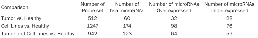
Table 1. Differential microRNAs among CC tissues and CC cell lines
Comparison Number of Probe set hsa-microRNAsNumber of Number of microRNAs Over-expressed Number of microRNAs Under-expressed
Tumor vs. Healthy 512 60 32 28
Cell Lines vs. Healthy 1247 174 98 76
Life Technologies, Carlsbad, CA, USA) and ana-lysed in a 3100 ABI PRISM Genetic Analyzer (Applied Biosystems). The sequences were aligned and compared with National Center for Biotechnology Information databases.
RNA isolation and quality control
The tissue samples were disrupted with TissueLyser (Qiagen) from 1 to 3 min. Total RNA
was obtained with the miRVana Isolation Kit
(Ambion, Life Technologies, CA, USA) according to the manufacturer’s instructions. RNA quan-tity and purity was measured using Nanodrop ND-1000 Spectrophotometer and RNA quality was analysed with Agilent RNA 6000 Nano in Agilent 2100 Bioanalyzer (Agilent Technologies, Santa Clara, CA, USA). In this study, we used only total RNA with RNA Integrated Number (RIN) >8.
Affymetrix miRNA microarray assay
We used the GeneChip miRNA Array (Affymetrix, Santa Clara, CA, USA). This chip contains
miR-NAs and pre-miRmiR-NAs of the Sanger miRNA database v11 of 71 organisms, such as: human, mouse, rat, dog, and monkey, among others. Each array contains 45,930 probes comprising of 7,788 miRNAs probe sets as quadruplicates and remaining probes as inter-nal controls. The assay was started with 1 µg of
total RNA. Total RNA samples were first extend -ed with the Poly (A)-Tailing Reaction follow-ed by the Biotin Labelling Reaction employing
FlashTag Biotin HSR RNA Labelling Kit (Genisphere, Hatfield, PA, USA). The labelling
process was checked with Enzyme Linked Oligosorbent Assay QC (ELOSA). Biotin-labelled total RNA samples were hybridized to the arrays, washed, and stained according to the manufacturer’s instructions. Microarrays were scanned using GeneChip Scanner 3000 7G
(Affymetrix). Median fluorescent intensity was
obtained with Affymetrix GeneChip Command Console software through Command Console Library File miRNA-1_0_2Xgain.
miRNA microarray analysis and target predic-tor analysis of miRNA
miRNA microarray data analysis was performed in three stages. First, the data were analysed for quality control purposes using free miRNA QC Tool software (Affymetrix). In this analysis, the background correction was adjusted by means of Robust Multi-chip Average (RMA). Subsequently, the data were log2-transformed, summarized, and normalized with Median-polish and Quantile, respectively. In the second stage, microarray analysis was achieved by
means of CEL files of Partek Genomics Suite
v6.6 software. Probe set were summarized by means of Median Polish, Quantile normaliza-tion. Background noises correction was
per-formed by means of RMA, finally the data were
transformed by Log2. The next step was inspec-tion of miRNA expression by means of Principal
Component Analysis (PCA). The final stage con -sisted of identifying differential microRNA expression in the samples by mean of Analysis of Variance (ANOVA) with the healthy cervical tissues as baseline. Moreover, CC cell lines, and CC tumors were compared also against healthy cervical tissue by geometric least
squares model. The significant microRNA
expressed were selected by means of cut-off based on fold change >2, <-2, and a False Discovery Ration (FDR) <0.05 (Table 1). Finally,
[image:4.612.91.306.96.386.2]the hierarchical cluster of significant expressed
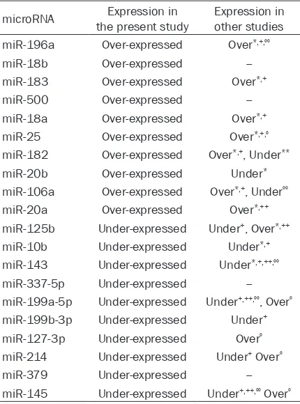
Table 2. miRNAs reported as over-expressed and under-expressed in cervical cells
microRNA the present studyExpression in Expression in other studies
miR-196a Over-expressed Over*,+,ºº
miR-18b Over-expressed
--miR-183 Over-expressed Over*,+
miR-500 Over-expressed
--miR-18a Over-expressed Over*,+
miR-25 Over-expressed Over*,+,º
miR-182 Over-expressed Over*,+, Under**
miR-20b Over-expressed Under*
miR-106a Over-expressed Over*,+, Underºº
miR-20a Over-expressed Over*,++
miR-125b Under-expressed Under+, Over*,++
miR-10b Under-expressed Under*,+
miR-143 Under-expressed Under*,+,++,ºº
miR-337-5p Under-expressed
--miR-199a-5p Under-expressed Under+,++,ºº, Overº
miR-199b-3p Under-expressed Under+
miR-127-3p Under-expressed Overº
miR-214 Under-expressed Under+ Overº
miR-379 Under-expressed
--miR-145 Under-expressed Under+,++,ºº Overº
mL of 100 mM dNTP 1.0 mL, MultiScribe Reverse Transcriptase 50 U/mL, 0.19 mL RNAase Inhibitor 20 U/mL, and free RNase water. The mix reaction was incubated for 30 min at 16°C, 30 min at 42°C, and for 5 min at 85°C. A 10-mL mix reaction of real-time PCR contained 1.33 mL RT product, 0.5 mL TaqMan Small RNA Assay (20X), 5.0 mL TaqMan Universal Master Mix II with UNG and 3.17 mL free RNase water. The mix reaction was incu-bated for 2 min at 50°C, 10 min at 95°C, and
40 amplification cycles for 15 sec at 95°C and for 60 sec at 60°C. Relative quantification was
calculated by the 2e(-DDCt) method, where DCt = Ct(miR-196a) - Ct(RNU6B) and DDCt = DCt(Cell line) -
DCt(Average Normal) [21].
Immunodetection of HOXC8 in cervical cancer cells
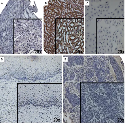
Detection of HOXC8 was performed in CC tis-sues and in cervical cell lines. For detection of cervical cancer tissues, a tissue microarray was constructed with 24 CC cases and normal cervical tissues by using a tissue microarrayer (Chemicon Co., Billerica, MA, USA). The immu-nostaining steps comprised antigen exposure with Trilogy solution, which was incubated for 45 min at 96°C. After blocking of endogenous peroxidase with 3% H2O2, the slide was incu-bated with Bovine Serum Albumin (BSA) for 30 min at room temperature. Next, the slides were incubated with polyclonal anti-HOXC8 antibody (ab86236; Abcam, Cambridge, MA, USA) over-night at 4°C (1:200 dilution in 1% BSA). Later, the slides were treated with Streptavidin-Biotin Complex Peroxidase Mouse/Rabbit Method (Dako, Glostup, Denmark) for 1 h at room tem-perature. Signal development was obtained with 0.05% 3,3-diaminobenzidine tetrahydro-chloride, 0.01% H2O2, and counterstained with haematoxylin. HOXC8 expression was evaluat-ed with 10X and 20X microscope as negative and positive staining. Healthy kidney or healthy bladder tissue were used as positive and nega-tive controls, respecnega-tively.
Results
Differential expression of cellular microRNAs in cervical cancer cell lines
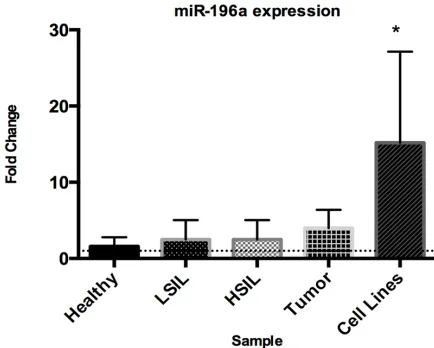
[image:5.612.88.305.71.245.2]microRNA expression was determined from 7,788 microRNAs in four healthy cervical tis-sues, four cervical cancer tissues and twelve Figure 2. miR-196a increased expression in cervical cells.
Tiny expression change of miR-196a in LSIL and HSIL with respect to healthy tissues, while a tendency of increased expression of miR-196 in cervical carcinomas was shown. As expected, a significant over-expression of miR-196a in CC cell lines was observed. X-axis corresponds to the sam-ples, Y-axis to the fold change of miR-196a in arbitrary units. (*) indicates that the expression of miR-196a level was significantly higher in CC cell lines.
microRNAs was obtained by means of Euclidian method (dissimilarity of samples).
For target predictor analysis of miR-196a we used different database as Diana-microT v5.0, TarBase v6.0, MirTarget2 v2, TargetScan v6.2, microRNA.org, PicTar and miRecords for pre-dicted miRNA-196a potential target. In addi-tion, we used GeneGo MetaCore (Thompson Reuters) to build the microRNA network to known predicted targets and regulator genes of miR-196a. We employed 50 interactions to generate a network; thus we were able to iden-tify genes upstream and downstream of micro-RNA to know the features and network of
spe-cific microRNA and target microRNA. For a more
detailed GeneGo MetaCore analysis, go to http://www.genego.com/.
miRNA quantitive RT-PCR
mRNA was subject to quantitative PCR using
the TaqMan miRNA Reverse Transcription Kit
cervical cancer cell lines using GeneChip miRNA microarray, Affymetrix. Selection of tis-sue for microarray study was performed using a
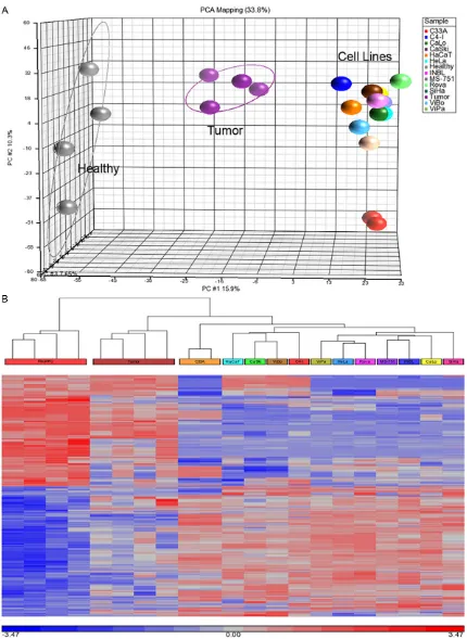
systematic workflow as described in the meth -ods. The overall average number of probe sets detected in the samples was 31.3% (2,439 of 7,788 for each chip) for healthy cervical tissue, 34% (2,720 of 7,788) for cervical tumor and 28.4% (2,213 of 7,788) for the 12 cell lines. Pearson correlation of 0.90-0.98 for quality controls by QC Tool was obtained. Therefore, the microarray data of healthy, tumor and cell lines were comparable for expression analysis.
The data clearly showed overall clustering of microRNA expression among the three studies group, indicating differences in microRNA expression by tumor tissues and CC cell lines compared with healthy cervical tissues (Figure 1A). Identification of altered microRNA in CC
[image:6.612.94.523.76.492.2]es were obtained out of which 518 were over-expressed and 424 under-over-expressed. The heat map displayed microRNA expression in the samples, where the tumor tissues showed a transition of microRNAs expression between CC cell lines and healthy tissue (Figure 1B). From initial list (815 microRNA sequences of 942 in total), only 123 sequences correspond to human sequences, representing 14.5% of altered expression of human microRNAs in CC cell lines. Most of these microRNA sequences are commonly found in different mammalian species such as rat, mouse, chimpanzee, goril-la, and dog, suggesting that microRNA group is highly conserved among species. Table 2
depicts some microRNAs that were identified in more than five species, including human, in
addition to their expression levels and other reports on CC.
Among the ten microRNAs, the most signifi -cantly over-expressed were: 196a, 18b, 183, 500, 18a, 25, miR-182, miR-20b, miR-106a, and miR-20a. On the
other hand, among significantly
under-exp-ressed were 145, 379, 214, 127-3p 199b 199a, 337-5p, miR-143, miR-10b, and miR-125b. All cell lines were cultured under the same growth conditions to avoid positive false in microRNA expression. In addition, a technical replica of C33A cell line in Principal Component Analysis (PCA) and heat map had similar result as those of the original analysis, revealing high reproducibility of results.
Increased expression of miR-196a in cervical cancer cell lines and cervical tissues
[image:7.612.90.306.103.713.2]One the most expressed microRNAs in tumor tissues and CC cells was miR-196a in microar-ray assay (p=4.75E-04, p=1.32E-07, respec-tively). To demonstrate over-expression of miR-196a, total RNA was subjected to quantitative RT-PCR assay to examine the relative expres-sion in LSIL (Low-grade Squamous Intraepi- thelial Lesion, n=10), HSIL (High-grade Squa- mous Intraepithelial Lesion, n=10), tumor tis-sues (n=15) and CC cell lines (n=12) compared with healthy cervical tissues (Figure 2). As expected, expression of miR-196a in LSIL and HSIL showed a tiny variation with respect to healthy tissues, whereas tumor tissues showed a slight increase in expression but not compa-rable to CC cell lines. Increase expression in all
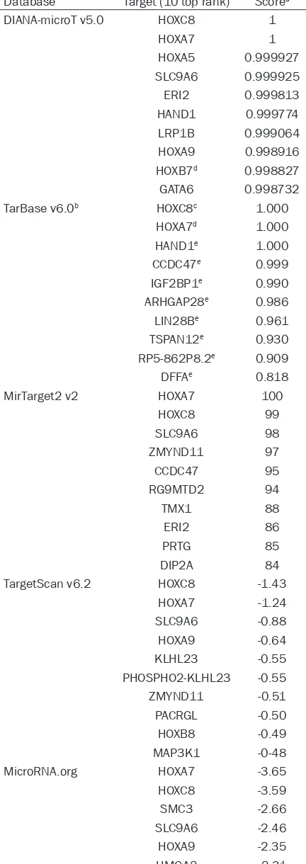
Table 3. Putative target of miR-196a in different database
Database Target (10 top rank) Scorea
DIANA-microT v5.0 HOXC8 1
HOXA7 1
HOXA5 0.999927
SLC9A6 0.999925
ERI2 0.999813
HAND1 0.999774
LRP1B 0.999064
HOXA9 0.998916
HOXB7d 0.998827
GATA6 0.998732
TarBase v6.0b HOXC8c 1.000
HOXA7d 1.000
HAND1e 1.000
CCDC47e 0.999
IGF2BP1e 0.990
ARHGAP28e 0.986
LIN28Be 0.961
TSPAN12e 0.930
RP5-862P8.2e 0.909
DFFAe 0.818
MirTarget2 v2 HOXA7 100
HOXC8 99
SLC9A6 98
ZMYND11 97
CCDC47 95
RG9MTD2 94
TMX1 88
ERI2 86
PRTG 85
DIP2A 84
TargetScan v6.2 HOXC8 -1.43
HOXA7 -1.24
SLC9A6 -0.88
HOXA9 -0.64
KLHL23 -0.55
PHOSPHO2-KLHL23 -0.55
ZMYND11 -0.51
PACRGL -0.50
HOXB8 -0.49
MAP3K1 -0-48
MicroRNA.org HOXA7 -3.65
HOXC8 -3.59
SMC3 -2.66
SLC9A6 -2.46
HOXA9 -2.35
CC tissues was not statistically significant,
whereas in CC cell lines, the increased
expres-sion of miR-196a was statistically significant
(p<0.0001) in comparison with healthy tiss- ues.
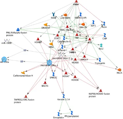
In order to find putative target genes of
miR-196a, we performed bioinformatics analysis. First analysis was performed to identify miR-196a network interaction using GeneGo Metacore Suite. This analysis showed down-stream and updown-stream interaction genes such as: embryonic development (HOX genes) and
cellular remodelling (Actin/Keratin) (Figure 3).
We identified indirect interaction genes such as
p53 and SMAD that are associated with cell
death [22]. Second, we identified miR-196a tar -get based on experimental data and/or bioin-formatics prediction such as: reporter gene assay, qPCR, western blot, sequencing, among others (Table 3). Target prediction analysis showed to HOXC8 as the best target of
miR-196a. Finally, in search of probable relationship in HOXC8 expression in CC, healthy cervical tis-sues and cervical carcinoma tistis-sues on tissue microarray were subjected to immunodetection with healthy bladder (negative tissue) and healthy kidney (positive tissue) as controls (Figure 4A, 4B), as well as CC cell lines (Figure 4C). Positive immunostaining was observed in nucleus of cervical epithelium cells (Figure 4D). In contrast, a negative immunoreaction was observed in the CC tissue and cell lines (Figure 4C and 4E).
Discussion
At present, CC is an important health problem worldwide. Despite the wide acceptance of CC screening programs in Mexico, more than 12,000 women are diagnosed with CC, and among these, 50% will die annually. It is accept-ed that CC is relataccept-ed to persistent HPV infec-tion; however, the knowledge on cervical car-cinogenesis is scarce. Most of the studies on CC are focused on HPV infection, gene expres-sion and currently on proteomic studies, all of which shade light on multiple hits presented in transformed cell [23]. During the last decade, microRNA studies in cancer research were
car-ried out define specific expression patterns and
those associated with diagnosis and prognosis [24].
In the present study, >100 human microRNAs were differentially expressed in CC tissues and CC cell lines in comparison with healthy cervi-ces. Among the microRNAs over-expressed were miR-25, -106b, -17-92, and -106a clusters that have been previously reported altered in several neoplasms [25-29]. These clusters are related to growth, and proliferation via Tumor Growth Factor-beta (TGF-ß), and inhibition of differentiation [30, 31]. We also observed an over-expression of miRNA-15b. Similar results have been previously reported in colorectal, lung, and pancreatic cancer [32-34], correlat-ing this over-expression with prognosis. These data support the fact that these microRNA clus-ters are commonly altered in human cancer and that they are probably associated with the process of carcinogenesis. Moreover, under-expressed has also been reported in miR-143/145 cluster in several cancer types includ-ing CC [15, 35]. This cluster is associated with angiogenesis and metastasis pathways [35-37]. The agreement of our results with previous data suggests that miRNA-145 could act as microRNA suppressor regulator in cancer.
CTBS -2.18
ZMYND11 -2.10
C1orf88 -2.07
C3orf88 -2.06
PicTar HOXC8 12.74
ZMYND11 6.56
CCNJ 5.08
MLR2 4.77
KIAA0685 4.64
HMGA2 4.32
PPP1R15B 4.27
FLJ46247 4.20
SLC9A6 4.18
DDX19 4.08
miRecords HOXC8
-GAN
-RBM26
-CDKN1B
-EPHA7
-ERG
-HOXB7
-CDYL
-ZNF385B
-RSPO2
To know whether all tumor tissues and cell lines exhibited similar behaviour in terms of microR-NA expression, the PCA and differential expres-sion analysis of the cells were carried out. As expected, the samples were grouped based on miRNA expression. In addition, heat map showed a transition of microRNAs expression to tumor tissues between healthy tissue and cell lines. Interestingly, all cell lines were clus-tered in PCA, except C33A cell line. It is known that C33A presents mutated pRb and p53 genes. These molecular events could explain our PCA results.
In order to know whether the presence of HPV
could influences cellular microRNA expression,
the analysis was performed in positive HPV and
negative HPV cell lines without finding any dif -ferential expression of microRNAs with regard to HPV infection. Furthermore, two of the three negative HPV cell lines (ViBo, and HaCaT cells) were clustered positive HPV. These data
sug-gest that the presence of specific HPV do not influence cellular microRNA expression in cell
[image:9.612.90.524.71.500.2]RNA expression [38]. This finding could support
partially the present results.
Furthermore, the most significantly
over-expressed microRNA in the tumor tissues and CC cell lines was miR-196a, this microRNA is a redundant sequence in the genome. MIR196A1 and MIR196A2 genes are transcribed and pro-cessed in microRNA biogenesis pathway to generate mature miR-196a, which are located on 17q21.32 (-) (in the RP11 gene) and 12q13.13 (+) (intron 1 of the HOXC5 and HOXC6 genes) (see Figure 5). Recent findings of our
group in comparative genomic hybridization analysis showed a gain of copy number of RP11 gene in CC samples (In preparation). In this situ-ation, we hypothesize that over-expression of miR-196a could derive in part from MIR196A1
gene amplification.
At present, there is scarce knowledge on expression of this microRNA in cancer, however several studies have reported its over-expres-sion in melanoma, oesophageal, lung, and pan-creatic carcinomas [39-42]. We found
statisti-cally significant over-expression of miR-196a in
CC cell lines in compared healthy tissues, whereas miR-196 expression in tumor tissues showed a slight increase but not comparable to CC cell lines, probably the low expression in CC tumors could be by cellular heterogeneity while
cell lines are clonal cells, as well as lesion pre-cancerous cervix (LSIL and HSIL).
In order to acquire some additional information
on potential target, first analysis consisted in a
network regulation of miR-196a based on GeneGo Metacore which showed that the potential targets of miR-196a were genes involved in embryonic development and cellular remodelling such as: HOXs (HOXC8, HOXA7,
HOXD8, HOXA9, HOXB8) and Actin/Keratin.
Second analysis, seven targets predictor
data-bases were used in the first analysis with all of
them depicting HOXC8 as the best target of miR-196a (Table 3), bioinformatics approaches were used and this revealed a potential regula-tor target of miR-196a to HOXC8. Moreover, HOXC8 has been validated as target of miR-196a in different studies [41, 43-45].
Recent works showed that the absence of HOXC8 expression in some tumors is related with progression and metastasis [43-45]. In CC, several member of the family of HOX genes have been evaluated, however did not show change in the level of expression of HOXA9, HOXB1, HOXB6, HOXB7 and HOXB8 [46, 47]. In additional, two studies has reported a contro-versial mRNA expression of HOXC8 [48, 49].
According to our bioinformatics finding and
[image:10.612.91.524.70.258.2]assay was performed in healthy cervical epi-thelium, CC tissues, and CC cell lines. The result of the assay indicated absence of HOXC8 protein in CC samples, suggesting that HOXC8 could be a gene involved in cervical cancer
pro-cess. This finding is in agreement with previous
study by Alami et al., [48]. We suggest that miR-196a could be an oncogenic element and an important factor in CC cells.
In summary, our present data is showing a het-erogeneity microRNAs expression in CC cells, suggesting that development of cancer in cervi-cal cells is an extremely complex event due to high complexity of microRNA expression among
other factors. We could not provide a specific microRNA expression profile related with the
HPV infection, which could suggest the
pres-ence of fine molecular events making to the CC
as more complex disease. Finally, we also sug-gest that miR-196a could be another oncogen-ic element in cervoncogen-ical cancer cells. However, we suggest that more studies be carried out to ascertain the results of the present study.
Acknowledgements
This work was supported in part by grant Fondos Sectoriales 69719 and 87244 from CONACYT Mexico. This paper constitutes a
par-tial fulfilment of the Graduate Program in PhD
Biological Sciences of National Autonomous University of México (UNAM) for Villegas-Ruiz V.
She acknowledges the scholarship and finan -cial support provided by CONACYT, UNAM and IMSS. We thanks to MD J. Ortiz and H. Serna for providing samples; Miss R. Coronel for helpful ELOSA microarrays standardization. Dra P. Piña for technical help for tissue microarrays, and PhD L Padilla for his critical review of this manuscript.
Disclosure of conflict of interest
The authors declare no interest conflicts. Address correspondence to: Dr. Mauricio Salcedo, Laboratorio de Oncologia Genómica, Unidad de Investigación Médicas en Enferemedades Oncologí- cas, Hospital de Oncologia, Centro Medico Nacional Siglo XXI, IMSS, Av Cuahtémoc 330, Col Doctores, Mexico, D.F, 06720. Tel: (01) 55 562 769 00 ext 2796; E-mail: maosal89@yahoo.com
References
[1] Gregory RI and Shiekhattar R. MicroRNA bio-genesis and cancer. Cancer Res 2005; 65: 3509-3512.
[2] Meltzer PS. Cancer genomics: small RNAs with big impacts. Nature 2005; 435: 745-746. [3] Mendell JT. MicroRNAs: critical regulators of
development, cellular physiology and malig-nancy. Cell Cycle 2005; 4: 1179-1184. [4] Fabbri M, Croce CM and Calin GA. MicroRNAs.
Cancer J 2008; 14: 1-6.
[5] Lytle JR, Yario TA and Steitz JA. Target mRNAs are repressed as efficiently by microRNA-bind -ing sites in the 5’ UTR as in the 3’ UTR. Proc Natl Acad Sci U S A 2007; 104: 9667-9672. [6] Ouellet DL, Perron MP, Gobeil LA, Plante P and
Provost P. MicroRNAs in gene regulation: when the smallest governs it all. J Biomed Biotechnol 2006; 2006: 69616.
[7] Calin GA and Croce CM. MicroRNA signatures in human cancers. Nat Rev Cancer 2006; 6: 857-866.
[8] Esquela-Kerscher A and Slack FJ. Oncomirs - microRNAs with a role in cancer. Nat Rev Can-cer 2006; 6: 259-269.
[9] Lu J, Getz G, Miska EA, Alvarez-Saavedra E, Lamb J, Peck D, Sweet-Cordero A, Ebert BL, Mak RH, Ferrando AA, Downing JR, Jacks T, Horvitz HR and Golub TR. MicroRNA expres-sion profiles classify human cancers. Nature 2005; 435: 834-838.
[10] Volinia S, Calin GA, Liu CG, Ambs S, Cimmino A, Petrocca F, Visone R, Iorio M, Roldo C, Ferracin M, Prueitt RL, Yanaihara N, Lanza G, Scarpa A, Vecchione A, Negrini M, Harris CC and Croce CM. A microRNA expression signature of hu-man solid tumors defines cancer gene targets. Proc Natl Acad Sci U S A 2006; 103: 2257-2261.
[11] World Health Organization. http://www.who. int/mediacentre/factsheets/fs380/en/. [12] Moody CA and Laimins LA. Human
papillomavi-rus oncoproteins: pathways to transformation. Nat Rev Cancer 2010; 10: 550-560.
[13] Hu X, Schwarz JK, Lewis JS Jr, Huettner PC, Rader JS, Deasy JO, Grigsby PW and Wang X. A microRNA expression signature for cervical cancer prognosis. Cancer Res 2010; 70: 1441-1448.
[14] Lee JW, Choi CH, Choi JJ, Park YA, Kim SJ, Hwang SY, Kim WY, Kim TJ, Lee JH, Kim BG and Bae DS. Altered MicroRNA expression in cervi-cal carcinomas. Clin Cancer Res 2008; 14: 2535-2542.
[15] Lui WO, Pourmand N, Patterson BK and Fire A. Patterns of known and novel small RNAs in hu-man cervical cancer. Cancer Res 2007; 67: 6031-6043.
[17] Muralidhar B, Goldstein LD, Ng G, Winder DM, Palmer RD, Gooding EL, Barbosa-Morais NL, Mukherjee G, Thorne NP, Roberts I, Pett MR and Coleman N. Global microRNA profiles in cervical squamous cell carcinoma depend on Drosha expression levels. J Pathol 2007; 212: 368-377.
[18] Pereira PM, Marques JP, Soares AR, Carreto L and Santos MA. MicroRNA expression variabil-ity in human cervical tissues. PLoS One 2010; 5: e11780.
[19] Wang X, Tang S, Le SY, Lu R, Rader JS, Meyers C and Zheng ZM. Aberrant expression of onco-genic and tumor-suppressive microRNAs in cervical cancer is required for cancer cell growth. PLoS One 2008; 3: e2557.
[20] Schmitt M, Dondog B, Waterboer T and Pawlita M. Homogeneous amplification of genital hu -man alpha papillomaviruses by PCR using novel broad-spectrum GP5+ and GP6+ prim-ers. J Clin Microbiol 2008; 46: 1050-1059. [21] Livak KJ and Schmittgen TD. Analysis of rela
-tive gene expression data using real-time quantitative PCR and the 2(-Delta Delta C(T)) Method. Methods 2001; 25: 402-408. [22] Atfi A and Baron R. p53 brings a new twist to
the Smad signaling network. Sci Signal 2008; 1: pe33.
[23] Higareda-Almaraz JC, Enriquez-Gasca Mdel R, Hernandez-Ortiz M, Resendis-Antonio O and Encarnacion-Guevara S. Proteomic patterns of cervical cancer cell lines, a network perspec-tive. BMC Syst Biol 2011; 5: 96.
[24] Schoof CR, Botelho EL, Izzotti A and Vasques Ldos R. MicroRNAs in cancer treatment and prognosis. Am J Cancer Res 2012; 2: 414-433. [25] Ambs S, Prueitt RL, Yi M, Hudson RS, Howe
TM, Petrocca F, Wallace TA, Liu CG, Volinia S, Calin GA, Yfantis HG, Stephens RM and Croce CM. Genomic profiling of microRNA and mes -senger RNA reveals deregulated microRNA ex-pression in prostate cancer. Cancer Res 2008; 68: 6162-6170.
[26] Diosdado B, van de Wiel MA, Terhaar Sive Droste JS, Mongera S, Postma C, Meijerink WJ, Carvalho B and Meijer GA. MiR-17-92 cluster is associated with 13q gain and c-myc expres-sion during colorectal adenoma to adenocarci-noma progression. Br J Cancer 2009; 101: 707-714.
[27] Kim YK, Yu J, Han TS, Park SY, Namkoong B, Kim DH, Hur K, Yoo MW, Lee HJ, Yang HK and Kim VN. Functional links between clustered microRNAs: suppression of cell-cycle inhibitors by microRNA clusters in gastric cancer. Nucleic Acids Res 2009; 37: 1672-1681.
[28] Motoyama K, Inoue H, Takatsuno Y, Tanaka F, Mimori K, Uetake H, Sugihara K and Mori M. Over- and under-expressed microRNAs in
hu-man colorectal cancer. Int J Oncol 2009; 34: 1069-1075.
[29] Pichiorri F, Suh SS, Ladetto M, Kuehl M, Pa -lumbo T, Drandi D, Taccioli C, Zanesi N, Alder H, Hagan JP, Munker R, Volinia S, Boccadoro M, Garzon R, Palumbo A, Aqeilan RI and Croce CM. MicroRNAs regulate critical genes associ-ated with multiple myeloma pathogenesis. Proc Natl Acad Sci U S A 2008; 105: 12885-12890.
[30] Jevnaker AM, Khuu C, Kjole E, Bryne M and Os -mundsen H. Expression of members of the miRNA17-92 cluster during development and in carcinogenesis. J Cell Physiol 2011; 226: 2257-2266.
[31] Li L, Shi JY, Zhu GQ and Shi B. MiR-17-92 clus-ter regulates cell proliferation and collagen synthesis by targeting TGFB pathway in mouse palatal mesenchymal cells. J Cell Biochem 2012; 113: 1235-1244.
[32] Miko E, Czimmerer Z, Csanky E, Boros G, Bus-lig J, Dezso B and Scholtz B. Differentially ex-pressed microRNAs in small cell lung cancer. Exp Lung Res 2009; 35: 646-664.
[33] Xi Y, Formentini A, Chien M, Weir DB, Russo JJ, Ju J, Kornmann M and Ju J. Prognostic Values of microRNAs in Colorectal Cancer. Biomark Insights 2006; 2: 113-121.
[34] Zhang Y, Li M, Wang H, Fisher WE, Lin PH, Yao Q and Chen C. Profiling of 95 microRNAs in pancreatic cancer cell lines and surgical speci-mens by real-time PCR analysis. World J Surg 2009; 33: 698-709.
[35] Akao Y, Nakagawa Y and Naoe T. MicroRNAs 143 and 145 are possible common onco-mi-croRNAs in human cancers. Oncol Rep 2006; 16: 845-850.
[36] Takagi T, Iio A, Nakagawa Y, Naoe T, Tanigawa N and Akao Y. Decreased expression of mi-croRNA-143 and -145 in human gastric can-cers. Oncology 2009; 77: 12-21.
[37] Wang CJ, Zhou ZG, Wang L, Yang L, Zhou B, Gu J, Chen HY and Sun XF. Clinicopathological sig-nificance of microRNA-31, -143 and -145 ex -pression in colorectal cancer. Dis Markers 2009; 26: 27-34.
[38] Greco D, Kivi N, Qian K, Leivonen SK, Auvinen P and Auvinen E. Human papillomavirus 16 E5 modulates the expression of host microRNAs. PLoS One 2011; 6: e21646.
[39] Braig S, Mueller DW, Rothhammer T and Bosserhoff AK. MicroRNA miR-196a is a cen -tral regulator of HOX-B7 and BMP4 expression in malignant melanoma. Cell Mol Life Sci 2010; 67: 3535-3548.
Albar-racin CT. MicroRNA-196a targets annexin A1: a microRNA-mediated mechanism of annexin A1 downregulation in cancers. Oncogene 2008; 27: 6667-6678.
[41] Schimanski CC, Frerichs K, Rahman F, Berger M, Lang H, Galle PR, Moehler M and Gockel I. High miR-196a levels promote the oncogenic phenotype of colorectal cancer cells. World J Gastroenterol 2009; 15: 2089-2096.
[42] Liu XH, Lu KH, Wang KM, Sun M, Zhang EB, Yang JS, Yin DD, Liu ZL, Zhou J, Liu ZJ, De W and Wang ZX. MicroRNA-196a promotes non-small cell lung cancer cell proliferation and in-vasion through targeting HOXA5. BMC Cancer 2012; 12: 348.
[43] Li Y, Zhang M, Chen H, Dong Z, Ganapathy V, Thangaraju M and Huang S. Ratio of miR-196s to HOXC8 messenger RNA correlates with breast cancer cell migration and metastasis. Cancer Res 2010; 70: 7894-7904.
[44] Mueller DW and Bosserhoff AK. MicroRNA miR-196a controls melanoma-associated ge- nes by regulating HOX-C8 expression. Int J Can-cer 2011; 129: 1064-1074.
[45] Adwan H, Zhivkova-Galunska M, Georges R, Eyol E, Kleeff J, Giese NA, Friess H, Bergmann F and Berger MR. Expression of HOXC8 is in-versely related to the progression and metas-tasis of pancreatic ductal adenocarcinoma. Br J Cancer 2011; 105: 288-295.
[46] Lopez R, Garrido E, Pina P, Hidalgo A, Lazos M, Ochoa R and Salcedo M. HOXB homeobox gene expression in cervical carcinoma. Int J Gynecol Cancer 2006; 16: 329-335.
[47] Lopez R, Garrido E, Vazquez G, Pina P, Perez C, Alvarado I and Salcedo M. A subgroup of HOX Abd-B gene is differentially expressed in cervi-cal cancer. Int J Gynecol Cancer 2006; 16: 1289-1296.
[48] Alami Y, Castronovo V, Belotti D, Flagiello D and Clausse N. HOXC5 and HOXC8 expression are selectively turned on in human cervical cancer cells compared to normal keratinocytes. Bio-chem Biophys Res Commun 1999; 257: 738-745.