research communications
Acta Cryst.(2017). E73, 905–907 https://doi.org/10.1107/S2056989017007642
905
Received 17 May 2017Accepted 23 May 2017
Edited by W. T. A. Harrison, University of Aberdeen, Scotland
Keywords:crystal structure; N-heterocyclic phosphine; NHP; 2-azido-1,3,2-diazaphospho-lidine.
CCDC reference:1551849
Supporting information:this article has supporting information at journals.iucr.org/e
Crystal structure of
2-azido-1,3-bis(2,6-diiso-propylphenyl)-1,3,2-diazaphospholidine
Alex J. Veinot, Amber D. Blair and Jason D. Masuda*
Department of Chemistry, Saint Mary’s University, 923 Robie St., Halifax, Nova Scotia, B3H 3C3, Canada. *Correspondence e-mail: jason.masuda@smu.ca
The title compound, C26H38N5P, was synthesized by reacting
2-chloro-1,3-bis(2,6-diisopropylphenyl)-1,3,2-diazaphospholidine with sodium azide and a catalytic amount of lithium chloride in tetrahydrofuran. The title compound is the first structurally characterized 2-azido-1,3,2-diazaphospholidine and exhibits a P atom in a trigonal pyramidal geometry. The azide P—N bond length of 1.8547 (16) A˚ is significantly longer than the P—N separations for the chelating diamine [P—N = 1.6680 (15) and 1.6684 (14) A˚ ]. The sterically hindered 2,6-diisopropylphenyl groups twist away from the central heterocycle, with dihedral angles between the central heteocyclic ring and benzene rings of 76.17 (10) and 79.74 (9). In the crystal, a weak C—H N link to the terminal N atom of the
azide group leads to [100] chains.
1. Chemical context
Phosphine azides possess at least one azide group attached to phosphorus and display a broad range of reactivity that is directly dependent on the other substituents attached to the P atom. One of the most interesting properties of these mol-ecules is that both free and coordinated alkyl and aryl deri-vatives are much more reactive than their corresponding amino derivatives, as demonstrated by their lower thermal and photochemical stability. For example, the phosphinoazide complex Ph2P(N3)–Cr(CO)5 readily undergoes photolysis
under UV light to produce the phosphino–isocyanate complex Ph2P(NCO)–Cr(CO)5(Ocandoet al., 1985), while the related
bis(diisopropylamino) complex (iPr2N)2P(N3)–Cr(CO)5 does
not (Cowley et al., 1995). The crystal structure of the title compound is the first reported example of a structurally characterized 2-azido-1,3,2-diazaphospholidine; however, a few closely related compounds are known, such as those derived from 1,3,2-diazaphospholenes.
2. Structural commentary
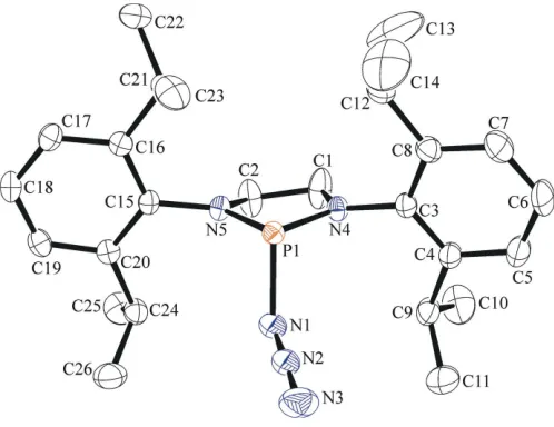
[image:1.610.190.420.578.703.2]The molecular structure of the title compound is shown in Fig. 1. It crystallizes in the monoclinic space groupP21/nwith
one molecule in the asymmetric unit. The bond lengths between the P atom and its flanking N atoms are similar [P1— N4 = 1.6680 (15) A˚ , P1—N5 = 1.6684 (14) A˚ and N4—P1— N5 = 91.14 (7)], while the phosphorus centre adopts a
trigonal pyramidal geometry, with the sum of the angles at phosphorus equal to 294.14 (7). The azide group is
quasi-linear [N3—N2—N1 = 176.6 (2)], with similar N—N bond
lengths [N1—N2 = 1.168 (2) A˚ and N2—N3 = 1.155 (2) A˚]. The phosphorus–azide bond length (P1—N1) is significantly longer [1.8547 (16) A˚ ] than found for atoms N4 and N5. The average sum of the bond angles at the N4 and N5 positions is 359.87 (12), very close to an ideal trigonal planar geometry.
This is a strong indication that the nominal lone pairs of atoms N4 and N5 participate in N—P interactions and, when coupled with the significantly longer P1—N1 bond length, suggests a partial ionic character similar to earlier reports in acyclic structures (Cowley et al., 1995). The overall confor-mation of the C1/C2/N4/N5/P1 ring is well described as an envelope, with atom N5 deviating from the other atoms (r.m.s. deviation = 0.030 A˚ ) by0.274 (2) A˚ . The steric demands of the bulky 2,6-diisopropylphenyl groups cause the aromatic rings to twist away from the central five-membered ring, with torsion angles of 103.69 (18) and 101.83 (17) for P1—N1—
C3—C4 and P1—N2—C15—C20, respectively. The isopropyl groups are oriented away from the central five-membered
ring, but the ‘congested’ nature of the molecule results in intramolecular short contacts between all four of the methine H atoms (H9, H12, H21 and H24) and atoms N4 and N5 (Table 1).
3. Supramolecular features
The only significant directional interaction in the crystal of the title compound is a long [2.69 (3) A˚ ] C—H N hydrogen bond to the terminal N atom of the azide group, which results in [100] chains in the crystal (Fig. 2).
4. Database survey
A search of the Cambridge Structural Database (Groomet al., 2016) indicated that no other 2-azido-1,3,2-(diarylamino)-phospholidine derivatives have been structurally character-ized. Some structurally similar compounds were identified, however, namely 2-azido-1,3-bis(2,6-diisopropyllphenyl)-1,3,2-diazaphospholene (CSD refcode CILBAC; Gedigaet al., 2014) and its corresponding 2,6-dimethylphenyl derivative (GOFHAL; Burcket al., 2008). Acyclic derivatives featuring bis(diisopropylamino) (PIJZAJ; Englert et al., 1993) and bis(dicylohexylamino) (ZABCEK; Cowleyet al., 1995) ligands are known, and also 1-azido-N,N0-bis(2,4,6-tri-tert
-butyl-phenyl)phosphinediamine (YABVUV; Niegeret al., 2016).
5. Synthesis and crystallization
The synthesis of the title compound was achieved using a similar method as reported in the literature for 2-azido-1,3-bis(2,6-diisopropyllphenyl)-1,3,2-diazaphospholene (Gediga et al., 2014). In a 20 ml scintillation vial, 0.102 g (0.229 mmol, 1 eq.) of colourless 2-chloro-1,3-bis(2,6-diisopropylphenyl)-1,3,2-diazaphospholidine were dissolved in 1 ml of THF producing a colourless solution. To this solution, 0.016 g
906
Veinotet al. C26H38N5P Acta Cryst.(2017). E73, 905–907
[image:2.610.313.564.69.256.2]research communications
Table 1
Hydrogen-bond geometry (A˚ ,).
D—H A D—H H A D A D—H A
C9—H9 N4 1.00 2.43 2.926 (2) 110 C12—H12 N4 1.00 2.44 2.913 (2) 109 C21—H21 N5 1.00 2.49 2.932 (2) 106 C24—H24 N1 1.00 2.66 3.443 (3) 136 C24—H24 N5 1.00 2.46 2.955 (2) 110 C22—H22C N3i 0.98 2.69 3.669 (3) 174
Symmetry code: (i)x1;y;z.
Figure 2
[image:2.610.42.298.93.169.2] [image:2.610.46.295.525.717.2]The packing of the title compound, showing intermolecular C—H N interactions as dashed lines, which result in [100] chains.
Figure 1
(0.246 mmol, 1.1 eq.) of colourless sodium azide and a spatula tip (<1 mg) of lithium chloride were added to solution immediately producing a colourless mixture. The reaction mixture was left to stir for 1 d and monitored using 31P{1H} NMR spectroscopy, and once the starting material was completely consumed the reaction mixture was driedin vacuo. Extraction of the colourless residue with cold pentane, followed by filtration through Celite produced a colourless solution, which afforded 0.060 g (60%) of the title compound as a colourless powder after removal of the solvent. Crystals of the product were obtained by concentrating the filtrate and storing in a 238 K freezer overnight.1H NMR (CDCl3):7.31
(t, 3JHH= 7.6 Hz, 2H, p-Dipp), 7.24–7.17 (m, 4H, m-Dipp),
3.88–3.82 (pseudo-q, 2H, NHC-CH2), 3.74 (sept, 3
JHH= 6.8 Hz,
2H, iPr-CH), 3.48–3.39 (m, 4H, iPr-CH, NHC-CH2), 1.33–1.25
(m, 3JHH= 6.8 Hz, 24H, iPr-CH3).13C{1H} NMR (CDCl3):
150.3, 148.4, 136.2, 128.1, 124.7, 124.2, 54.4, 29.0, 25.3, 24.9, 24.5.31P{1H} NMR (CDCl
3):129.8. IR (KBr pellet):3062
(w), 2963 (s), 2926 (m), 2867 (m), 2500 (w), 2125 (m), 2085 (s, N N N), 1678 (w), 1584 (w), 1462 (s), 1445 (s), 1383 (m), 1363 (m), 1324 (m), 1323 (m), 1257 (s), 1211 (w), 1185 (w), 1106 (m), 1075 (s), 1056 (w), 1043 (w), 980 (w), 946 (w), 935 (w), 852 (w), 806 (s), 761 (s), 730 (w), 688 (w), 651 (w), 602 (w), 583 (w), 550 (w), 542 (w), 470 (s), 437 cm1 (w). M.p. (K): 415.4–417.6 (decomposes, gas was released).
6. Refinement
Crystal data, data collection and structure refinement details are summarized in Table 2. H atoms were included in geometrically idealized positions and refined using a riding model. Dihedral angles for the methyl H atoms were allowed to refine freely. The atomic displacement parameters of atoms N1 and N2 were constrained to be approximately equal using an EADP command.
Acknowledgements
We thank the Natural Sciences and Engineering Research Council of Canada (through the Discovery Grants Program to JDM). JDM also acknowledges support from the Canadian Foundation for Innovation, the Nova Scotia Research and Innovation Trust Fund and Saint Mary’s University.
References
Bruker (2008). APEX2, SAINT and SADABS. Bruker AXS Inc., Madison, Wisconsin, USA.
Burck, S., Gudat, D., Nieger, M., Schalley, C. A. & Weilandt, T. (2008).Dalton Trans.pp. 3478–3485.
Cowley, A. H., Gabbaı¨, F. P., Bertrand, G., Carrano, C. J. & Bond, M. R. (1995).J. Organomet. Chem.493, 95–99.
Englert, U., Paetzold, P. & Eversheim, E. (1993).Z. Kristallogr.208, 307–309.
Farrugia, L. J. (2012).J. Appl. Cryst.45, 849–854.
Gediga, M., Burck, S., Bender, J., Fo¨rster, D., Nieger, M. & Gudat, D. (2014).Eur. J. Inorg. Chem.pp. 1818–1825.
Groom, C. R., Bruno, I. J., Lightfoot, M. P. & Ward, S. C. (2016).Acta Cryst.B72, 171–179.
Nieger, M., Niecke, E. & Larbig, M. (2016). Private communication (refcode YABVUV). CCDC, Cambridge, England.
Ocando, E., Majid, S., Pierre Majoral, J., Baceiredo, A. & Bertrand, G. (1985).Polyhedron,4, 1667–1668.
Sheldrick, G. M. (2015a).Acta Cryst.A71, 3–8. Sheldrick, G. M. (2015b).Acta Cryst.C71, 3–8. Spek, A. L. (2009).Acta Cryst.D65, 148–155. Westrip, S. P. (2010).J. Appl. Cryst.43, 920–925.
research communications
Acta Cryst.(2017). E73, 905–907 Veinotet al. C
[image:3.610.45.289.93.368.2]26H38N5P
907
Table 2
Experimental details.
Crystal data
Chemical formula C26H38N5P
Mr 451.58
Crystal system, space group Monoclinic,P21/n
Temperature (K) 150
a,b,c(A˚ ) 10.0148 (12), 17.343 (2), 15.6270 (19)
(
) 105.948 (2)
V(A˚3) 2609.7 (5)
Z 4
Radiation type MoK
(mm1) 0.13
Crystal size (mm) 0.390.350.27
Data collection
Diffractometer Siemens/Bruker APEXII Absorption correction Multi-scan (SADABS; Bruker,
2008) Tmin,Tmax 0.718, 0.746
No. of measured, independent and observed [I> 2(I)] reflections
29851, 5708, 4350
Rint 0.047
(sin /)max(A˚1) 0.639
Refinement
R[F2> 2(F2)],wR(F2),S 0.046, 0.123, 1.02
No. of reflections 5708 No. of parameters 291
H-atom treatment H-atom parameters constrained
max,min(e A˚3) 0.36,0.39
supporting information
sup-1
Acta Cryst. (2017). E73, 905-907
supporting information
Acta Cryst. (2017). E73, 905-907 [https://doi.org/10.1107/S2056989017007642]
Crystal structure of
2-azido-1,3-bis(2,6-diisopropylphenyl)-1,3,2-diazaphospho-lidine
Alex J. Veinot, Amber D. Blair and Jason D. Masuda
Computing details
Data collection: APEX2 (Bruker, 2008); cell refinement: SAINT (Bruker, 2008); data reduction: SAINT (Bruker, 2008);
program(s) used to solve structure: SHELXT2014 (Sheldrick, 2015a); program(s) used to refine structure: SHELXL2016
(Sheldrick, 2015b); molecular graphics: ORTEP-3 for Windows (Farrugia, 2012) and PLATON (Spek, 2009); software
used to prepare material for publication: publCIF (Westrip, 2010).
2-Azido-1,3-bis(2,6-diisopropylphenyl)-1,3,2-diazaphospholidine
Crystal data
C26H38N5P
Mr = 451.58 Monoclinic, P21/n
a = 10.0148 (12) Å
b = 17.343 (2) Å
c = 15.6270 (19) Å
β = 105.948 (2)°
V = 2609.7 (5) Å3
Z = 4
F(000) = 976
Dx = 1.149 Mg m−3
Mo Kα radiation, λ = 0.71073 Å Cell parameters from 5265 reflections
θ = 2.2–25.2°
µ = 0.13 mm−1
T = 150 K Block, colourless 0.39 × 0.35 × 0.27 mm
Data collection
Siemens/Bruker APEXII diffractometer
Detector resolution: 66 pixels mm-1
φ and ω scans
Absorption correction: multi-scan (SADABS; Bruker, 2008)
Tmin = 0.718, Tmax = 0.746 29851 measured reflections
5708 independent reflections 4350 reflections with I > 2σ(I)
Rint = 0.047
θmax = 27.0°, θmin = 1.8°
h = −12→12
k = −22→22
l = −19→19
Refinement
Refinement on F2 Least-squares matrix: full
R[F2 > 2σ(F2)] = 0.046
wR(F2) = 0.123
S = 1.02 5708 reflections 291 parameters 0 restraints
Primary atom site location: structure-invariant direct methods
Hydrogen site location: inferred from neighbouring sites
H-atom parameters constrained
w = 1/[σ2(F
o2) + (0.0515P)2 + 1.4293P] where P = (Fo2 + 2Fc2)/3
supporting information
sup-2
Acta Cryst. (2017). E73, 905-907 Special details
Geometry. All esds (except the esd in the dihedral angle between two l.s. planes) are estimated using the full covariance matrix. The cell esds are taken into account individually in the estimation of esds in distances, angles and torsion angles; correlations between esds in cell parameters are only used when they are defined by crystal symmetry. An approximate (isotropic) treatment of cell esds is used for estimating esds involving l.s. planes.
Fractional atomic coordinates and isotropic or equivalent isotropic displacement parameters (Å2)
x y z Uiso*/Ueq
supporting information
sup-3
Acta Cryst. (2017). E73, 905-907
H14B 0.305181 0.567885 0.031406 0.123* H14C 0.164104 0.576421 0.059862 0.123* C15 0.58170 (17) 0.61147 (10) 0.42027 (11) 0.0207 (3) C16 0.46359 (18) 0.65928 (10) 0.40491 (11) 0.0219 (4) C17 0.46834 (19) 0.72263 (10) 0.46045 (12) 0.0274 (4) H17 0.389882 0.755641 0.450633 0.033* C18 0.5844 (2) 0.73839 (11) 0.52929 (12) 0.0305 (4) H18 0.585062 0.781626 0.566744 0.037* C19 0.6994 (2) 0.69150 (10) 0.54386 (12) 0.0281 (4) H19 0.779039 0.703016 0.591430 0.034* C20 0.70149 (18) 0.62737 (10) 0.49017 (11) 0.0231 (4) C21 0.33327 (18) 0.64485 (11) 0.32984 (12) 0.0277 (4) H21 0.331695 0.589036 0.313540 0.033* C22 0.2000 (2) 0.66235 (13) 0.35634 (17) 0.0453 (6) H22A 0.191522 0.718159 0.363168 0.068* H22B 0.203732 0.636691 0.412819 0.068* H22C 0.119651 0.643396 0.309992 0.068* C23 0.3355 (2) 0.69207 (13) 0.24749 (14) 0.0412 (5) H23A 0.254521 0.678656 0.198252 0.062* H23B 0.420602 0.680639 0.230557 0.062* H23C 0.332647 0.747141 0.260929 0.062* C24 0.83013 (19) 0.57663 (11) 0.51126 (12) 0.0285 (4) H24 0.815047 0.535143 0.465149 0.034* C25 0.8521 (2) 0.53809 (13) 0.60211 (14) 0.0442 (5) H25A 0.769090 0.508486 0.602823 0.066* H25B 0.869085 0.577684 0.648599 0.066* H25C 0.932307 0.503398 0.613113 0.066* C26 0.9599 (2) 0.62141 (13) 0.50874 (16) 0.0432 (5) H26A 0.946650 0.644086 0.449550 0.065* H26B 1.039822 0.586419 0.521593 0.065* H26C 0.976794 0.662518 0.553468 0.065*
Atomic displacement parameters (Å2)
U11 U22 U33 U12 U13 U23
supporting information
sup-4
Acta Cryst. (2017). E73, 905-907
C8 0.0265 (9) 0.0280 (10) 0.0285 (10) 0.0009 (7) 0.0029 (8) −0.0011 (8) C9 0.0325 (11) 0.0347 (11) 0.0301 (10) 0.0094 (8) 0.0027 (8) −0.0077 (8) C10 0.0661 (17) 0.0387 (13) 0.0538 (15) 0.0155 (12) −0.0002 (13) 0.0041 (11) C11 0.0322 (12) 0.0669 (17) 0.0542 (15) 0.0099 (11) 0.0081 (10) −0.0126 (12) C12 0.0262 (10) 0.0320 (11) 0.0448 (12) 0.0055 (8) −0.0022 (9) −0.0070 (9) C13 0.073 (2) 0.065 (2) 0.192 (4) 0.0142 (17) 0.086 (3) 0.009 (2) C14 0.098 (3) 0.0568 (19) 0.082 (2) 0.0371 (17) 0.0091 (19) 0.0182 (16) C15 0.0260 (9) 0.0196 (8) 0.0177 (8) −0.0012 (7) 0.0080 (7) 0.0004 (6) C16 0.0245 (9) 0.0198 (8) 0.0223 (9) −0.0013 (7) 0.0079 (7) 0.0033 (7) C17 0.0320 (10) 0.0206 (9) 0.0304 (10) 0.0035 (7) 0.0100 (8) 0.0000 (7) C18 0.0400 (11) 0.0231 (9) 0.0285 (10) −0.0017 (8) 0.0093 (8) −0.0069 (8) C19 0.0321 (10) 0.0280 (10) 0.0219 (9) −0.0041 (8) 0.0035 (7) −0.0014 (7) C20 0.0261 (9) 0.0222 (9) 0.0212 (8) −0.0006 (7) 0.0068 (7) 0.0025 (7) C21 0.0247 (9) 0.0230 (9) 0.0329 (10) −0.0002 (7) 0.0036 (8) −0.0010 (8) C22 0.0250 (11) 0.0445 (13) 0.0645 (15) −0.0016 (9) 0.0090 (10) −0.0103 (11) C23 0.0414 (12) 0.0395 (12) 0.0344 (11) 0.0035 (9) −0.0038 (9) 0.0086 (9) C24 0.0284 (10) 0.0263 (9) 0.0272 (10) 0.0026 (8) 0.0015 (7) −0.0014 (8) C25 0.0451 (13) 0.0438 (13) 0.0366 (12) 0.0078 (10) −0.0009 (10) 0.0118 (10) C26 0.0291 (11) 0.0429 (13) 0.0576 (14) 0.0040 (9) 0.0120 (10) 0.0013 (11)
Geometric parameters (Å, º)
supporting information
sup-5
Acta Cryst. (2017). E73, 905-907
C9—C11 1.528 (3) C23—H23C 0.9800 C9—H9 1.0000 C24—C26 1.524 (3) C10—H10A 0.9800 C24—C25 1.529 (3) C10—H10B 0.9800 C24—H24 1.0000 C10—H10C 0.9800 C25—H25A 0.9800 C11—H11A 0.9800 C25—H25B 0.9800 C11—H11B 0.9800 C25—H25C 0.9800 C11—H11C 0.9800 C26—H26A 0.9800 C12—C14 1.503 (3) C26—H26B 0.9800 C12—C13 1.516 (4) C26—H26C 0.9800
supporting information
sup-6
Acta Cryst. (2017). E73, 905-907
C8—C7—H7 119.4 H22A—C22—H22B 109.5 C7—C8—C3 117.90 (17) C21—C22—H22C 109.5 C7—C8—C12 119.86 (17) H22A—C22—H22C 109.5 C3—C8—C12 122.18 (17) H22B—C22—H22C 109.5 C4—C9—C10 110.70 (18) C21—C23—H23A 109.5 C4—C9—C11 111.68 (17) C21—C23—H23B 109.5 C10—C9—C11 110.99 (19) H23A—C23—H23B 109.5 C4—C9—H9 107.8 C21—C23—H23C 109.5 C10—C9—H9 107.8 H23A—C23—H23C 109.5 C11—C9—H9 107.8 H23B—C23—H23C 109.5 C9—C10—H10A 109.5 C20—C24—C26 112.19 (16) C9—C10—H10B 109.5 C20—C24—C25 110.59 (16) H10A—C10—H10B 109.5 C26—C24—C25 109.90 (17) C9—C10—H10C 109.5 C20—C24—H24 108.0 H10A—C10—H10C 109.5 C26—C24—H24 108.0 H10B—C10—H10C 109.5 C25—C24—H24 108.0 C9—C11—H11A 109.5 C24—C25—H25A 109.5 C9—C11—H11B 109.5 C24—C25—H25B 109.5 H11A—C11—H11B 109.5 H25A—C25—H25B 109.5 C9—C11—H11C 109.5 C24—C25—H25C 109.5 H11A—C11—H11C 109.5 H25A—C25—H25C 109.5 H11B—C11—H11C 109.5 H25B—C25—H25C 109.5 C14—C12—C13 109.7 (2) C24—C26—H26A 109.5 C14—C12—C8 112.9 (2) C24—C26—H26B 109.5 C13—C12—C8 110.17 (19) H26A—C26—H26B 109.5 C14—C12—H12 108.0 C24—C26—H26C 109.5 C13—C12—H12 108.0 H26A—C26—H26C 109.5 C8—C12—H12 108.0 H26B—C26—H26C 109.5 C12—C13—H13A 109.5
supporting information
sup-7
Acta Cryst. (2017). E73, 905-907
P1—N4—C3—C8 −77.2 (2) C17—C18—C19—C20 −0.2 (3) C8—C3—C4—C5 0.6 (3) C18—C19—C20—C15 −0.2 (3) N4—C3—C4—C5 179.73 (18) C18—C19—C20—C24 −178.33 (17) C8—C3—C4—C9 −178.35 (18) C16—C15—C20—C19 0.3 (2) N4—C3—C4—C9 0.8 (3) N5—C15—C20—C19 −176.51 (15) C3—C4—C5—C6 0.1 (3) C16—C15—C20—C24 178.31 (16) C9—C4—C5—C6 179.1 (2) N5—C15—C20—C24 1.5 (2) C4—C5—C6—C7 −0.6 (4) C17—C16—C21—C23 84.6 (2) C5—C6—C7—C8 0.4 (4) C15—C16—C21—C23 −94.5 (2) C6—C7—C8—C3 0.3 (3) C17—C16—C21—C22 −38.6 (2) C6—C7—C8—C12 −177.1 (2) C15—C16—C21—C22 142.23 (18) C4—C3—C8—C7 −0.8 (3) C19—C20—C24—C26 −58.9 (2) N4—C3—C8—C7 −179.90 (18) C15—C20—C24—C26 123.11 (19) C4—C3—C8—C12 176.48 (18) C19—C20—C24—C25 64.2 (2) N4—C3—C8—C12 −2.6 (3) C15—C20—C24—C25 −113.80 (19) C5—C4—C9—C10 −65.8 (3)
Hydrogen-bond geometry (Å, º)
D—H···A D—H H···A D···A D—H···A
C9—H9···N4 1.00 2.43 2.926 (2) 110 C12—H12···N4 1.00 2.44 2.913 (2) 109 C21—H21···N5 1.00 2.49 2.932 (2) 106 C24—H24···N1 1.00 2.66 3.443 (3) 136 C24—H24···N5 1.00 2.46 2.955 (2) 110 C22—H22C···N3i 0.98 2.69 3.669 (3) 174