Received 28 November 2019 Accepted 2 December 2019
Edited by W. T. A. Harrison, University of Aberdeen, Scotland
Keywords:crystal structure; diamide; hydrogen bonding; Hirshfeld surface analysis; computa-tional chemistry.
CCDC reference:1969282
Supporting information:this article has supporting information at journals.iucr.org/e
N
,
N
000-Bis(pyridin-3-ylmethyl)ethanediamide
mono-hydrate: crystal structure, Hirshfeld surface analysis
and computational study
Sang Loon Tan and Edward R. T. Tiekink*
Research Centre for Crystalline Materials, School of Science and Technology, Sunway University, 47500 Bandar Sunway, Selangor Darul Ehsan, Malaysia. *Correspondence e-mail: edwardt@sunway.edu.my
The molecular structure of the title bis-pyridyl substituted diamide hydrate, C14H14N4O2H2O, features a central C2N2O2 residue (r.m.s. deviation =
0.0205 A˚ ) linked at each end to 3-pyridyl rings through methylene groups. The pyridyl rings lie to the same side of the plane, i.e. have asyn-periplanar relationship, and form dihedral angles of 59.71 (6) and 68.42 (6) with the central plane. An almost orthogonal relationship between the pyridyl rings is indicated by the dihedral angle between them [87.86 (5)]. Owing to an anti disposition between the carbonyl-O atoms in the core, two intramolecular amide-N—H O(carbonyl) hydrogen bonds are formed, each closing anS(5) loop. Supramolecular tapes are formed in the crystal via amide-N— H O(carbonyl) hydrogen bonds and ten-membered { HNC2O}2 synthons.
Two symmetry-related tapes are linked by a helical chain of hydrogen-bonded water molecules via water-O—H N(pyridyl) hydrogen bonds. The resulting aggregate is parallel to theb-axis direction. Links between these,via methylene-C—H O(water) and methylene-C—H (pyridyl) interactions, give rise to a layer parallel to (101); the layers stack without directional interactions between them. The analysis of the Hirshfeld surfaces point to the importance of the specified hydrogen-bonding interactions, and to the significant influence of the water molecule of crystallization upon the molecular packing. The analysis also indicates the contribution of methylene-C—H O(carbonyl) and pyridyl-C— H C(carbonyl) contacts to the stability of the inter-layer region. The calculated interaction energies are consistent with importance of significant electrostatic attractions in the crystal.
1. Chemical context
Having both amide and pyridyl functionality, bis(pyridin-n -ylmethyl)ethanediamide molecules of the general formulan -NC5H4CH2N(H)C( O)C( O)CH2C5H4N-n, forn= 2, 3 and
4, hereafter nLH2, are attractive co-crystal coformers via
conventional hydrogen bonding. In the same way, complexa-tion to metals may also be envisaged. It is therefore not surprising that there is now a wealth of structural information for these molecules occurring in co-crystals, salts and metal complexes, as has been reviewed recently (Tiekink, 2017). Complementing hydrogen-bonding interactions, the nLH2
2019; Tan & Tiekink, 2019a,b,c) and complexation to zinc(II) 1,1-dithiolates (Armanet al., 2018; Tiekink, 2018; Tan, Chunet al., 2019), that the title compound, 3LH2H2O, (I), was
isolated. Herein, the crystal and molecular structures of (I) are described along with a detailed analysis of the molecular packing by means of an analysis of the calculated Hirshfeld surfaces, two-dimensional fingerprint plots and the calculation of energies of interaction.
2. Structural commentary
The molecular structures of the two constituents comprising the crystallographic asymmetric unit of (I) are shown in Fig. 1. The 3LH2 molecule lacks crystallographic symmetry and
comprises a central C2N2O2residue connected at either side to
two 3-pyridyl residues viamethylene links. The six atoms of the central residue are almost co-planar as seen in their r.m.s. deviation of 0.0205 A˚ : the maximum deviations above and below the plane are 0.0291 (9) A˚ for N3 and 0.0321 (11) A˚ for C8. The N1- and N3-pyridyl rings form dihedral angles of 59.71 (6) and 68.42 (6), respectively, with the central plane and lie to the same side of the plane, having asyn-periplanar relationship. The dihedral angle formed between the pyridyl rings is 87.86 (5), indicating an almost edge-to-face relation-ship. The carbonyl-O atoms have anantidisposition enabling the formation of intramolecular amide-N—H O(carbonyl) hydrogen bonds that closeS(5) loops, Table 1.
3. Supramolecular features
Significant conventional hydrogen bonding is noted in the crystal of (I) with the geometric parameters characterizing these included in Table 1. The most striking feature of the supramolecular association is the formation of tapes via
amide-N—H O(carbonyl) hydrogen bonds leading to a sequence of inter-connected ten-membered { HNC2O}2
synthons. Two such tapes are connected by hydrogen bonds provided by the water molecule of crystallization. Thus, alternating water molecules in helical chains of hydrogen-bonded water molecules, being aligned along the b-axis direction and propagated by 21symmetry, connect to3LH2via
water-O—H N(pyridyl) hydrogen bonds to form the one-dimensional aggregate shown in Fig. 2(a). The presence of methylene-C—H O(water) and methylene-C—H
(pyridyl) contacts stabilizes a layer lying parallel to (101). The layers stack without directional interactions between them, Fig. 2(b).
26
Tan and Tiekink C14H14N4O2H2O Acta Cryst.(2020). E76, 25–31
[image:2.610.79.261.174.269.2]research communications
Figure 1
[image:2.610.316.563.242.619.2]The molecular structure of the constituents of (I) showing the atom-labelling scheme and displacement ellipsoids at the 70% probability level. The water-O—H N(pyridyl) hydrogen bond is indicated by the dashed line.
Figure 2
Molecular packing in the crystal of (I): (a) one-dimensional chain whereby tapes sustained by amide-N—H O(carbonyl) hydrogen bonds and ten-membered { HNC2O}2synthons are connected,viawater-O—
[image:2.610.44.295.609.698.2]4. Hirshfeld surface analysis
The calculations of the Hirshfeld surfaces and two-dimen-sional fingerprint plots were performed on the crystal-lographic asymmetric unit shown in Fig. 1, using Crystal Explorer 17(Turneret al., 2017) and based on the procedures as described previously (Tan, Jotaniet al., 2019). The analysis identified a number of red spots on thednormsurface of
3
LH2
with varying degrees of intensity indicating the presence of interactions with contact distances shorter than the sum of the respective van der Waals radii (Spackman & Jayatilaka, 2009). Referring to the images of Fig. 3, the most intense red spots stem from the amide-N—H O(carbonyl) and water-O— H N(pyridyl) hydrogen bonds, Table 1. Some additional
contacts are detected through the Hirshfeld surface analysis for C1—H1 O1W, C5–H5 N4, C12—H12 C7, C6– H6A O2 and C7 O1 interactions with the red spots ranging from moderately to weakly intense. The data in Table 2 provide a succinct summary of interatomic contacts revealed in the above analysis; the O2 H6Aand C7 H12 contacts occur in the inter-layer region.
To verify the nature of the aforementioned interactions, the
3
LH2molecule in (I) was subjected to electrostatic potential
mapping. The results show that almost all of the interactions identified through the dnorm mapping are electrostatic in
nature as can be seen from the distinctive blue
(electro-Table 1
Hydrogen-bond geometry (A˚ ,).
D—H A D—H H A D A D—H A
N2—H2N O2 0.85 (2) 2.36 (2) 2.7279 (18) 107.0 (16) N3—H3N O1 0.86 (2) 2.299 (19) 2.6924 (18) 108.0 (15) O1W—H1W N1 0.95 (2) 1.86 (2) 2.7958 (18) 169 (2) O1W—H2W O1Wi 0.88 (2) 1.97 (2) 2.8364 (15) 166 (2) N2—H2N O1ii 0.85 (2) 2.03 (2) 2.8227 (18) 155.2 (18)
N3—H3N O2iii 0.86 (2) 2.02 (2) 2.8022 (18) 151.6 (17) C9—H9A O1Wiv 0.99 2.45 3.3772 (19) 156
C6—H6B Cg1iii 0.99 2.74 3.7043 (16) 166
Symmetry codes: (i) xþ3 2;yþ
1 2;zþ
3
2; (ii) x;yþ1;z; (iii) x;y1;z; (iv)
x1 2;yþ
1 2;z
[image:3.610.46.296.85.189.2]1 2.
Figure 3
Thednormmapping of the Hirshfeld surface for3LH2in (I) within the
[image:3.610.315.566.93.187.2]range of0.3259 to 1.0656 arbitrary units, showing the red spots for (a) N2—H2N O1 (intense, connected by green dashed line), N3— H3N O2 (intense, green dashed line) and C6—H6A O2 (diminutive, green dashed line) interactions, (b) O1W—H1W N1 (intense, yellow dashed line), C5—H5 N4 (moderately intense, yellow dashed line) and C7 O1 (diminutive, blue dashed line) interactions.
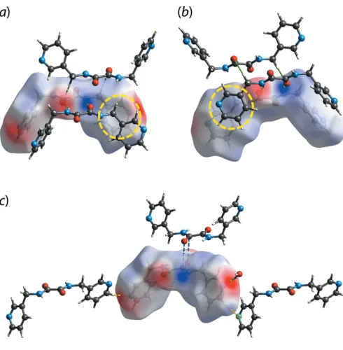
Figure 4
The electrostatic potential mapped onto the Hirshfeld surface within the isosurface value of 0.0964 to 0.1012 atomic units for 3LH
2 in (I),
showing the charge complementarity for (a) C6—H6A O2 (green dashed lines), (b) N2—H2N O1 and N3—H3N O2 (green dashed lines) and (c) C5—H5 N4 (yellow dashed line), O1W—H1W N1 (yellow dashed line) and C7 O1 (blue dashed lines) interactions. The yellow circles in (a) and (b) highlight the dispersive nature of the methylene-C—H (pyridyl) interaction with no charge complemen-tarity.
Table 2
Summary of short interatomic contacts (A˚ ) in (I)a.
Contact Distance Symmetry operation
O2 H3N 1.89 x, 1 +y,z
O1 H2N 1.89 x,1 +y,z
O2 H6A 2.57 1x, 1y, 1z
N4 H5 2.52 1
2+x, 3 2y,
1 2+z
C7 H12 2.64 x,y, 1z
O1W H1 2.55 3
2x, 1 2+y,
3 2z
C7 O1 3.16 1x,y, 1z
N1 H1W 1.83 x,y,z
[image:3.610.318.563.391.641.2] [image:3.610.46.287.421.673.2]positive) and red (electronegative) regions on the surface, albeit with varying intensity, Fig. 4. A notable exception is found for the methylene-C—H (pyridyl) interaction which is manifested in the pale regions in Fig. 4(a) and (b). This indicates no charge complementarity consistent with the interaction beings mainly dispersive in nature.
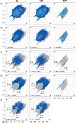
The quantification of the close contacts to the Hirshfeld surface was performed through the analysis of the two-dimensional fingerprint plots for (I) as well as for the indivi-dual molecular components. As shown in Fig. 5(a), the overall fingerprint plot of (I) exhibits a bug-like profile with a pair of symmetric spikes. This is in contrast to the asymmetric profile of3LH2, with splitting of the spike in the internal region due to
the formation of the O—H N hydrogen bond, Fig. 5(e), suggesting a prominent role played by the water molecule in influencing the overall contacts in (I). The observation is very
different to that of the benzene solvate of4LH2in which the
overall surface contacts for4LH2are not very much influenced
by the benzene molecule as demonstrated by the similar profiles for the solvate and individual 4LH2 molecule (Tan,
Halcovitch et al., 2019). The decomposition of the overall profile of (I) shows that the most significant contacts are primarily H H contacts (43.5%), followed by O H/H O (21.1%), C H/H C (19.6%) and N H/H N (9.8%) contacts, with all of these interactions havingdi+dedistances
less than the respective sums of van der Waals radii (vdW),i.e.
H H 2.26 A˚ [(vdW) = 2.40 A˚ ], O H/H O 1.88 A˚ [(vdW) = 2.72 A˚ ], C H/H C2.62 A˚ [(vdW) = 2.90 A˚ ] and N H/H N2.50 A˚ [(vdW) = 2.75 A˚ ].
As for the individual 3LH2 molecule, the dominance of
these contacts follows the order H H (41.1%;di+de2.33 A˚ ),
C H/H C (21.2%;di+de2.60 A˚ ), O H/H O (17.9%;di
+de1.88 A˚ ) and N H/H N (13.5%;di+de1.80 A˚ ). While
the aforementioned interactions are almost evenly distributed between the internal and external contacts for (I), some contacts for3LH
2are found to either to be inclined towards
the internal or external contact region compared with (I), such as that displayed by (internal)-O H-(external) (8.4%)versus
(internal)-H O-(external) (9.5%) and (internal)-N H-(external) (8.8%)versus (internal)-H N-(external) (4.6%), respectively, Fig. 5(c)–(e).
The hydrate molecule exhibits a completely different fingerprint profile, which is dominated by three major contacts, namely H H (46.9%;di+de2.26 A˚ ), O H/H O
(39.4%;di+de1.88 A˚ ) and H N (13.7%;di+de1.80 A˚ ). In
particular, the second most dominant contacts are found to be heavily inclined toward (internal)-O H-(external) (30.5%) as compared to (internal)-H O-(external) (8.9%), presum-ably due to relatively large contact surface area.
5. Computational chemistry
All associations between molecules in (I), as described in
Hirshfeld surface analysis, were subjected to the calculation of the interaction energy usingCrystal Explorer 17(Turneret al., 2017) based on the method described previously (Tan, Jotani
et al., 2019) to evaluate the strength of each interaction, Table 3. Among those close contacts, the (3LH2)2 dimer
28
Tan and Tiekink C14H14N4O2H2O Acta Cryst.(2020). E76, 25–31
[image:4.610.315.567.92.198.2]research communications
Figure 5
(a) The overall two-dimensional fingerprint plots for (I) and for the individual 3LH2 and water molecules, and those delineated into (b)
H H, (c) H O/O H, (d) H C/C H and (e) H N/N H contacts. The percentage contributions to the surfaces are indicated therein.
Table 3
Summary of interaction energies (kJ mol1) calculated for (I).
Contact Eele Epol Edis Erep Etot
N2—H2N O1i+
N3—H3N O2i 68.5 15.0 49.2 86.4 73.0
C12—H12 C7ii 6.7 2.0 46.1 26.0 32.7
C6—H6A O2iii 12.9 2.9 28.2 13.5 32.0
O1W—H1W N1iv 51.9 11.2 6.5 65.1 28.6
O1W—H2W O1Wv 36.9 7.1 3.5 34.3 26.2
C7 O1vi 2.3 3.0 31.4 18.4 20.7
C5—H5 N4vii 9.4 2.0 8.1 8.7 13.0
C1—H1 O1Wviii 8.1 1.3 3.9 3.9 10.5
Symmetry operations: (i)x, 1 +y,z; (ii)x,y, 1z; (iii) 1x, 1y, 1z; (iv)x,y,z; (v)3
[image:4.610.46.298.277.684.2]connected by a ten-membered { HNC2O}2synthon has the
greatestEintenergy of73.0 kJ mol
1
which is comparable in energy to the classical eight-membered { HOCO}2synthon
(Tan & Tiekink, 2019a). Perhaps unexpectedly, the C12– H12 C7 contact which also sustains a pair of3LH2molecules
constitutes the second strongest interaction with Eint =
32.7 kJ mol1, and this is followed by the C6—H6A O2 (32.0 kJ mol1), O1W—H1W N1 (28.6 kJ mol1), O1W—H2W O1W (26.2 kJ mol1), C7 O1 (20.7 kJ mol1), C5—H5 N4 (13.0 kJ mol1) and C1— H1 O1W (10.5 kJ mol1) interactions. As expected, the N2—H2N O1, N3—H3N O2, O1W—H1W N1 and O1W—H2W O1Winteractions are associated with distinct electropositive and electronegative sites and therefore, are mainly governed by electrostatic forces, while the rest of the close contacts are dispersive in nature. The relatively stable nature of the C12—H12 C7 and C6—H6A O2 inter-actions as compared to the O1W—H1W N1 and O1W— H2W O1Winteractions could be due to the presence of low repulsion energies in the former as compared to the latter.
The crystal of (I) is mainly sustained by electrostatic forces owing to the strong N2—H2N O1/ N3—H3N O2, O1W— H1W N1 and O1W—H2W O1W hydrogen bonding leading to a barricade-like electrostatic energy framework parallel to (101), as shown in Fig. 6(a). This is further stabilized by the dispersion forces arising from other supporting inter-actions which result in another barricade-like dispersion energy framework parallel to (100), Fig. 6(b). The overall energy framework for (I) is shown in Fig. 6(c).
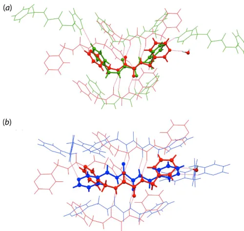
A comparison of the distribution of contacts on the Hirshfeld surfaces between the3LH2molecule in (I) and in its
two polymorphic forms,i.e. Form I and Form II (Jotaniet al., 2016), with latter having two independent molecules, was performed. This analysis returned the data shown in Table 4 and indicates that3LH2in (I) is relatively closer to Form I as
compared to the independent molecules comprising Form II. This conclusion is consistent with the analysis of the packing similarity in which a comparison of (I) and Form I exhibits an r.m.s. deviation of 0.895 A˚ while a comparison with Form II exhibits an r.m.s. deviation of 1.581 A˚ , despite only one out of 20 molecules displaying some similarity with the reference
3
LH2 molecule in (I), Fig. 7. The packing analysis was
[image:5.610.44.564.72.212.2]performed using Mercury (Macrae et al., 2006), with the analysis criteria being set that only molecules within the 20%
Figure 6
[image:5.610.312.562.465.702.2] [image:5.610.44.295.673.750.2]Perspective views of the energy framework of (I), showing the (a) electrostatic force, (b) dispersion force and (c) total energy diagram. The cylindrical radius is proportional to the relative strength of the corresponding energies and they were adjusted to the same scale factor of 100 with a cut-off value of 8 kJ mol1within 212 unit cells.
Figure 7
A comparison of the molecular packing of3LH
2: (a) (I) (red) and Form I
(green) and (b) (I) (red) and Form II (blue), showing the differences in terms of molecular connectivity of3LH
2with r.m.s. deviations of 0.895
and 1.581 A˚ , respectively. Table 4
A comparison of the distribution of contacts (%) to the calculated Hirshfeld surfaces for (I) and for Forms I and II (Jotaniet al., 2016).
Contact (I) Form I Form IIa Form IIb
H H 41.1 44.1 35.8 36.9
C H/H C 21.2 16.7 31.4 22.4
O H/H O 17.9 15.7 14.2 19.6
N H/H N 13.5 16.7 18.0 19.5
C O/O C 2.3 2.1 0.1 0.1
tolerance for both distances and angles were included in the calculation while molecules with a variation >20% were discarded, and that molecular inversions were allowed during calculation. It is therefore also apparent through this analysis that the water molecules in (I) play a crucial role in influencing the packing of3LH2in (I).
6. Database survey
The3LH2molecule has been characterized in two polymorphs
(Jotaniet al., 2016) and in a number of (neutral) co-crystals. A characteristic of these structures is a long central C—C bond and conformational flexibility in terms of the relative dispo-sition of the 3-pyridyl substituents with respect to the central C2N2O2chromophore (Tiekink, 2017). Indeed, the relatively
long length of the central C—C bonds often attracts a level C alert inPLATON(Spek, 2009). Of the data included in Table 5 [for the chemical diagrams of (II) and (III), see Scheme 2], the shorter of the C—C bonds is 1.515 (3) A˚ , found in the co-crystal of 3LH2 with HO2CCH2N(H)C( O)N(H)CH2CO2H
(Nguyenet al., 2001) and the longest bond of 1.550 (17) A˚ is found in the co-crystal of3LH2with (III) (Jinet al., 2013). In
terms of conformational flexibility, the two polymorphs of
3
LH2highlight this characteristic of these molecules (Jotaniet
al., 2016). In Form I, the pyridyl rings lie to the same side of the central C2N2O2 and therefore, have a syn-periplanar
relationship, or, more simply, a U-shape. In Form II, comprising two independent molecules, each is disposed about a centre of inversion so the relationship isanti-periplanar, or S-shaped. DFT calculations revealed that the difference in energy between the two conformations is less than 1 kcal1 (Jotani et al., 2016). Despite this result, most of the 3LH2
molecules are centrosymmetric, S-shaped. For the U-shaped molecules, the dihedral angles between the central plane and pyridyl rings range from 59.71 (6) to 84.61 (9). The compar-able range for the S-shaped molecules, for which both dihedral angles are identical from symmetry, is 64.2 (3) to 84.79 (18).
7. Synthesis and crystallization
The precursor, N,N0-bis(pyridin-3-ylmethyl)oxalamide, was prepared according to the literature (Schauer et al., 1997). Crystallization of the precursor in a DMF (1 ml) and ethanol (1 ml) mixture resulted in the isolation of the title hydrate, (I); m.p.: 409.4–410.7 K. IR (cm1): 3578(O—H), 3321(N—H), 3141–2804(C—H), 1687–1649(C O), 1524–1482(C C), 1426(C—N), 710(C C).
30
Tan and Tiekink C14H14N4O2H2O Acta Cryst.(2020). E76, 25–31
[image:6.610.47.566.101.286.2] [image:6.610.339.537.322.621.2]research communications
Table 5
Geometric data,i.e.central C—C bond lengths (A˚ ) and dihedral angles (), for3
LH2in its polymorphs, solvates and (neutral) co-crystals; see Scheme 2
for the chemical diagrams of (II) and (III).
Compound Symmetry Conformation C—C C2N2O2/(3-py) (3-py)/(3-py) REFCODE Reference
Polymorphs
Form I – U 1.544 (4) 74.98 (10), 84.61 (9) 88.40 (7) OWOHAL Jotaniet al.(2016)
Form IIa
1 S 1.5383 (16) 77.29 (4) 0 OWOHAL01 Jotaniet al.(2016)
1 S 1.5460 (16) 75.93 (3) 0
Solvate
(I) – U 1.541 (2) 59.71 (6), 68.42 (6) 87.86 (5) – This work
Co-crystals of3LH 2with
HO2CCH2N(H)C( O)N(H)CH2CO2H 1 S 1.515 (3) 81.41 (7) 0 CAJQEK Nguyenet al.(2001)
HO2CCH2N(H)C( O)C( O)N(H)CH2CO2H 1 S 1.532 (19) 64.2 (3) 0 CAJQAG Nguyenet al.(2001)
2-NH2C6H4CO2H 1 S 1.543 (2) 74.64 (4), 74.64 (4) 0 DIDZAT Armanet al.(2012)
(II) 1 S 1.533 (3) 79.50 (6) 0 EMACIG Suzukiet al.(2016)
C6F4I2 1 S 1.544 (4) 70.72 (9) 0 IPOSIP Hursthouseet al.(2003)
2-HO2CC6H4SSC6H4CO2-2 – U 1.543 (3) 61.22 (5), 69.43 (5) 72.12 (8) KUZSOO Armanet al.(2010)
4-NO2C6H4CO2H 1 S 1.530 (2) 78.20 (4) 0 PAGFIP Syedet al.(2016)
(III) 1 S 1.550 (17) 80.5 (4) 0 REWVUM Jinet al.(2013)
I—C C—C C—I 1 S 1.542 (10) 76.6 (2) 0 WANNOP Goroffet al.(2005)
I—C C—C C—C C—I 1 S 1.548 (11) 84.7 (2) 0 WANPIL Goroffet al.(2005)
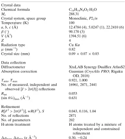
8. Refinement
Crystal data, data collection and structure refinement details are summarized in Table 6. The carbon-bound H atoms were placed in calculated positions (C—H = 0.95–0.99 A˚ ) and were included in the refinement in the riding-model approximation, withUiso(H) set to 1.2–1.5Ueq(C). The oxygen- and
nitrogen-bound H atoms were located in a difference-Fourier map and refined with O—H = 0.840.01 A˚ and N—H = 0.880.01 A˚ , respectively, and withUiso(H) set to 1.5Ueq(O) or 1.2Ueq(N).
Owing to poor agreement, one reflection, i.e. (551), was omitted from the final cycles of refinement.
Funding information
Crystallographic research at Sunway University is supported by Sunway University Sdn Bhd (grant No. STR-RCTR-RCCM-001-2019).
References
Arman, H. D., Miller, T., Poplaukhin, P. & Tiekink, E. R. T. (2010).
Acta Cryst.E66, o2590–o2591.
Arman, H. D., Miller, T. & Tiekink, E. R. T. (2012).Z. Kristallogr. Cryst. Mater.227, 825–830.
Arman, H. D., Poplaukhin, P. & Tiekink, E. R. T. (2018). Z. Kristallogr. New Cryst. Struct.233, 159–161.
Brandenburg, K. (2006).DIAMOND. Crystal Impact GbR, Bonn, Germany.
Farrugia, L. J. (2012).J. Appl. Cryst.45, 849–854.
Goroff, N. S., Curtis, S. M., Webb, J. A., Fowler, F. W. & Lauher, J. W. (2005).Org. Lett.7, 1891–1893.
Hursthouse, M. B., Gelbrich, T. & Plater, M. J. (2003). Private communication (refcode: IPOSIP). CCDC, Cambridge, England. Jin, H., Plonka, A. M., Parise, J. B. & Goroff, N. S. (2013).
CrystEngComm,15, 3106–3110.
Jin, H., Young, C. N., Halada, G. P., Phillips, B. L. & Goroff, N. S. (2015).Angew. Chem. Int. Ed.54, 14690–14695.
Jotani, M. M., Zukerman-Schpector, J., Madureira, L. S., Poplaukhin, P., Arman, H. D., Miller, T. & Tiekink, E. R. T. (2016). Z. Kristallogr. Cryst. Mater.231, 415–425.
Macrae, C. F., Edgington, P. R., McCabe, P., Pidcock, E., Shields, G. P., Taylor, R., Towler, M. & van de Streek, J. (2006).J. Appl. Cryst.39, 453–457.
Nguyen, T. L., Fowler, F. W. & Lauher, J. W. (2001).J. Am. Chem. Soc. 123, 11057–11064.
Rigaku OD (2018). CrysAlis PRO. Oxford Diffraction, Yarnton, England.
Schauer, C. L., Matwey, E., Fowler, F. W. & Lauher, J. W. (1997).J. Am. Chem. Soc.119, 10245–10246.
Sheldrick, G. M. (2015a).Acta Cryst.A71, 3–8. Sheldrick, G. M. (2015b).Acta Cryst.C71, 3–8.
Spackman, M. A. & Jayatilaka, D. (2009).CrystEngComm,11, 19–32. Spek, A. L. (2009).Acta Cryst.D65, 148–155.
Suzuki, M., Kotyk, J. F. K., Khan, S. I. & Rubin, Y. (2016).J. Am. Chem. Soc.138, 5939–5956.
Syed, S., Halim, S. N. A., Jotani, M. M. & Tiekink, E. R. T. (2016).
Acta Cryst.E72, 76–82.
Tan, S. L., Halcovitch, N. R. & Tiekink, E. R. T. (2019).Acta Cryst.
E75, 1133–1139.
Tan, S. L., Jotani, M. M. & Tiekink, E. R. T. (2019).Acta Cryst.E75, 308–318.
Tan, S. L. & Tiekink, E. R. T. (2019a).Acta Cryst.E75, 1–7. Tan, S. L. & Tiekink, E. R. T. (2019b).Z. Kristallogr. New Cryst.
Struct.234, 1113–1116.
Tan, S. L. & Tiekink, E. R. T. (2019c). Z. Kristallogr. New Cryst. Struct.234, 1117–1119.
Tan, Y. S., Chun, H. Z., Jotani, M. M. & Tiekink, E. R. T. (2019).Z. Kristallogr. Cryst. Mater.234, 165–175.
Tiekink, E. R. T. (2017). Multi-Component Crystals: Synthesis, Concepts, Function, edited by E. R. T. Tiekink & J. Schpector-Zukerman, pp. 289–319. Singapore: De Gruyter.
Tiekink, E. R. T. (2018).Crystals,8, article No. 18 (29 pages). Turner, M. J., Mckinnon, J. J., Wolff, S. K., Grimwood, D. J.,
Spackman, P. R., Jayatilaka, D. & Spackman, M. A. (2017).Crystal Explorer 17. The University of Western Australia.
Westrip, S. P. (2010).J. Appl. Cryst.43, 920–925.
[image:7.610.312.566.90.372.2]Wilhelm, C., Boyd, S. A., Chawda, S., Fowler, F. W., Goroff, N. S., Halada, G. P., Grey, C. P., Lauher, J. W., Luo, L., Martin, C. D., Parise, J. B., Tarabrella, C. & Webb, J. A. (2008).J. Am. Chem. Soc. 130, 4415–4420.
Table 6
Experimental details.
Crystal data
Chemical formula C14H14N4O2H2O
Mr 288.31
Crystal system, space group Monoclinic,P21/n
Temperature (K) 100
a,b,c(A˚ ) 12.4784 (4), 5.0247 (1), 22.2410 (6)
() 90.170 (3)
V(A˚3) 1394.51 (6)
Z 4
Radiation type CuK
(mm1) 0.82
Crystal size (mm) 0.090.070.03
Data collection
Diffractometer XtaLAB Synergy Dualflex AtlasS2
Absorption correction Gaussian (CrysAlis PRO; Rigaku OD, 2018)
Tmin,Tmax 0.921, 1.000
No. of measured, independent and observed [I> 2(I)] reflections
16961, 2871, 2441
Rint 0.053
(sin/ )max(A˚
1) 0.631
Refinement
R[F2> 2(F2)],wR(F2),S 0.043, 0.116, 1.04
No. of reflections 2871
No. of parameters 202
H-atom treatment H atoms treated by a mixture of independent and constrained refinement
max,min(e A˚
3) 0.30,0.24
Computer programs:CrysAlis PRO(Rigaku OD, 2018),SHELXS(Sheldrick, 2015a),
SHELXL2017(Sheldrick, 2015b),ORTEP-3 for Windows(Farrugia, 2012),DIAMOND
supporting information
sup-1
Acta Cryst. (2020). E76, 25-31
supporting information
Acta Cryst. (2020). E76, 25-31 [https://doi.org/10.1107/S2056989019016153]
N
,
N
′
-Bis(pyridin-3-ylmethyl)ethanediamide monohydrate: crystal structure,
Hirshfeld surface analysis and computational study
Sang Loon Tan and Edward R. T. Tiekink
Computing details
Data collection: CrysAlis PRO (Rigaku OD, 2018); cell refinement: CrysAlis PRO (Rigaku OD, 2018); data reduction:
CrysAlis PRO (Rigaku OD, 2018); program(s) used to solve structure: SHELXS (Sheldrick, 2015a); program(s) used to refine structure: SHELXL2017 (Sheldrick, 2015b); molecular graphics: ORTEP-3 for Windows (Farrugia, 2012) and
DIAMOND (Brandenburg, 2006); software used to prepare material for publication: publCIF (Westrip, 2010).
N,N′-Bis(pyridin-3-ylmethyl)ethanediamide monohydrate
Crystal data
C14H14N4O2·H2O
Mr = 288.31
Monoclinic, P21/n
a = 12.4784 (4) Å
b = 5.0247 (1) Å
c = 22.2410 (6) Å
β = 90.170 (3)°
V = 1394.51 (6) Å3
Z = 4
F(000) = 608
Dx = 1.373 Mg m−3
Cu Kα radiation, λ = 1.54184 Å Cell parameters from 5162 reflections
θ = 4.0–75.9°
µ = 0.82 mm−1
T = 100 K Prism, colourless 0.09 × 0.07 × 0.03 mm
Data collection
XtaLAB Synergy Dualflex AtlasS2 diffractometer
Detector resolution: 5.2558 pixels mm-1
ω scans
Absorption correction: gaussian (Crysalis PRO; Rigaku OD, 2018)
Tmin = 0.921, Tmax = 1.000
16961 measured reflections
2871 independent reflections 2441 reflections with I > 2σ(I)
Rint = 0.053
θmax = 76.7°, θmin = 4.0°
h = −14→15
k = −6→6
l = −27→28
Refinement
Refinement on F2
Least-squares matrix: full
R[F2 > 2σ(F2)] = 0.043
wR(F2) = 0.116
S = 1.04 2871 reflections 202 parameters 0 restraints
Primary atom site location: structure-invariant direct methods
Secondary atom site location: difference Fourier map
Hydrogen site location: mixed
H atoms treated by a mixture of independent and constrained refinement
w = 1/[σ2(F
o2) + (0.0553P)2 + 0.7659P]
where P = (Fo2 + 2Fc2)/3
(Δ/σ)max < 0.001
Δρmax = 0.30 e Å−3
sup-2
Acta Cryst. (2020). E76, 25-31
Special details
Geometry. All esds (except the esd in the dihedral angle between two l.s. planes) are estimated using the full covariance matrix. The cell esds are taken into account individually in the estimation of esds in distances, angles and torsion angles; correlations between esds in cell parameters are only used when they are defined by crystal symmetry. An approximate (isotropic) treatment of cell esds is used for estimating esds involving l.s. planes.
Fractional atomic coordinates and isotropic or equivalent isotropic displacement parameters (Å2)
x y z Uiso*/Ueq
O1 0.39488 (9) −0.1106 (2) 0.53440 (5) 0.0211 (3)
O2 0.28502 (10) 0.4170 (2) 0.45056 (5) 0.0245 (3)
N1 0.51280 (11) 0.8092 (3) 0.72982 (6) 0.0230 (3)
N2 0.40224 (11) 0.3337 (3) 0.55256 (6) 0.0178 (3)
H2N 0.3890 (16) 0.486 (4) 0.5378 (9) 0.021*
N3 0.26914 (11) −0.0297 (3) 0.43753 (6) 0.0176 (3)
H3N 0.2848 (16) −0.182 (4) 0.4529 (8) 0.021*
N4 −0.08573 (13) 0.1419 (4) 0.36223 (9) 0.0434 (5)
C1 0.52700 (13) 0.6284 (3) 0.68624 (7) 0.0205 (3)
H1 0.598157 0.573079 0.677719 0.025*
C2 0.44417 (12) 0.5164 (3) 0.65271 (7) 0.0176 (3)
C3 0.34062 (13) 0.6008 (3) 0.66496 (7) 0.0202 (3)
H3 0.281622 0.530908 0.642955 0.024*
C4 0.32438 (13) 0.7884 (3) 0.70976 (7) 0.0224 (3)
H4 0.254145 0.849326 0.718705 0.027*
C5 0.41200 (13) 0.8860 (3) 0.74135 (7) 0.0227 (3)
H5 0.400111 1.012319 0.772402 0.027*
C6 0.47006 (13) 0.3104 (3) 0.60569 (7) 0.0202 (3)
H6A 0.545984 0.329839 0.593668 0.024*
H6B 0.461045 0.130847 0.623270 0.024*
C7 0.37291 (12) 0.1213 (3) 0.52134 (7) 0.0163 (3)
C8 0.30359 (12) 0.1859 (3) 0.46578 (7) 0.0170 (3)
C9 0.20743 (13) −0.0186 (3) 0.38182 (7) 0.0196 (3)
H9A 0.228818 −0.169871 0.355973 0.024*
H9B 0.225732 0.147561 0.360270 0.024*
C10 0.08770 (13) −0.0283 (3) 0.39089 (7) 0.0199 (3)
C11 0.02169 (15) 0.1432 (4) 0.35990 (9) 0.0370 (5)
H11 0.054734 0.272529 0.334924 0.044*
C12 −0.13026 (14) −0.0379 (4) 0.39790 (8) 0.0304 (4)
H12 −0.206194 −0.042293 0.400717 0.036*
C13 −0.07261 (17) −0.2165 (5) 0.43067 (11) 0.0486 (6)
H13 −0.107827 −0.342052 0.455703 0.058*
C14 0.03821 (16) −0.2127 (5) 0.42700 (10) 0.0450 (6)
H14 0.079722 −0.336857 0.449325 0.054*
O1W 0.71328 (9) 0.9787 (2) 0.77119 (5) 0.0217 (3)
H1W 0.642 (2) 0.942 (4) 0.7593 (9) 0.033*
supporting information
sup-3
Acta Cryst. (2020). E76, 25-31
Atomic displacement parameters (Å2)
U11 U22 U33 U12 U13 U23
O1 0.0241 (6) 0.0137 (5) 0.0254 (6) 0.0006 (4) −0.0029 (4) 0.0011 (4)
O2 0.0323 (7) 0.0139 (5) 0.0272 (6) 0.0018 (5) −0.0065 (5) 0.0007 (4)
N1 0.0189 (7) 0.0242 (7) 0.0261 (7) −0.0010 (5) −0.0026 (5) −0.0033 (5)
N2 0.0207 (7) 0.0122 (6) 0.0206 (6) 0.0009 (5) −0.0012 (5) 0.0013 (5)
N3 0.0192 (7) 0.0131 (6) 0.0205 (6) 0.0005 (5) −0.0011 (5) −0.0001 (5)
N4 0.0187 (8) 0.0514 (11) 0.0600 (11) 0.0000 (7) −0.0009 (7) 0.0268 (9)
C1 0.0160 (7) 0.0205 (8) 0.0248 (8) 0.0001 (6) −0.0022 (6) −0.0008 (6)
C2 0.0178 (7) 0.0156 (7) 0.0195 (7) −0.0013 (6) −0.0013 (6) 0.0023 (5)
C3 0.0161 (7) 0.0219 (8) 0.0227 (7) −0.0035 (6) −0.0016 (6) 0.0002 (6)
C4 0.0170 (8) 0.0263 (8) 0.0239 (7) −0.0002 (6) 0.0023 (6) −0.0018 (6)
C5 0.0213 (8) 0.0243 (8) 0.0226 (7) −0.0008 (6) −0.0003 (6) −0.0034 (6)
C6 0.0192 (8) 0.0175 (7) 0.0239 (7) 0.0014 (6) −0.0037 (6) −0.0018 (6)
C7 0.0151 (7) 0.0140 (7) 0.0198 (7) −0.0001 (5) 0.0032 (6) 0.0010 (5)
C8 0.0168 (7) 0.0151 (7) 0.0192 (7) 0.0013 (6) 0.0028 (6) 0.0003 (5)
C9 0.0195 (8) 0.0196 (7) 0.0197 (7) −0.0003 (6) −0.0004 (6) −0.0012 (6)
C10 0.0206 (8) 0.0193 (7) 0.0198 (7) −0.0013 (6) 0.0000 (6) −0.0023 (6)
C11 0.0198 (9) 0.0432 (11) 0.0481 (11) −0.0018 (8) −0.0005 (8) 0.0262 (9)
C12 0.0187 (8) 0.0370 (10) 0.0355 (9) −0.0035 (7) 0.0031 (7) 0.0028 (8)
C13 0.0268 (10) 0.0584 (14) 0.0605 (14) −0.0057 (10) 0.0068 (9) 0.0343 (12)
C14 0.0241 (10) 0.0509 (13) 0.0601 (13) 0.0014 (9) −0.0001 (9) 0.0345 (11)
O1W 0.0186 (6) 0.0205 (6) 0.0261 (6) −0.0009 (5) −0.0027 (4) 0.0012 (5)
Geometric parameters (Å, º)
O1—C7 1.2313 (18) C4—H4 0.9500
O2—C8 1.2314 (18) C5—H5 0.9500
N1—C5 1.341 (2) C6—H6A 0.9900
N1—C1 1.341 (2) C6—H6B 0.9900
N2—C7 1.3244 (19) C7—C8 1.541 (2)
N2—C6 1.456 (2) C9—C10 1.509 (2)
N2—H2N 0.85 (2) C9—H9A 0.9900
N3—C8 1.323 (2) C9—H9B 0.9900
N3—C9 1.4579 (19) C10—C14 1.374 (2)
N3—H3N 0.86 (2) C10—C11 1.376 (2)
N4—C12 1.326 (2) C11—H11 0.9500
N4—C11 1.342 (2) C12—C13 1.361 (3)
C1—C2 1.392 (2) C12—H12 0.9500
C1—H1 0.9500 C13—C14 1.386 (3)
C2—C3 1.388 (2) C13—H13 0.9500
C2—C6 1.507 (2) C14—H14 0.9500
C3—C4 1.387 (2) O1W—H1W 0.95 (2)
C3—H3 0.9500 O1W—H2W 0.88 (2)
C4—C5 1.387 (2)
sup-4
Acta Cryst. (2020). E76, 25-31
C7—N2—C6 121.32 (13) O1—C7—C8 120.84 (13)
C7—N2—H2N 118.1 (13) N2—C7—C8 113.84 (13)
C6—N2—H2N 119.8 (13) O2—C8—N3 125.51 (14)
C8—N3—C9 122.84 (13) O2—C8—C7 121.59 (13)
C8—N3—H3N 117.8 (13) N3—C8—C7 112.89 (13)
C9—N3—H3N 119.4 (13) N3—C9—C10 113.95 (12)
C12—N4—C11 116.55 (16) N3—C9—H9A 108.8
N1—C1—C2 124.18 (15) C10—C9—H9A 108.8
N1—C1—H1 117.9 N3—C9—H9B 108.8
C2—C1—H1 117.9 C10—C9—H9B 108.8
C3—C2—C1 117.51 (14) H9A—C9—H9B 107.7
C3—C2—C6 123.21 (14) C14—C10—C11 116.47 (16)
C1—C2—C6 119.28 (14) C14—C10—C9 123.13 (15)
C2—C3—C4 119.13 (14) C11—C10—C9 120.31 (15)
C2—C3—H3 120.4 N4—C11—C10 125.07 (17)
C4—C3—H3 120.4 N4—C11—H11 117.5
C5—C4—C3 119.17 (15) C10—C11—H11 117.5
C5—C4—H4 120.4 N4—C12—C13 123.24 (17)
C3—C4—H4 120.4 N4—C12—H12 118.4
N1—C5—C4 122.66 (15) C13—C12—H12 118.4
N1—C5—H5 118.7 C12—C13—C14 119.05 (18)
C4—C5—H5 118.7 C12—C13—H13 120.5
N2—C6—C2 112.50 (12) C14—C13—H13 120.5
N2—C6—H6A 109.1 C10—C14—C13 119.61 (18)
C2—C6—H6A 109.1 C10—C14—H14 120.2
N2—C6—H6B 109.1 C13—C14—H14 120.2
C2—C6—H6B 109.1 H1W—O1W—H2W 103.3 (19)
H6A—C6—H6B 107.8
C5—N1—C1—C2 −0.1 (2) O1—C7—C8—O2 −176.62 (15)
N1—C1—C2—C3 0.8 (2) N2—C7—C8—O2 4.9 (2)
N1—C1—C2—C6 −178.75 (14) O1—C7—C8—N3 2.8 (2)
C1—C2—C3—C4 −0.5 (2) N2—C7—C8—N3 −175.72 (13)
C6—C2—C3—C4 178.98 (14) C8—N3—C9—C10 −94.71 (17)
C2—C3—C4—C5 −0.3 (2) N3—C9—C10—C14 −48.5 (2)
C1—N1—C5—C4 −0.8 (2) N3—C9—C10—C11 135.08 (17)
C3—C4—C5—N1 1.0 (3) C12—N4—C11—C10 0.8 (3)
C7—N2—C6—C2 −146.60 (14) C14—C10—C11—N4 −0.4 (3)
C3—C2—C6—N2 37.4 (2) C9—C10—C11—N4 176.3 (2)
C1—C2—C6—N2 −143.09 (14) C11—N4—C12—C13 −0.6 (3)
C6—N2—C7—O1 3.0 (2) N4—C12—C13—C14 0.0 (4)
C6—N2—C7—C8 −178.56 (13) C11—C10—C14—C13 −0.3 (3)
C9—N3—C8—O2 3.0 (2) C9—C10—C14—C13 −176.9 (2)
supporting information
sup-5
Acta Cryst. (2020). E76, 25-31
Hydrogen-bond geometry (Å, º)
D—H···A D—H H···A D···A D—H···A
N2—H2N···O2 0.85 (2) 2.36 (2) 2.7279 (18) 107.0 (16)
N3—H3N···O1 0.86 (2) 2.299 (19) 2.6924 (18) 108.0 (15)
O1W—H1W···N1 0.95 (2) 1.86 (2) 2.7958 (18) 169 (2)
O1W—H2W···O1Wi 0.88 (2) 1.97 (2) 2.8364 (15) 166 (2)
N2—H2N···O1ii 0.85 (2) 2.03 (2) 2.8227 (18) 155.2 (18)
N3—H3N···O2iii 0.86 (2) 2.02 (2) 2.8022 (18) 151.6 (17)
C9—H9A···O1Wiv 0.99 2.45 3.3772 (19) 156
C6—H6B···Cg1iii 0.99 2.74 3.7043 (16) 166