Poly[dimethylammonium [(
l
2-benzene-1,2-dicarboxylato-
j
2O
1:O
3)[
l
2-3-(pyri-din-4-yl)-1H-pyrazol-1-ido-
j
2N
1:N
3]-cuprate(II)]]
Liu Na
Department of Chemistry, Hengshui University, Heng Shui 053000, People’s Republic of China
Correspondence e-mail: 281828541@qq.com
Received 3 June 2013; accepted 12 June 2013
Key indicators: single-crystal X-ray study;T= 293 K; mean(C–C) = 0.006 A˚; some non-H atoms missing;Rfactor = 0.046;wRfactor = 0.124; data-to-parameter ratio = 14.7.
In the title complex, {(C2H8N)[Cu(C8H4O4)(C8H6N3)]}n, there are two CuIIcations (each located on a centre of inversion), one benzene-1,2-dicarboxylate dianion, one 3-(pyridin-4-yl)-1H-pyrazol-1-ide anion and one dimethylammonium cation in the asymmetric unit. The dimethylammonium cation was highly disordered and was treated with the SQUEEZE routine
in PLATON [Spek (2009). Acta Cryst. D65, 148–155]; the
crystallographic data takes into account the presence of the cation. Each CuIIcation exhibits a square-planar coordination geometry. A benzene-1,2-dicarboxylate dianion bridges two CuIIcations, building a linear chain along [001]. The chains are connected by 3-(pyridin-4-yl)-1H-pyrazol-1-ide anions, constructing a layer parallel to (101). The layers are assembled into a three-dimensional supramolecular network through C—H interactions.
Related literature
For background to complexes derived from 4-(1H -pyrazol-3-yl)pyridine, see: Davies et al. (2005); Tan et al. (2011); For background to complexes derived from benzene-1,2-dicarb-oxylic acid, see: Guo (2010); Yanet al.(2012).
Experimental
Crystal data
(C2H8N)[Cu(C8H4O4)(C8H6N3)]
Mr= 417.91
Triclinic,P1
a= 8.0978 (16) A˚
b= 9.7244 (19) A˚
c= 11.694 (2) A˚
= 89.26 (3)
= 89.12 (3)
= 89.64 (3) V= 920.7 (3) A˚3
Z= 2
MoKradiation
= 1.22 mm1
T= 293 K
0.240.220.21 mm
Data collection
Rigaku SCXmini diffractometer Absorption correction: multi-scan
(ABSCOR; Higashi, 1995)
Tmin= 0.759,Tmax= 0.784
8051 measured reflections 3236 independent reflections 2486 reflections withI> 2(I)
Rint= 0.044
Refinement
R[F2> 2(F2)] = 0.046
wR(F2) = 0.124
S= 1.06 3236 reflections
220 parameters
H-atom parameters constrained max= 0.50 e A˚
3
min=0.30 e A˚
[image:1.610.313.566.586.617.2]3
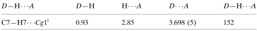
Table 1
Hydrogen-bond geometry (A˚ ,).
Cg1 is the centroid of the N2,N3,C9–C11 ring.
D—H A D—H H A D A D—H A
C7—H7 Cg1i
0.93 2.85 3.698 (5) 152
Symmetry code: (i)x;yþ1;z.
Data collection: CrystalClear (Rigaku, 2005); cell refinement: CrystalClear; data reduction:CrystalClear; program(s) used to solve structure:SHELXS97(Sheldrick, 2008); program(s) used to refine structure: SHELXL97 (Sheldrick, 2008); molecular graphics: ORTEPII (Johnson, 1976) and DIAMOND (Brandenburg, 1999); software used to prepare material for publication:SHELXL97.
The author acknowledges Hengshui University for
supporting this work.
metal-organic compounds
m400
Liu Na doi:10.1107/S1600536813016334 Acta Cryst.(2013). E69, m400–m401Acta Crystallographica Section E
Structure Reports
Online
Supplementary data and figures for this paper are available from the IUCr electronic archives (Reference: TK5232).
References
Brandenburg, K. (1999).DIAMOND. Crystal Impact GbR, Bonn, Germany. Davies, G. M., Adams, H. & Ward, M. D. (2005).Acta Cryst.C61, m485–m487.
Higashi, T. (1995).ABSCOR. Rigaku Corporation, Tokyo, Japan.
Johnson, C. K. (1976).ORTEPII. Report ORNL-5138, Oak Ridge National Laboratory, Tennessee, USA.
Rigaku (2005).CrystalClear. Rigaku Corporation, Tokyo, Japan. Sheldrick, G. M. (2008).Acta Cryst.A64, 112–122.
Spek, A. L. (2009).Acta Cryst.D65, 148–155.
supporting information
sup-1
Acta Cryst. (2013). E69, m400–m401
supporting information
Acta Cryst. (2013). E69, m400–m401 [https://doi.org/10.1107/S1600536813016334]
Poly[dimethylammonium [(
µ
2-benzene-1,2-dicarboxylato-
κ
2O
1:
O
3)
[
µ
2-3-(pyridin-4-yl)-1
H
-pyrazol-1-ido-
κ
2N
1:
N
3]cuprate(II)]]
Liu Na
S1. Comment
The immense research in coordination polymers is due to their potential applications and the diversity in topological
architectures. The choice of organic linker is crucial for constructing coordination polymers. Polycarboxylate linkers and
N-containing ligands are popular for building novel architectures. Among various polycarboxylate linkers,
benzene-1,2-dicarboxylic acid (1,2-H2bdc) exhibits rich coordination modes to metal centers owing to its rigidity and polycarboxylate
groups (Guo, 2010; Yan et al., 2012). On the other hand, among the N-containing ligands, 4-(1H-pyrazol-3-yl)pyridine
(L) processes molecular recognition sites for C—H···π and π-π interactions to form interesting supramolecular structures
(Davies et al., 2005; Tan et al., 2011). Herein, we report the synthesis and structure of a novel Cu(II) coordination
polymer with 1,2-H2bdc and L.
As illustrated in Fig. 1, there are two types of CuII cations in the asymmetric unit. Cu(1) exhibits a square planar
coordination sphere, defined by two N atoms from two pyrazole rings and two O atoms from two different carboxylate
ligands. Cu(2) also shows a square planar coordination geometry with two N atoms and two O atoms. However, two N
atoms come from the pyridine rings from L. Benzene-1,2-dicarboxylic acids adopt only a single µ2-(η1,η1)
bis-monodentate coordination mode connecting two CuII ions and leads to a one-dimensional linear chain. The chains are
connected by L to construct a two-dimensional layer (Fig. 2). The layers are further self-assembled into a
three-dimensional supramolecular network through C—H···π interactions (Fig. 3 & Table 1).
S2. Experimental
A mixture of copper nitrate (0.2 mmol), 4-(1H-pyrazol-3-yl)pyridine (0.2 mmol), benzene-1,2-dicarboxylic acid (0.2
mmol) were dissolved in a DMAC/Ethanol/H2O solvent mixture (5 ml, v:v:v = 1:1:1), and placed in a capped vial (10
ml), which was heated to 373 K for three days and then cooled to room temperature. The crystals obtained were washed
with water and dried in air. Element analysis, calculated for C18H18CuN4O4: C 51.69, H 4.31, N 13.40%; found: C 51.74,
H 4.24, N 13.44%.
S3. Refinement
Carbon-bound H atoms were placed at calculated positions and were treated as riding on the parent atoms with C—H =
0.93 Å, and with Uiso(H) = 1.2 Ueq(C). The dimethylammonium cation was highly disordered and was treated with the
Figure 1
The structure of the title compound, showing the atomic numbering scheme with 30% probability displacement
ellipsoids; disordered cation omitted. [Symmetry codes: (i) 2 - x, 1 - y, 1 - z, (ii) 1 + x, y, z, (iii) 1 - x, 1 - y, - z, (iv) 2 - x, 1
- y, - z.]
Figure 2
[image:4.610.114.477.480.622.2]supporting information
sup-3
[image:5.610.131.474.73.404.2]Acta Cryst. (2013). E69, m400–m401 Figure 3
A view of the three-dimensional constructed by C—H···π; disordered cation omitted.
Poly[dimethylammonium [(µ2-benzene-1,2-dicarboxylato-κ2O1:O3)[µ2-3-(pyridin-4-yl)-1H
-pyrazol-1-ido-κ2N1:N3]cuprate(II)]]
Crystal data
(C2H8N)[Cu(C8H4O4)(C8H6N3)] Mr = 417.91
Triclinic, P1 Hall symbol: -P 1 a = 8.0978 (16) Å b = 9.7244 (19) Å c = 11.694 (2) Å α = 89.26 (3)° β = 89.12 (3)° γ = 89.64 (3)° V = 920.7 (3) Å3
Z = 2 F(000) = 430 Dx = 1.507 Mg m−3
Mo Kα radiation, λ = 0.71073 Å Cell parameters from 8598 reflections θ = 3.0–27.6°
µ = 1.22 mm−1 T = 293 K Block, blue
0.24 × 0.22 × 0.21 mm
Data collection
Rigaku SCXmini diffractometer
Radiation source: fine-focus sealed tube Graphite monochromator
ω scans
θmax = 25.0°, θmin = 3.0° h = −9→9
l = −13→13
Refinement
Refinement on F2 Least-squares matrix: full R[F2 > 2σ(F2)] = 0.046 wR(F2) = 0.124 S = 1.06 3236 reflections 220 parameters 0 restraints
Primary atom site location: structure-invariant direct methods
Secondary atom site location: difference Fourier map
Hydrogen site location: inferred from neighbouring sites
H-atom parameters constrained w = 1/[σ2(Fo2) + (0.0557P)2 + 0.9233P]
where P = (Fo2 + 2Fc2)/3 (Δ/σ)max < 0.001
Δρmax = 0.50 e Å−3 Δρmin = −0.30 e Å−3
Special details
Geometry. All e.s.d.'s (except the e.s.d. in the dihedral angle between two l.s. planes) are estimated using the full covariance matrix. The cell e.s.d.'s are taken into account individually in the estimation of e.s.d.'s in distances, angles and torsion angles; correlations between e.s.d.'s in cell parameters are only used when they are defined by crystal symmetry. An approximate (isotropic) treatment of cell e.s.d.'s is used for estimating e.s.d.'s involving l.s. planes.
Refinement. Refinement of F2 against ALL reflections. The weighted R-factor wR and goodness of fit S are based on F2, conventional R-factors R are based on F, with F set to zero for negative F2. The threshold expression of F2 > σ(F2) is used only for calculating R-factors(gt) etc. and is not relevant to the choice of reflections for refinement. R-factors based on F2 are statistically about twice as large as those based on F, and R- factors based on ALL data will be even larger.
Fractional atomic coordinates and isotropic or equivalent isotropic displacement parameters (Å2)
x y z Uiso*/Ueq
Cu1 1.0000 0.5000 0.5000 0.0276 (2)
Cu2 1.0000 0.5000 0.0000 0.0269 (2)
O1 0.9981 (3) 0.6272 (3) 0.3683 (2) 0.0309 (6)
O2 0.8655 (4) 0.7485 (3) 0.4981 (3) 0.0519 (9)
O3 0.8239 (3) 0.5948 (3) 0.1664 (2) 0.0329 (6)
O4 1.0087 (3) 0.6914 (3) 0.0509 (2) 0.0318 (6)
N1 0.1845 (4) 0.4575 (3) 0.1055 (3) 0.0304 (8)
N2 0.7841 (4) 0.4230 (3) 0.4546 (3) 0.0310 (8)
N3 0.7012 (4) 0.4575 (3) 0.3568 (3) 0.0284 (7)
C1 0.9180 (5) 0.7341 (4) 0.3997 (3) 0.0320 (9)
C2 0.8933 (5) 0.8487 (4) 0.3141 (3) 0.0299 (9)
C3 0.8962 (5) 0.8307 (4) 0.1954 (3) 0.0287 (9)
C4 0.9107 (5) 0.6958 (4) 0.1364 (3) 0.0270 (8)
C5 0.8805 (5) 0.9467 (4) 0.1244 (4) 0.0387 (10)
H5 0.8842 0.9362 0.0455 0.046*
C6 0.8600 (6) 1.0754 (5) 0.1688 (4) 0.0504 (12)
H6 0.8488 1.1511 0.1200 0.061*
C7 0.8559 (7) 1.0929 (5) 0.2847 (4) 0.0539 (13)
H7 0.8424 1.1803 0.3150 0.065*
C8 0.8719 (6) 0.9800 (5) 0.3561 (4) 0.0445 (11)
H8 0.8683 0.9924 0.4349 0.053*
supporting information
sup-5
Acta Cryst. (2013). E69, m400–m401
H9 0.7116 0.2992 0.5839 0.042*
C10 0.5388 (5) 0.3190 (4) 0.4571 (3) 0.0357 (10)
H10 0.4502 0.2649 0.4812 0.043*
C11 0.5517 (5) 0.3955 (4) 0.3579 (3) 0.0287 (9)
C12 0.4293 (4) 0.4167 (4) 0.2673 (3) 0.0283 (9)
C13 0.2849 (5) 0.3433 (5) 0.2711 (4) 0.0451 (12)
H13 0.2666 0.2781 0.3287 0.054*
C14 0.1682 (5) 0.3670 (5) 0.1896 (4) 0.0447 (12)
H14 0.0715 0.3160 0.1939 0.054*
C15 0.3227 (6) 0.5277 (6) 0.1017 (4) 0.0567 (14)
H15 0.3367 0.5935 0.0440 0.068*
C16 0.4478 (6) 0.5087 (5) 0.1790 (4) 0.0566 (15)
H16 0.5450 0.5587 0.1710 0.068*
Atomic displacement parameters (Å2)
U11 U22 U33 U12 U13 U23
Cu1 0.0263 (4) 0.0308 (4) 0.0260 (4) −0.0010 (3) −0.0127 (3) 0.0035 (3)
Cu2 0.0247 (4) 0.0338 (4) 0.0226 (3) −0.0005 (3) −0.0098 (3) −0.0011 (3)
O1 0.0306 (14) 0.0326 (16) 0.0298 (14) 0.0011 (12) −0.0102 (12) 0.0046 (12)
O2 0.068 (2) 0.055 (2) 0.0314 (17) 0.0063 (17) 0.0068 (16) 0.0069 (15)
O3 0.0355 (15) 0.0350 (16) 0.0283 (14) −0.0081 (12) −0.0066 (12) 0.0024 (12)
O4 0.0318 (15) 0.0367 (16) 0.0271 (14) −0.0033 (12) −0.0019 (12) −0.0018 (12)
N1 0.0264 (17) 0.038 (2) 0.0272 (17) −0.0013 (14) −0.0066 (14) 0.0004 (15)
N2 0.0301 (18) 0.037 (2) 0.0261 (17) −0.0010 (15) −0.0137 (14) 0.0061 (15)
N3 0.0259 (17) 0.0328 (19) 0.0269 (17) 0.0009 (14) −0.0125 (14) 0.0000 (14)
C1 0.033 (2) 0.037 (2) 0.027 (2) −0.0043 (18) −0.0089 (18) 0.0011 (18)
C2 0.033 (2) 0.029 (2) 0.028 (2) 0.0001 (17) −0.0050 (17) 0.0006 (17)
C3 0.027 (2) 0.032 (2) 0.027 (2) 0.0014 (16) −0.0038 (16) 0.0019 (17)
C4 0.0250 (19) 0.032 (2) 0.024 (2) 0.0002 (17) −0.0112 (17) 0.0000 (16)
C5 0.045 (3) 0.042 (3) 0.029 (2) 0.001 (2) −0.0041 (19) 0.0073 (19)
C6 0.070 (3) 0.029 (3) 0.052 (3) 0.006 (2) 0.000 (3) 0.010 (2)
C7 0.081 (4) 0.027 (2) 0.054 (3) 0.007 (2) −0.001 (3) −0.004 (2)
C8 0.059 (3) 0.041 (3) 0.033 (2) 0.000 (2) −0.005 (2) −0.006 (2)
C9 0.036 (2) 0.041 (3) 0.028 (2) −0.0007 (19) −0.0116 (18) 0.0113 (18)
C10 0.027 (2) 0.047 (3) 0.033 (2) −0.0069 (18) −0.0058 (18) 0.0040 (19)
C11 0.024 (2) 0.032 (2) 0.030 (2) −0.0009 (16) −0.0063 (17) −0.0014 (17)
C12 0.024 (2) 0.036 (2) 0.0245 (19) −0.0001 (16) −0.0068 (16) −0.0030 (17)
C13 0.039 (3) 0.062 (3) 0.034 (2) −0.014 (2) −0.011 (2) 0.019 (2)
C14 0.031 (2) 0.062 (3) 0.041 (3) −0.016 (2) −0.013 (2) 0.015 (2)
C15 0.038 (3) 0.076 (4) 0.056 (3) −0.016 (2) −0.024 (2) 0.036 (3)
C16 0.033 (2) 0.073 (4) 0.064 (3) −0.025 (2) −0.027 (2) 0.036 (3)
Geometric parameters (Å, º)
Cu1—O1 1.963 (3) C3—C4 1.494 (5)
Cu1—O1i 1.963 (3) C5—C6 1.370 (6)
Cu2—O4 1.964 (3) C6—H6 0.9300
Cu2—O4ii 1.964 (3) C7—C8 1.378 (6)
Cu2—N1iii 1.990 (3) C7—H7 0.9300
Cu2—N1iv 1.990 (3) C8—H8 0.9300
O1—C1 1.276 (5) C9—C10 1.387 (6)
O2—C1 1.230 (5) C9—H9 0.9300
O3—C4 1.254 (4) C10—C11 1.373 (5)
O4—C4 1.268 (4) C10—H10 0.9300
N1—C15 1.315 (5) C11—C12 1.474 (5)
N1—C14 1.317 (5) C12—C16 1.365 (6)
N1—Cu2v 1.990 (3) C12—C13 1.373 (6)
N2—C9 1.324 (5) C13—C14 1.369 (6)
N2—N3 1.372 (4) C13—H13 0.9300
N3—C11 1.355 (5) C14—H14 0.9300
C1—C2 1.503 (5) C15—C16 1.378 (6)
C2—C8 1.384 (6) C15—H15 0.9300
C2—C3 1.400 (5) C16—H16 0.9300
C3—C5 1.399 (5)
O1—Cu1—O1i 180.000 (1) C3—C5—H5 119.3
O1—Cu1—N2i 89.29 (12) C7—C6—C5 120.1 (4)
O1i—Cu1—N2i 90.71 (12) C7—C6—H6 119.9
O1—Cu1—N2 90.71 (12) C5—C6—H6 119.9
O1i—Cu1—N2 89.29 (12) C6—C7—C8 119.4 (4)
N2i—Cu1—N2 180.00 (17) C6—C7—H7 120.3
O4—Cu2—O4ii 180.00 (14) C8—C7—H7 120.3
O4—Cu2—N1iii 88.12 (12) C7—C8—C2 121.9 (4)
O4ii—Cu2—N1iii 91.88 (12) C7—C8—H8 119.1
O4—Cu2—N1iv 91.88 (12) C2—C8—H8 119.1
O4ii—Cu2—N1iv 88.12 (12) N2—C9—C10 111.0 (3)
N1iii—Cu2—N1iv 180.00 (19) N2—C9—H9 124.5
C1—O1—Cu1 106.8 (2) C10—C9—H9 124.5
C4—O4—Cu2 104.6 (2) C11—C10—C9 105.5 (3)
C15—N1—C14 116.5 (3) C11—C10—H10 127.3
C15—N1—Cu2v 121.6 (3) C9—C10—H10 127.3
C14—N1—Cu2v 121.6 (3) N3—C11—C10 107.7 (3)
C9—N2—N3 106.2 (3) N3—C11—C12 123.1 (3)
C9—N2—Cu1 128.4 (3) C10—C11—C12 129.1 (3)
N3—N2—Cu1 125.2 (2) C16—C12—C13 116.6 (4)
C11—N3—N2 109.6 (3) C16—C12—C11 123.9 (3)
O2—C1—O1 122.5 (4) C13—C12—C11 119.5 (3)
O2—C1—C2 119.0 (4) C14—C13—C12 119.5 (4)
O1—C1—C2 118.5 (3) C14—C13—H13 120.2
C8—C2—C3 118.7 (4) C12—C13—H13 120.2
C8—C2—C1 117.3 (4) N1—C14—C13 124.0 (4)
C3—C2—C1 124.0 (3) N1—C14—H14 118.0
supporting information
sup-7
Acta Cryst. (2013). E69, m400–m401
C5—C3—C4 116.0 (3) N1—C15—C16 123.3 (4)
C2—C3—C4 125.4 (3) N1—C15—H15 118.4
O3—C4—O4 122.1 (4) C16—C15—H15 118.4
O3—C4—C3 121.5 (3) C12—C16—C15 120.1 (4)
O4—C4—C3 116.3 (3) C12—C16—H16 120.0
C6—C5—C3 121.3 (4) C15—C16—H16 120.0
C6—C5—H5 119.3
Symmetry codes: (i) −x+2, −y+1, −z+1; (ii) −x+2, −y+1, −z; (iii) x+1, y, z; (iv) −x+1, −y+1, −z; (v) x−1, y, z.
Hydrogen-bond geometry (Å, º)
Cg1 is the centroid of the N2,N3,C9–C11 ring.
D—H···A D—H H···A D···A D—H···A
C7—H7···Cg1vi 0.93 2.85 3.698 (5) 152