research communications
Acta Cryst.(2015).E71, 523–527 doi:10.1107/S2056989015006994
523
Received 2 April 2015 Accepted 7 April 2015
Edited by H. Stoeckli-Evans, University of Neuchaˆtel, Switzerland
†
Keywords:crystal structure; carbamoylmethyl-phosphane oxide (CMPO);-bromoketone; isopropoxydiphenylphosphane; C—H O hydrogen bonds; C—H interactions
CCDC references:1058397; 1012828
Supporting information:this article has supporting information at journals.iucr.org/e
Crystal structures of
1-(4-chlorophenyl)-2-(di-phenylphosphoryl)ethan-1-one and
1-(diphenyl-phosphoryl)-3,3-dimethylbutan-2-one
Erin G. Leach,aAlyssa A. Kulesza,aRichard J. Staplesb and Shannon M. Birosa*
a
Department of Chemistry, Grand Valley State University, 1 Campus Dr., Allendale, MI 49401, USA, andbCenter for Crystallographic Research, Department of Chemistry, Michigan State University, 578 S. Shaw Lane, East Lansing, MI 48824, USA. *Correspondence e-mail: biross@gvsu.edu
The title compounds, C20H16ClO2P, (I), and C18H21O2P, (II), were synthesized
viaan Arbuzov reaction between an-bromoketone and isopropoxydiphenyl-phosphane. In the crystals of both compounds, molecules are linked via
bifurcated C—H (O,O) hydrogen bonds, forming chains propagating along [100] for (I) and along [010] for (II). The chains are linked via C—H
interactions, leading to the formation of sheets lying parallel to (010) for (I) and (001) for (II). The absolute structure of compound (II) was determined by resonant scattering [Flack parameter = 0.088 (14)].
1. Chemical context
The luminescent properties of lanthanide metals continue to gain attention from researchers interested in the coordination chemistry off-block elements. Direct excitation of lanthanides is difficult due to the parity forbiddenf–ftransitions required and relatively low molar absorptivities, but fortunately this excitation can be sensitized with an appropriate organic ligand. The ligand acts as an antenna by harvesting the exci-tation energy and transferring this energy to the metal emit-ting state (Weissman, 1942). The resulemit-ting emission bands have peak widths less than 10 nm, with a color characteristic of each lanthanide ion. As such, lanthanide metals have found uses in both material and biological applications (de Betten-court-Dias, 2007; Thibon & Pierre, 2009; Eliseeva & Bu¨nzli, 2010).
Recently, the carbamoylmethylphosphane oxide (CMPO) group has been shown to be an effective ligand for the sensitization of lanthanide luminescence (Sharovaet al., 2012; Rosario-Amorin et al., 2013; Sartainet al., 2015). We under-took this work to investigate the role of the aryl carbonyl group on the ability of the CMPO moiety to act as an antenna in this process. Tuning the structure of these organic ligands may be tantamount to potential improvements in the absorption, transfer, and emission of energy by the resultant lanthanide–ligand complex. We report herein on the synthesis and crystal structure of two new CMPO ligands.
2. Structural commentary
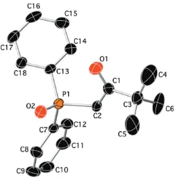
The molecular structures of compounds (I) and (II) are shown in Figs. 1 and 2, respectively. While compound (I) crystallized in the orthorhombic centrosymmetric space group Pbca, compound (II) crystallized in the chiral monoclinic space group P21. In compound (I), the two phenyl rings (C9–C14
and C15–C20) are inclined to one another by 75.53 (8), and to the chlorobenzene ring (C3–C8) by 47.98 (8) and 62.16 (8), respectively. Atom P1 has a distorted tetrahedral geometry with the C—P O bond angles varying from 112.02 (7) to 114.35 (7), while the C—P—C angles vary from 105.04 (7) to 106.60 (7). The carbonyl group (C1 O1) and the phosphoryl group (P1 O2) are anti to one another, most probably to minimize unfavourable dipole–dipole interactions. In compound (II), the two phenyl rings (C7–C12 and C13–C18) are inclined to one another by 86.4 (2). Atom P1 also has a distorted tetrahedral geometry with the C—P O bond angles varying from 111.47 (16) to 115.06 (16), while the C—P—C bond angles vary from 101.84 (15) to 109.21 (16). Here the
carbonyl group (C1 O1) and the phosphoryl group
(P1 O2) aresynto one another.
3. Supramolecular features
In the crystal of (I), the phosphoryl groups are aligned with the a axis, and as the individual molecules stack in this direction they appear to rotate around the chlorine atom that lies close to the twofold screw axis, creating a pinwheel arrangement of molecules (Fig. 3). The molecules are linked
viabifurcated C—H (O,O) hydrogen bonds, forming chains propagating along [100]; see Fig. 3 and Table 1. The chains are linked via C—H interactions (Table 1), forming sheets lying parallel to (010).
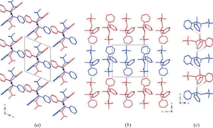
Compound (II) packs in a similar arrangement to (I) in the solid state, although subtle differences result in the formation of a chiral crystal from an achiral compound (Fig. 4). For compound (II), the phosphoryl groups are again aligned in one direction (along thebaxis), but in this case, the P1—C2 bond in the center of the molecule lies about a twofold screw axis and acts as the pivot point for the pinwheel arrangement rather than the terminal chlorine atom as seen in the crystal of
524
Leachet al. C20H16ClO2P and C18H21O2P Acta Cryst.(2015).E71, 523–527
[image:2.610.47.296.68.285.2]research communications
Figure 1
[image:2.610.313.565.108.168.2]A view of the molecular structure of compound (I), showing the atom labelling. Displacement ellipsoids are drawn at the 50% probability level. H atoms have been omitted for clarity.
Figure 2
A view of the molecular structure of compound (II), showing the atom labelling. Displacement ellipsoids are drawn at the 50% probability level. H atoms have been omitted for clarity.
Table 1
Hydrogen-bond geometry (A˚ ,) for (I).
Cg1 andCg3 are the centroids of rings C3–C8 and C15–C20, respectively.
D—H A D—H H A D A D—H A
C14—H14 O2i 0.95 2.30 3.1899 (19) 156
C20—H20 O2i 0.95 2.50 3.4487 (19) 176
C5—H5 Cg3ii 0.95 3.00 3.8873 (17) 156
C13—H13 Cg1i 0.95 2.90 3.5373 (19) 126
Symmetry codes: (i)x1 2;y;zþ
3
2; (ii)xþ1;yþ1;zþ2.
Table 2
Hydrogen-bond geometry (A˚ ,) for (II).
Cg1 is the centroid of ring C7–C12.
D—H A D—H H A D A D—H A
C2—H2B O2i 0.99 2.19 3.176 (5) 176
C12—H12 O2i 0.95 2.53 3.373 (5) 148
C17—H17 Cg1ii 0.95 2.80 3.721 (5) 164
Symmetry codes: (i)xþ1;y1
[image:2.610.313.565.236.287.2] [image:2.610.45.297.447.703.2]compound (I). The absence of an inversion center or mirror plane results in a chiral twist to the packing within this crystal. Here, molecules are also linkedviabifurcated C—H (O,O) hydrogen bonds, forming chains propagating along [010] (see Table 2 and Fig. 4) and the chains are linked viaC—H
interactions (Table 2), forming sheets parallel to (001).
4. Database Survey
The Cambridge Structural Database (CSD, Version 5.36, November 2014; Groom & Allen, 2014) contains 11 structures with a -ketodiphenylphosphoryl moiety. Three of these
structures are related to the title compounds, but have either an alkyl group bonded to the keto function or branching at the
-carbon, viz. E-(5SR,6SR )-3,6-dimethyl-5-diphenylphos-phinoyl-7-triphenylmethoxyhept-2-en-4-one acetone solvate (SUGWOG; Doyle et al., 1993), anti-(2S,4S)-2-(N,N -di-benzylamino)-4-diphenylphosphinoyl-1-phenylpentan-3-one monohydrate (RIZCEI; O’Brienet al., 1997) and (4R,5R )-4,5- dihydroxy-1,5-diphenyl-2-(diphenylphosphinoyl)pentan-1-one) (FODBUW: Boesen et al., 2005). The last compound (FODBUW) crystallizes in a chiral space group (P212121), as
does compound (II). The phenyl rings of the diphenyl-phosphinoyl group in each of these three compounds are
research communications
Acta Cryst.(2015).E71, 523–527 Leachet al. C
[image:3.610.111.493.74.264.2]20H16ClO2P and C18H21O2P
525
Figure 4
The crystal packing diagram of compound (II) (drawn as purple and pink sticks) viewed along: (a) thebaxis (the center of the P1—C1 bond that coincides with the twofold screw axis is denoted with a grey dot); (b) theaaxis; (c) along thebaxis with the bifurcated hydrogen bonds shown as dashed lines (see Table 2 for details). H atoms have been omitted for clarity in parts (a) and (b) and only those involved in hydrogen bonding are shown in (c). Figure 3
[image:3.610.122.491.490.712.2]inclined to one another by ca 67.97, 73.25 and 68.24, respectively, similar to the arrangement in compound (I).
5. Synthesis and crystallization
The title compounds, (I) and (II), were prepared following slightly modified literature procedures (Arnaud-Neu et al., 1996; Schuster et al., 2009) by the Arbuzov reaction of isopropoxydiphenylphosphane (Shintou et al., 2003) with 2-bromo-40-chloroacetophenone for (I) and 1-bromo-pinacolone for (II). For both compounds, crystals suitable for X-ray diffraction analysis were grown by slow evaporation of a solution of the compound in CDCl3.
6. Refinement
Crystal data, data collection and structure refinement details are summarized in Table 3. The hydrogen atoms were placed in calculated positions and refined as riding atoms: C—H = 0.95–0.99 A˚ withUiso(H)= 1.5Ueq(C) for methyl H atoms and
1.2Ueq(C) for other H atoms.
Acknowledgements
We thank Grand Valley State University (Weldon Fund, OURS, CSCE) for financial support of this work. We are grateful to the NSF for financial support (REU-1062944) and NMR instrumentation (300 MHz Jeol, CCLI-0087655), as well as Pfizer, Inc. for the generous donation of a Varian Inova 400 FT NMR. The CCD-based X-ray diffractometers at Michigan State University were upgraded and/or replaced by departmental funds. We also thank Professor James Krikke and Professor William Winchester (GVSU) for help with instrumentation.
References
Arnaud-Neu, F., Bo¨hmer, V., Dozol, J.-F., Gru¨ttner, C., Jakobi, R. A., Kraft, D., Mauprivez, O., Rouquette, H., Schwing-Weill, M.-J., Simon, N. & Vogt, W. (1996).J. Chem. Soc. Perkin Trans. 2, pp. 1175–1182.
Bettencourt-Dias, A. de (2007).Curr. Org. Chem.11, 1460–1480. Boesen, T., Fox, D. J., Galloway, W., Pedersen, D. S., Tyzack, C. R. &
Warren, S. (2005).Org. Biomol. Chem.3, 630–637.
Bourhis, L. J., Dolomanov, O. V., Gildea, R. J., Howard, J. A. K. & Puschmann, H. (2015).Acta Cryst.A71, 59–75.
526
Leachet al. C20H16ClO2P and C18H21O2P Acta Cryst.(2015).E71, 523–527
[image:4.610.47.553.92.412.2]research communications
Table 3
Experimental details.
(I) (II)
Crystal data
Chemical formula C20H16ClO2P C18H21O2P
Mr 354.75 300.32
Crystal system, space group Orthorhombic,Pbca Monoclinic,P21
Temperature (K) 173 173
a,b,c(A˚ ) 11.7380 (2), 14.4453 (3), 19.9515 (3) 8.3416 (2), 10.5161 (2), 10.2790 (2)
,,(
) 90, 90, 90 90, 112.212 (1), 90
V(A˚3) 3382.95 (10) 834.77 (3)
Z 8 2
Radiation type CuK CuK
(mm1) 2.97 1.47
Crystal size (mm) 0.360.170.13 0.430.140.08
Data collection
Diffractometer Bruker APEXII CCD Bruker SMARTAPEXCCD area detector
Absorption correction Multi-scan (SADABS; Bruker, 2013) Multi-scan (SADABS; Bruker, 2013)
Tmin,Tmax 0.599, 0.754 0.631, 0.754
No. of measured, independent and observed [I> 2(I)] reflections
17900, 3297, 2880 7043, 3006, 2774
Rint 0.033 0.042
(sin/ )max(A˚1) 0.617 0.617
Refinement
R[F2> 2(F2)],wR(F2),S 0.032, 0.089, 1.04 0.042, 0.118, 1.13
No. of reflections 3297 3006
No. of parameters 217 193
No. of restraints 0 1
H-atom treatment H-atom parameters constrained H-atom parameters constrained
max,min(e A˚3) 0.32,0.39 0.49,0.38
Absolute structure – Flackxdetermined using 1090 quotients
[(I+)(I)]/[(I+)+(I)] (Parsonset al., 2013)
Absolute structure parameter – 0.088 (14)
Bruker (2013).APEX2,SAINT,XPREPandSADABS. Bruker AXS Inc., Madison, Wisconsin, USA.
Dolomanov, O. V., Bourhis, L. J., Gildea, R. J., Howard, J. A. K. & Puschmann, H. (2009).J. Appl. Cryst.42, 339–341.
Doyle, M. J., Hall, D., Raithby, P. R., Skelton, N. & Warren, S. (1993).
J. Chem. Soc. Perkin Trans. 1, pp. 517–523.
Eliseeva, S. V. & Bu¨nzli, J. G. (2010).Chem. Soc. Rev.39, 189–227. Groom, C. R. & Allen, F. H. (2014).Angew. Chem. Int. Ed.53, 662–
671.
O’Brien, P., Powell, H. R., Raithby, P. R. & Warren, S. (1997). J. Chem. Soc. Perkin Trans. 1, pp. 1031–1040.
Palmer, D. (2007). CrystalMaker. CrystalMaker Software, Bicester, England.
Parsons, S., Flack, H. D. & Wagner, T. (2013).Acta Cryst.B69, 249– 259.
Rosario-Amorin, D., Ouizem, S., Dickie, D. A., Wen, Y., Paine, R. T., Gao, J., Grey, J. K., de Bettencourt-Dias, A., Hay, B. P. & Delmau, L. H. (2013).Inorg. Chem.52, 3063–3083.
Sartain, H. T., McGraw, S. N., Lawrence, C. L., Werner, E. J. & Biros, S. M. (2015).Inorg. Chim. Acta,426, 126–135.
Schuster, E. M., Nisnevich, G., Botoshansky, M. & Gandelman, M. (2009).Organometallics,28, 5025–5031.
Sharova, E. V., Artyushin, O. I., Turanov, A. N., Karandashev, V. K., Meshkova, S. B., Topilova, Z. M. & Odinets, I. L. (2012).Cent. Eur. J. Chem.10, 146–156.
Sheldrick, G. M. (2008).Acta Cryst.A64, 112–122. Sheldrick, G. M. (2015).Acta Cryst.C71, 3–8.
Shintou, T., Kikuchi, W. & Mukaiyama, T. (2003).Bull. Chem. Soc. Jpn,76, 1645–1667.
Thibon, A. & Pierre, V. C. (2009).Anal. Bioanal. Chem.394, 107–120. Weissman, S. I. (1942).J. Chem. Phys.10, 214–217.
research communications
Acta Cryst.(2015).E71, 523–527 Leachet al. C
supporting information
sup-1 Acta Cryst. (2015). E71, 523-527
supporting information
Acta Cryst. (2015). E71, 523-527 [https://doi.org/10.1107/S2056989015006994]
Crystal structures of 1-(4-chlorophenyl)-2-(diphenylphosphoryl)ethan-1-one and
1-(diphenylphosphoryl)-3,3-dimethylbutan-2-one
Erin G. Leach, Alyssa A. Kulesza, Richard J. Staples and Shannon M. Biros
Computing details
For both compounds, data collection: APEX2 (Bruker, 2013); cell refinement: APEX2 and SAINT (Bruker, 2013); data
reduction: SAINT and XPREP (Bruker, 2013); program(s) used to solve structure: SHELXS97 (Sheldrick, 2008);
program(s) used to refine structure: SHELXL2014 (Sheldrick, 2015); molecular graphics: CrystalMaker (Palmer, 2007);
software used to prepare material for publication: OLEX2 (Dolomanov et al., 2009; Bourhis et al., 2015).
(I) 1-(4-Chlorophenyl)-2-(diphenylphosphoryl)ethan-1-one
Crystal data
C20H16ClO2P Mr = 354.75
Orthorhombic, Pbca a = 11.7380 (2) Å
b = 14.4453 (3) Å
c = 19.9515 (3) Å
V = 3382.95 (10) Å3 Z = 8
F(000) = 1472
Dx = 1.393 Mg m−3
Cu Kα radiation, λ = 1.54178 Å Cell parameters from 9053 reflections
θ = 4.4–72.0°
µ = 2.97 mm−1 T = 173 K Needle, colourless 0.36 × 0.17 × 0.13 mm
Data collection
Bruker APEXII CCD diffractometer
Graphite monochromator
φ and ω scans
Absorption correction: multi-scan (SADABS; Bruker, 2013)
Tmin = 0.599, Tmax = 0.754 17900 measured reflections
3297 independent reflections 2880 reflections with I > 2σ(I)
Rint = 0.033
θmax = 72.2°, θmin = 4.4° h = −14→14
k = −17→17
l = −24→23
Refinement
Refinement on F2 Least-squares matrix: full
R[F2 > 2σ(F2)] = 0.032 wR(F2) = 0.089 S = 1.04 3297 reflections 217 parameters 0 restraints
Primary atom site location: structure-invariant direct methods
Hydrogen site location: inferred from neighbouring sites
H-atom parameters constrained
w = 1/[σ2(F
o2) + (0.0502P)2 + 1.0258P] where P = (Fo2 + 2Fc2)/3
supporting information
sup-2 Acta Cryst. (2015). E71, 523-527
Special details
Geometry. All e.s.d.'s (except the e.s.d. in the dihedral angle between two l.s. planes) are estimated using the full covariance matrix. The cell e.s.d.'s are taken into account individually in the estimation of e.s.d.'s in distances, angles and torsion angles; correlations between e.s.d.'s in cell parameters are only used when they are defined by crystal symmetry. An approximate (isotropic) treatment of cell e.s.d.'s is used for estimating e.s.d.'s involving l.s. planes.
Fractional atomic coordinates and isotropic or equivalent isotropic displacement parameters (Å2)
x y z Uiso*/Ueq
supporting information
sup-3 Acta Cryst. (2015). E71, 523-527
H20 0.2591 0.6286 0.7803 0.034*
Atomic displacement parameters (Å2)
U11 U22 U33 U12 U13 U23
Cl1 0.0357 (2) 0.0428 (3) 0.0521 (3) 0.00128 (18) −0.01410 (19) 0.00816 (19) P1 0.0163 (2) 0.0199 (2) 0.0265 (2) −0.00157 (13) −0.00033 (13) −0.00008 (13) O1 0.0337 (6) 0.0397 (7) 0.0364 (6) 0.0080 (5) 0.0075 (5) −0.0014 (5) O2 0.0181 (5) 0.0310 (6) 0.0386 (6) −0.0002 (5) −0.0015 (4) 0.0010 (5) C1 0.0251 (7) 0.0207 (7) 0.0311 (8) −0.0041 (6) 0.0033 (6) −0.0002 (6) C2 0.0215 (7) 0.0207 (7) 0.0307 (8) −0.0029 (6) −0.0001 (6) 0.0011 (6) C3 0.0264 (7) 0.0203 (7) 0.0289 (7) −0.0046 (6) 0.0017 (6) 0.0012 (6) C4 0.0330 (8) 0.0259 (8) 0.0292 (8) −0.0031 (7) 0.0059 (6) 0.0006 (6) C5 0.0416 (9) 0.0299 (9) 0.0269 (8) −0.0052 (7) −0.0015 (7) 0.0036 (6) C6 0.0291 (8) 0.0255 (8) 0.0395 (9) −0.0037 (7) −0.0057 (7) 0.0052 (7) C7 0.0303 (8) 0.0305 (9) 0.0365 (9) 0.0029 (7) 0.0014 (7) −0.0023 (7) C8 0.0314 (8) 0.0279 (8) 0.0275 (7) 0.0008 (6) −0.0002 (6) −0.0012 (6) C9 0.0231 (7) 0.0207 (7) 0.0269 (7) −0.0003 (6) 0.0003 (6) −0.0011 (5) C10 0.0281 (8) 0.0333 (9) 0.0342 (8) −0.0055 (7) 0.0053 (7) −0.0028 (7) C11 0.0454 (10) 0.0409 (10) 0.0310 (8) −0.0048 (8) 0.0106 (7) 0.0015 (7) C12 0.0508 (11) 0.0371 (10) 0.0273 (8) 0.0020 (8) −0.0037 (7) 0.0012 (7) C13 0.0328 (9) 0.0356 (10) 0.0357 (9) −0.0013 (7) −0.0088 (7) −0.0015 (7) C14 0.0241 (8) 0.0299 (8) 0.0308 (8) −0.0027 (6) −0.0008 (6) 0.0011 (6) C15 0.0242 (7) 0.0207 (7) 0.0240 (7) −0.0016 (6) 0.0001 (6) 0.0007 (5) C16 0.0277 (8) 0.0277 (8) 0.0315 (8) −0.0037 (6) −0.0027 (6) −0.0001 (6) C17 0.0440 (9) 0.0269 (9) 0.0307 (8) −0.0075 (7) −0.0060 (7) −0.0030 (6) C18 0.0511 (10) 0.0234 (8) 0.0291 (8) 0.0056 (7) −0.0006 (7) −0.0021 (6) C19 0.0341 (9) 0.0302 (9) 0.0393 (9) 0.0088 (7) 0.0007 (7) −0.0002 (7) C20 0.0258 (7) 0.0256 (8) 0.0345 (8) 0.0008 (6) −0.0026 (6) 0.0008 (6)
Geometric parameters (Å, º)
supporting information
sup-4 Acta Cryst. (2015). E71, 523-527
C6—C7 1.384 (2) C18—H18 0.9500 C7—H7 0.9500 C18—C19 1.382 (3) C7—C8 1.388 (2) C19—H19 0.9500 C8—H8 0.9500 C19—C20 1.384 (2) C9—C10 1.396 (2) C20—H20 0.9500
O2—P1—C2 114.35 (7) C14—C9—C10 119.68 (14) O2—P1—C9 112.64 (7) C9—C10—H10 120.2 O2—P1—C15 112.02 (7) C11—C10—C9 119.67 (16) C9—P1—C2 105.04 (7) C11—C10—H10 120.2 C9—P1—C15 106.60 (7) C10—C11—H11 119.9 C15—P1—C2 105.54 (7) C12—C11—C10 120.30 (16) O1—C1—C2 119.05 (14) C12—C11—H11 119.9 O1—C1—C3 120.86 (14) C11—C12—H12 119.9 C3—C1—C2 120.09 (13) C13—C12—C11 120.22 (16) P1—C2—H2A 108.9 C13—C12—H12 119.9 P1—C2—H2B 108.9 C12—C13—H13 119.8 C1—C2—P1 113.43 (10) C12—C13—C14 120.44 (16) C1—C2—H2A 108.9 C14—C13—H13 119.8 C1—C2—H2B 108.9 C9—C14—H14 120.2 H2A—C2—H2B 107.7 C13—C14—C9 119.69 (15) C4—C3—C1 117.63 (14) C13—C14—H14 120.2 C8—C3—C1 123.27 (14) C16—C15—P1 118.74 (12) C8—C3—C4 119.09 (15) C16—C15—C20 119.81 (15) C3—C4—H4 119.4 C20—C15—P1 121.42 (12) C5—C4—C3 121.18 (15) C15—C16—H16 119.9 C5—C4—H4 119.4 C15—C16—C17 120.17 (15) C4—C5—H5 120.8 C17—C16—H16 119.9 C4—C5—C6 118.47 (15) C16—C17—H17 120.0 C6—C5—H5 120.8 C18—C17—C16 119.95 (15) C5—C6—Cl1 119.11 (13) C18—C17—H17 120.0 C7—C6—Cl1 119.12 (13) C17—C18—H18 120.1 C7—C6—C5 121.75 (15) C19—C18—C17 119.88 (16) C6—C7—H7 120.4 C19—C18—H18 120.1 C6—C7—C8 119.20 (15) C18—C19—H19 119.6 C8—C7—H7 120.4 C18—C19—C20 120.89 (16) C3—C8—H8 119.9 C20—C19—H19 119.6 C7—C8—C3 120.25 (15) C15—C20—H20 120.4 C7—C8—H8 119.9 C19—C20—C15 119.27 (15) C10—C9—P1 117.37 (12) C19—C20—H20 120.4 C14—C9—P1 122.92 (12)
supporting information
sup-5 Acta Cryst. (2015). E71, 523-527
O1—C1—C3—C4 −2.5 (2) C9—P1—C15—C16 −117.67 (13) O1—C1—C3—C8 178.73 (16) C9—P1—C15—C20 60.42 (14) O2—P1—C2—C1 65.44 (12) C9—C10—C11—C12 −1.1 (3) O2—P1—C9—C10 −25.46 (15) C10—C9—C14—C13 1.0 (2) O2—P1—C9—C14 156.68 (13) C10—C11—C12—C13 1.0 (3) O2—P1—C15—C16 5.96 (14) C11—C12—C13—C14 0.1 (3) O2—P1—C15—C20 −175.96 (12) C12—C13—C14—C9 −1.1 (3) C1—C3—C4—C5 −176.70 (15) C14—C9—C10—C11 0.1 (2) C1—C3—C8—C7 177.34 (15) C15—P1—C2—C1 −58.14 (12) C2—P1—C9—C10 −150.53 (13) C15—P1—C9—C10 97.79 (13) C2—P1—C9—C14 31.61 (15) C15—P1—C9—C14 −80.08 (14) C2—P1—C15—C16 130.99 (12) C15—C16—C17—C18 0.5 (2) C2—P1—C15—C20 −50.92 (14) C16—C15—C20—C19 1.1 (2) C2—C1—C3—C4 176.67 (14) C16—C17—C18—C19 1.2 (3) C2—C1—C3—C8 −2.1 (2) C17—C18—C19—C20 −1.7 (3) C3—C1—C2—P1 −81.86 (15) C18—C19—C20—C15 0.6 (3) C3—C4—C5—C6 −0.7 (2) C20—C15—C16—C17 −1.6 (2) C4—C3—C8—C7 −1.4 (2)
Hydrogen-bond geometry (Å, º)
Cg1 and Cg3 are the centroids of rings C3–C8 and C15–C20, respectively.
D—H···A D—H H···A D···A D—H···A
C14—H14···O2i 0.95 2.30 3.1899 (19) 156 C20—H20···O2i 0.95 2.50 3.4487 (19) 176 C5—H5···Cg3ii 0.95 3.00 3.8873 (17) 156 C13—H13···Cg1i 0.95 2.90 3.5373 (19) 126
Symmetry codes: (i) x−1/2, y, −z+3/2; (ii) −x+1, −y+1, −z+2.
(II) 1-(Diphenylphosphoryl)-3,3-dimethylbutan-2-one
Crystal data
C18H21O2P Mr = 300.32
Monoclinic, P21 a = 8.3416 (2) Å
b = 10.5161 (2) Å
c = 10.2790 (2) Å
β = 112.212 (1)°
V = 834.77 (3) Å3 Z = 2
F(000) = 320
Dx = 1.195 Mg m−3
Cu Kα radiation, λ = 1.54178 Å Cell parameters from 5567 reflections
θ = 4.7–72.0°
µ = 1.47 mm−1 T = 173 K Needle, colourless 0.43 × 0.14 × 0.08 mm
Data collection
Bruker SMART APEX CCD area-detector diffractometer
Radiation source: sealed tube Graphite monochromator
Detector resolution: 8 pixels mm-1 ω and φ scans
Absorption correction: multi-scan (SADABS; Bruker, 2013)
Tmin = 0.631, Tmax = 0.754 7043 measured reflections 3006 independent reflections 2774 reflections with I > 2σ(I)
supporting information
sup-6 Acta Cryst. (2015). E71, 523-527
θmax = 72.0°, θmin = 4.7° h = −9→10
k = −12→12
l = −12→12
Refinement
Refinement on F2 Least-squares matrix: full
R[F2 > 2σ(F2)] = 0.042 wR(F2) = 0.118 S = 1.13 3006 reflections 193 parameters 1 restraint
Primary atom site location: structure-invariant direct methods
Hydrogen site location: inferred from neighbouring sites
H-atom parameters constrained
w = 1/[σ2(F
o2) + (0.0722P)2] where P = (Fo2 + 2Fc2)/3 (Δ/σ)max < 0.001
Δρmax = 0.49 e Å−3 Δρmin = −0.38 e Å−3
Absolute structure: Flack x determined using 1090 quotients [(I+)-(I-)]/[(I+)+(I-)] (Parsons et al., 2013)
Absolute structure parameter: 0.088 (14)
Special details
Geometry. All e.s.d.'s (except the e.s.d. in the dihedral angle between two l.s. planes) are estimated using the full covariance matrix. The cell e.s.d.'s are taken into account individually in the estimation of e.s.d.'s in distances, angles and torsion angles; correlations between e.s.d.'s in cell parameters are only used when they are defined by crystal symmetry. An approximate (isotropic) treatment of cell e.s.d.'s is used for estimating e.s.d.'s involving l.s. planes.
Fractional atomic coordinates and isotropic or equivalent isotropic displacement parameters (Å2)
x y z Uiso*/Ueq
supporting information
sup-7 Acta Cryst. (2015). E71, 523-527
C10 0.9069 (5) 0.0207 (5) 0.9603 (4) 0.0485 (11) H10 0.9994 −0.0023 1.0449 0.058* C11 0.8157 (5) −0.0721 (5) 0.8669 (4) 0.0459 (10) H11 0.8455 −0.1590 0.8873 0.055* C12 0.6802 (4) −0.0389 (4) 0.7431 (4) 0.0341 (8) H12 0.6178 −0.1031 0.6790 0.041* C13 0.2784 (4) 0.0408 (4) 0.5431 (3) 0.0270 (6) C14 0.2209 (5) −0.0666 (4) 0.4600 (4) 0.0399 (9) H14 0.2786 −0.0945 0.4012 0.048* C15 0.0789 (6) −0.1332 (5) 0.4632 (5) 0.0486 (11) H15 0.0387 −0.2059 0.4051 0.058* C16 −0.0046 (5) −0.0952 (5) 0.5495 (5) 0.0462 (10) H16 −0.1014 −0.1416 0.5513 0.055* C17 0.0532 (5) 0.0105 (4) 0.6328 (4) 0.0422 (9) H17 −0.0035 0.0366 0.6930 0.051* C18 0.1936 (5) 0.0793 (4) 0.6298 (4) 0.0337 (7) H18 0.2320 0.1528 0.6870 0.040*
Atomic displacement parameters (Å2)
U11 U22 U33 U12 U13 U23
P1 0.0238 (4) 0.0270 (4) 0.0251 (3) −0.0011 (3) 0.0113 (3) 0.0003 (3) O1 0.0312 (12) 0.068 (2) 0.0324 (11) 0.0044 (14) 0.0085 (10) 0.0033 (14) O2 0.0398 (13) 0.0270 (14) 0.0428 (14) 0.0012 (10) 0.0200 (11) 0.0020 (10) C1 0.0345 (17) 0.037 (2) 0.0268 (15) −0.0019 (13) 0.0119 (13) −0.0008 (13) C2 0.0276 (15) 0.0364 (18) 0.0257 (15) 0.0004 (13) 0.0143 (12) −0.0003 (13) C3 0.043 (2) 0.054 (3) 0.0278 (16) 0.0021 (17) 0.0182 (16) 0.0016 (15) C4 0.068 (3) 0.218 (12) 0.027 (2) 0.022 (5) 0.018 (2) −0.004 (4) C5 0.138 (6) 0.084 (5) 0.094 (5) −0.045 (5) 0.096 (5) −0.025 (4) C6 0.092 (4) 0.080 (4) 0.067 (3) 0.029 (3) 0.059 (3) 0.011 (3) C7 0.0222 (13) 0.0427 (18) 0.0212 (14) −0.0043 (13) 0.0106 (12) −0.0009 (12) C8 0.0411 (19) 0.047 (2) 0.0348 (19) −0.0138 (17) 0.0173 (16) −0.0079 (15) C9 0.0411 (19) 0.079 (3) 0.0333 (17) −0.027 (2) 0.0120 (15) −0.016 (2) C10 0.0291 (17) 0.083 (3) 0.0281 (17) −0.006 (2) 0.0045 (14) 0.0044 (19) C11 0.0315 (18) 0.063 (3) 0.038 (2) 0.0070 (18) 0.0082 (16) 0.0108 (19) C12 0.0267 (16) 0.043 (2) 0.0292 (17) −0.0013 (14) 0.0066 (14) 0.0005 (14) C13 0.0200 (13) 0.0328 (16) 0.0276 (15) 0.0006 (12) 0.0083 (12) 0.0047 (13) C14 0.0336 (18) 0.042 (2) 0.047 (2) −0.0061 (16) 0.0181 (17) −0.0107 (16) C15 0.0392 (19) 0.041 (3) 0.065 (3) −0.012 (2) 0.019 (2) −0.0121 (19) C16 0.0262 (16) 0.052 (2) 0.062 (3) −0.0053 (18) 0.0184 (18) 0.011 (2) C17 0.0314 (17) 0.057 (3) 0.044 (2) 0.0000 (17) 0.0216 (16) 0.0036 (18) C18 0.0292 (16) 0.0431 (19) 0.0306 (16) 0.0007 (15) 0.0133 (14) −0.0011 (14)
Geometric parameters (Å, º)
supporting information
sup-8 Acta Cryst. (2015). E71, 523-527
P1—C13 1.807 (3) C9—H9 0.9500 O1—C1 1.207 (5) C9—C10 1.387 (8) C1—C2 1.520 (5) C10—H10 0.9500 C1—C3 1.533 (5) C10—C11 1.378 (7) C2—H2A 0.9900 C11—H11 0.9500 C2—H2B 0.9900 C11—C12 1.390 (5) C3—C4 1.507 (6) C12—H12 0.9500 C3—C5 1.509 (7) C13—C14 1.388 (6) C3—C6 1.525 (7) C13—C18 1.391 (5) C4—H4A 0.9800 C14—H14 0.9500 C4—H4B 0.9800 C14—C15 1.387 (6) C4—H4C 0.9800 C15—H15 0.9500 C5—H5A 0.9800 C15—C16 1.378 (7) C5—H5B 0.9800 C16—H16 0.9500 C5—H5C 0.9800 C16—C17 1.375 (7) C6—H6A 0.9800 C17—H17 0.9500 C6—H6B 0.9800 C17—C18 1.387 (5) C6—H6C 0.9800 C18—H18 0.9500 C7—C8 1.387 (5)
supporting information
sup-9 Acta Cryst. (2015). E71, 523-527
C3—C5—H5B 109.5 C15—C16—H16 120.3 C3—C5—H5C 109.5 C17—C16—C15 119.5 (4) H5A—C5—H5B 109.5 C17—C16—H16 120.3 H5A—C5—H5C 109.5 C16—C17—H17 119.7 H5B—C5—H5C 109.5 C16—C17—C18 120.6 (4) C3—C6—H6A 109.5 C18—C17—H17 119.7 C3—C6—H6B 109.5 C13—C18—H18 120.0 C3—C6—H6C 109.5 C17—C18—C13 120.0 (4) H6A—C6—H6B 109.5 C17—C18—H18 120.0 H6A—C6—H6C 109.5
P1—C7—C8—C9 −178.3 (3) C3—C1—C2—P1 158.4 (3) P1—C7—C12—C11 178.3 (3) C7—P1—C2—C1 −177.5 (3) P1—C13—C14—C15 179.1 (3) C7—P1—C13—C14 −102.8 (3) P1—C13—C18—C17 −178.4 (3) C7—P1—C13—C18 75.6 (3) O1—C1—C2—P1 −23.1 (5) C7—C8—C9—C10 0.0 (6) O1—C1—C3—C4 −11.3 (7) C8—C7—C12—C11 0.3 (5) O1—C1—C3—C5 107.4 (6) C8—C9—C10—C11 0.1 (6) O1—C1—C3—C6 −132.0 (5) C9—C10—C11—C12 −0.1 (6) O2—P1—C2—C1 −56.8 (3) C10—C11—C12—C7 −0.1 (6) O2—P1—C7—C8 −5.5 (3) C12—C7—C8—C9 −0.2 (5) O2—P1—C7—C12 176.4 (3) C13—P1—C2—C1 71.8 (3) O2—P1—C13—C14 135.4 (3) C13—P1—C7—C8 −128.5 (3) O2—P1—C13—C18 −46.2 (3) C13—P1—C7—C12 53.5 (3) C2—P1—C7—C8 117.7 (3) C13—C14—C15—C16 −1.0 (7) C2—P1—C7—C12 −60.4 (3) C14—C13—C18—C17 0.0 (6) C2—P1—C13—C14 5.8 (4) C14—C15—C16—C17 0.3 (7) C2—P1—C13—C18 −175.8 (3) C15—C16—C17—C18 0.5 (7) C2—C1—C3—C4 167.2 (5) C16—C17—C18—C13 −0.7 (6) C2—C1—C3—C5 −74.1 (5) C18—C13—C14—C15 0.8 (6) C2—C1—C3—C6 46.5 (5)
Hydrogen-bond geometry (Å, º)
Cg1 is the centroid of ring C7–C12.
D—H···A D—H H···A D···A D—H···A
C2—H2B···O2i 0.99 2.19 3.176 (5) 176 C12—H12···O2i 0.95 2.53 3.373 (5) 148 C17—H17···Cg1ii 0.95 2.80 3.721 (5) 164