N
,
N
,
N
000-Trimethyl-
N
000000-(4-nitrophenyl)-
N
000-phenylguanidine
Ioannis Tiritiris,aWolfgang Freyband Willi Kantlehnera*
a
Fakulta¨t Chemie/Organische Chemie, Hochschule Aalen, Beethovenstrasse 1, D-73430 Aalen, Germany, andb
Institut fu¨r Organische Chemie, Universita¨t Stuttgart, Pfaffenwaldring 55, 70569 Stuttgart, Germany
Correspondence e-mail: willi.kantlehner@htw-aalen.de
Received 12 March 2014; accepted 28 March 2014
Key indicators: single-crystal X-ray study;T= 293 K; mean(C–C) = 0.003 A˚; Rfactor = 0.053;wRfactor = 0.124; data-to-parameter ratio = 14.7.
The C—N bond lengths in the guanidine unit of the title compound, C16H18N4O2, are 1.298 (2), 1.353 (2) and
1.401 (3) A˚ , indicating double- and single-bond character. The N—C—N angles are 115.81 (16), 118.90 (18) and 125.16 (18), showing a deviation of the CN3 plane from an
ideal trigonal–planar geometry. In the crystal, C—H O hydrogen bonds are observed between the methyl- and aromatic-H atoms and nitro-O atoms. One H atom of the phenyl ring and of the NMe2group associate with the O atoms
of the nitro group, giving chains along the a- and b-axis directions. Cross-linking of these two chains results in a two-dimensional network alongbc.
Related literature
For the synthesis and characterization of compounds for blue OLEDs, see: Agarwalet al.(2011). For the crystal structures of
N-methylated diphenylguanidines, see: Tanataniet al.(1998). For non-classical hydrogen bonds, see: Desiraju & Steiner (1999). For the crystal structure ofN00 -(4-carbazol-9-yl-phen-yl)-N,N0-diethyl-N,N0-diphenylguanidine, see: Tiritiris & Kantlehner (2013), and of N00-(4-methoxyphenyl)-N,N,N0
-trimethyl-N0-phenylguanidine, see: Tiritiriset al.(2014).
Experimental
Crystal data
C16H18N4O2 Mr= 298.34
Monoclinic,C2=c a= 18.409 (2) A˚
b= 7.7140 (8) A˚
c= 22.493 (3) A˚ = 109.503 (7)
V= 3010.9 (6) A˚3
Z= 8
MoKradiation = 0.09 mm1 T= 293 K
0.350.250.20 mm
Data collection
Nicolet P3/F diffractometer 2974 measured reflections 2974 independent reflections 2237 reflections withI> 2(I)
3 standard reflections every 50 reflections
intensity decay: 3%
Refinement
R[F2> 2(F2)] = 0.053
wR(F2) = 0.124 S= 1.06 2974 reflections
203 parameters
H-atom parameters constrained
max= 0.18 e A˚
3
min=0.22 e A˚
3
Table 1
Hydrogen-bond geometry (A˚ ,).
D—H A D—H H A D A D—H A
C12—H12 O2i 0.93 2.49 3.416 (3) 173 C2—H2A O1ii
0.96 2.72 3.064 (3) 102
Symmetry codes: (i)x;y1;z; (ii)x;yþ1;z1 2.
Data collection: XSCANS (Siemens, 1996); cell refinement:
XSCANS; data reduction:SHELXTL(Sheldrick, 2008); program(s)
used to solve structure: SHELXS97(Sheldrick, 2008); program(s)
used to refine structure: SHELXL97 (Sheldrick, 2008); molecular
graphics:DIAMOND(Brandenburg & Putz, 2005); software used to
prepare material for publication:SHELXL97.
The authors thank Dr B. Iliev (IoLiTec GmbH) for the synthesis of the title compound.
Supporting information for this paper is available from the IUCr electronic archives (Reference: NR2049).
organic compounds
o516
Tiritiriset al. doi:10.1107/S160053681400693X Acta Cryst.(2014). E70, o516–o517Acta Crystallographica Section E Structure Reports
Online
References
Agarwal, N., Nayak, P. K., Ali, F., Patankar, M. P., Narasimhan, K. L. & Periasamy, N. (2011).Synth. Met.161, 466–473.
Brandenburg, K. & Putz, H. (2005).DIAMOND. Crystal Impact GbR, Bonn, Germany.
Desiraju, G. R. & Steiner, T. (1999).The Weak Hydrogen Bond In Structural Chemistry and Biology, ch. 2. Oxford University Press.
Sheldrick, G. M. (2008).Acta Cryst.A64, 112–122.
Siemens (1996). XSCANS. Siemens Analytical X-ray Instruments Inc., Madison, Wisconsin, USA.
Tanatani, A., Yamaguchi, K., Azumaya, I., Fukutomi, R., Shudo, K. & Kagechika, H. (1998).J. Am. Chem. Soc.120, 6433–6442.
supporting information
sup-1
Acta Cryst. (2014). E70, o516–o517
supporting information
Acta Cryst. (2014). E70, o516–o517 [doi:10.1107/S160053681400693X]
N
,
N
,
N
′
-Trimethyl-
N
′′
-(4-nitrophenyl)-
N
′
-phenylguanidine
Ioannis Tiritiris, Wolfgang Frey and Willi Kantlehner
S1. Comment
We were interested in the synthesis and characterization of aromatic guanidines to examine their suitability in OLEDs
(Agarwal et al., 2011). Because the crystal structure of the title compound was not known so far, it was decided to carry
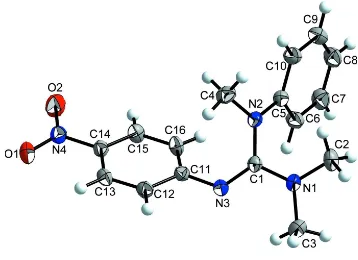
out an appropriate investigation. According to the structure analysis, the C1–N3 bond in the guanidine unit is 1.298 (2) Å,
indicating double bond character. The bond lengths C1–N2 = 1.401 (3) Å and C1–N1 = 1.353 (2) Å are elongated and
characteristic for C–N imine single bonds. The N–C1–N angles are 115.81 (16)° (N1–C1–N2), 125.16 (18)° (N2–C1–N3)
and 118.90 (18)° (N1–C1–N3), showing a deviation of the CN3 plane from an ideal trigonal planar geometry (Fig. 1).
Similar bond lengths and angles of the guanidine CN3 part have been found by structure analysis for
N′′-(4-Carbazol-9-yl-phenyl)- N,N′-diethyl-N,N′-diphenyl-guanidine (Tiritiris & Kantlehner, 2013), several N-methylated diphenylguanidines
(Tanatani et al., 1998) and N′′- (4-methoxyphenyl)-N,N,N′-trimethyl-N′- phenylguanidine (Tiritiris et al., 2014).
Non-classical C–H···O hydrogen bonds (Desiraju & Steiner, 1999) between methyl hydrogen atoms, aromatic hydrogen atoms
and oxygen atoms of the nitro groups are present [d(H···O) = 2.49 and 2.72 Å] (Tab. 1). One hydrogen atom of the phenyl
ring (H12) is associated with the oxygen atom (O2) of the nitro group, resulting in chains along the b axis. A second
hydrogen atom of the NMe2 group (H2A) is connected with O1, giving chains along the a axis. By crosslinking of both
chains, a two-dimensional network along bc results (Fig. 2).
S2. Experimental
One equivalent of N,N-dimethyl-N′,N′-methylphenyl- chloroformamidinium-chloride (synthesized from N,N-dimethyl-
N′,N′-methylphenylthiourea and phosgene) was reacted with one equivalent of 4-nitroaniline (Sigma-Aldrich) in
aceto-nitrile, in the presence of one equivalent triethylamine, at 273 K. The obtained mixture consisting of the guanidinium
chloride and triethylammonium chloride was reacted in the next step with an excess of an aqueous sodium hydroxide
solution at 273 K. After extraction of the guanidine with diethyl ether from the water phase, the solvent was evaporated
and the title compound was isolated in form of a colourless solid. Single crystals have been obtained by recrystallization
from a saturated acetonitrile solution at room temperature.
S3. Refinement
The hydrogen atoms of the methyl groups were allowed to rotate with a fixed angle around the C–N bond to best fit the
experimental electron density, with Uiso(H) set to 1.5Ueq(C) and d(C—H) = 0.96 Å. H atoms for Caromatic were positioned
Figure 1
[image:4.610.126.486.378.630.2]The structure of the title compound with atom labels and 50% probability displacement ellipsoids.
Figure 2
Packing diagram of the title compound. The C–H···O hydrogen bonds (indicated by dashed lines) are arranged in a
supporting information
sup-3
Acta Cryst. (2014). E70, o516–o517
N,N,N′-Trimethyl-N′′-(4-nitrophenyl)-N′-phenylguanidine
Crystal data
C16H18N4O2 Mr = 298.34 Monoclinic, C2/c Hall symbol: -C 2yc a = 18.409 (2) Å b = 7.7140 (8) Å c = 22.493 (3) Å β = 109.503 (7)° V = 3010.9 (6) Å3 Z = 8
F(000) = 1264 Dx = 1.316 Mg m−3
Mo Kα radiation, λ = 0.71073 Å Cell parameters from 35 reflections θ = 14–17°
µ = 0.09 mm−1 T = 293 K Plate, colorless 0.35 × 0.25 × 0.20 mm
Data collection
Nicolet P3/F diffractometer
Radiation source: sealed tube Graphite monochromator Wyckoff scan
2974 measured reflections 2974 independent reflections 2237 reflections with I > 2σ(I)
Rint = 0.000
θmax = 26.0°, θmin = 1.9° h = −22→21
k = 0→9 l = 0→27
3 standard reflections every 50 reflections intensity decay: 3%
Refinement
Refinement on F2 Least-squares matrix: full R[F2 > 2σ(F2)] = 0.053 wR(F2) = 0.124 S = 1.06 2974 reflections 203 parameters 0 restraints
Primary atom site location: structure-invariant direct methods
Secondary atom site location: difference Fourier map
Hydrogen site location: inferred from neighbouring sites
H-atom parameters constrained w = 1/[σ2(F
o2) + (0.0442P)2 + 2.9581P] where P = (Fo2 + 2Fc2)/3
(Δ/σ)max < 0.001 Δρmax = 0.18 e Å−3 Δρmin = −0.22 e Å−3
Extinction correction: SHELXL97 (Sheldrick, 2008), Fc*=kFc[1+0.001xFc2λ3/sin(2θ)]-1/4 Extinction coefficient: 0.0044 (4)
Special details
Geometry. All e.s.d.'s (except the e.s.d. in the dihedral angle between two l.s. planes) are estimated using the full
covariance matrix. The cell e.s.d.'s are taken into account individually in the estimation of e.s.d.'s in distances, angles and torsion angles; correlations between e.s.d.'s in cell parameters are only used when they are defined by crystal symmetry. An approximate (isotropic) treatment of cell e.s.d.'s is used for estimating e.s.d.'s involving l.s. planes.
Refinement. Refinement of F2 against ALL reflections. The weighted R-factor wR and goodness of fit S are based on F2,
conventional R-factors R are based on F, with F set to zero for negative F2. The threshold expression of F2 > σ(F2) is used only for calculating R-factors(gt) etc. and is not relevant to the choice of reflections for refinement. R-factors based on F2 are statistically about twice as large as those based on F, and R-factors based on ALL data will be even larger.
Fractional atomic coordinates and isotropic or equivalent isotropic displacement parameters (Å2)
x y z Uiso*/Ueq
C1 0.11956 (11) 0.3349 (3) 0.21285 (9) 0.0330 (4)
N1 0.09025 (10) 0.2129 (2) 0.16803 (8) 0.0384 (4)
N3 0.10725 (10) 0.3212 (2) 0.26620 (7) 0.0384 (4)
C2 0.12086 (14) 0.1763 (3) 0.11771 (10) 0.0493 (6)
H2A 0.0856 0.2179 0.0784 0.074*
H2B 0.1276 0.0534 0.1150 0.074*
H2C 0.1697 0.2333 0.1264 0.074*
C3 0.03122 (15) 0.0957 (3) 0.17362 (11) 0.0544 (6)
H3A 0.0551 −0.0061 0.1964 0.082*
H3B −0.0021 0.0632 0.1323 0.082*
H3C 0.0016 0.1525 0.1958 0.082*
C4 0.24120 (12) 0.4984 (3) 0.24453 (11) 0.0467 (5)
H4A 0.2483 0.4306 0.2818 0.070*
H4B 0.2445 0.6194 0.2552 0.070*
H4C 0.2805 0.4697 0.2269 0.070*
C5 0.13871 (12) 0.5590 (3) 0.14236 (9) 0.0351 (4)
C6 0.05972 (12) 0.5794 (3) 0.11112 (10) 0.0426 (5)
H6 0.0246 0.5297 0.1277 0.051*
C7 0.03365 (14) 0.6734 (3) 0.05569 (10) 0.0506 (6)
H7 −0.0191 0.6847 0.0349 0.061*
C8 0.08444 (15) 0.7502 (3) 0.03083 (10) 0.0529 (6)
H8 0.0665 0.8125 −0.0067 0.063*
C9 0.16233 (15) 0.7334 (3) 0.06238 (11) 0.0529 (6)
H9 0.1971 0.7867 0.0462 0.064*
C10 0.18983 (13) 0.6391 (3) 0.11746 (10) 0.0451 (5)
H10 0.2426 0.6291 0.1380 0.054*
C11 0.11811 (11) 0.4595 (3) 0.30767 (9) 0.0343 (4)
C12 0.14071 (12) 0.4207 (3) 0.37217 (9) 0.0381 (5)
H12 0.1473 0.3054 0.3849 0.046*
C13 0.15342 (12) 0.5484 (3) 0.41700 (9) 0.0389 (5)
H13 0.1689 0.5200 0.4596 0.047*
C14 0.14288 (11) 0.7195 (3) 0.39797 (9) 0.0359 (5)
C15 0.11713 (12) 0.7640 (3) 0.33462 (9) 0.0400 (5)
H15 0.1088 0.8795 0.3224 0.048*
C16 0.10416 (12) 0.6349 (3) 0.29011 (9) 0.0403 (5)
H16 0.0858 0.6639 0.2476 0.048*
N4 0.15796 (10) 0.8552 (2) 0.44528 (8) 0.0420 (4)
O1 0.17013 (11) 0.8141 (2) 0.50039 (7) 0.0594 (5)
O2 0.15764 (12) 1.0061 (2) 0.42853 (9) 0.0682 (6)
Atomic displacement parameters (Å2)
U11 U22 U33 U12 U13 U23
C1 0.0320 (10) 0.0350 (10) 0.0298 (9) 0.0008 (8) 0.0075 (8) 0.0000 (8)
N1 0.0432 (10) 0.0390 (9) 0.0322 (8) −0.0066 (8) 0.0116 (7) −0.0069 (7)
N2 0.0323 (8) 0.0417 (10) 0.0296 (8) −0.0062 (7) 0.0062 (7) −0.0008 (7)
N3 0.0466 (10) 0.0382 (10) 0.0318 (8) −0.0040 (8) 0.0148 (7) −0.0027 (7)
C2 0.0575 (14) 0.0557 (14) 0.0345 (11) 0.0041 (12) 0.0152 (10) −0.0095 (10) C3 0.0602 (15) 0.0503 (14) 0.0496 (13) −0.0181 (12) 0.0141 (11) −0.0088 (11)
supporting information
sup-5
Acta Cryst. (2014). E70, o516–o517
C5 0.0376 (10) 0.0360 (11) 0.0314 (9) −0.0010 (9) 0.0113 (8) −0.0025 (8)
C6 0.0363 (11) 0.0529 (13) 0.0386 (11) −0.0004 (10) 0.0124 (9) 0.0043 (10)
C7 0.0445 (13) 0.0609 (15) 0.0408 (12) 0.0045 (11) 0.0068 (10) 0.0038 (11)
C8 0.0671 (16) 0.0527 (14) 0.0349 (11) −0.0012 (12) 0.0118 (11) 0.0083 (11) C9 0.0603 (15) 0.0551 (15) 0.0477 (13) −0.0103 (12) 0.0237 (12) 0.0064 (11) C10 0.0412 (12) 0.0511 (13) 0.0430 (12) −0.0064 (10) 0.0142 (9) 0.0027 (10)
C11 0.0327 (10) 0.0400 (11) 0.0318 (10) −0.0023 (8) 0.0127 (8) −0.0015 (9)
C12 0.0458 (12) 0.0339 (11) 0.0343 (10) 0.0012 (9) 0.0128 (9) 0.0033 (9)
C13 0.0430 (11) 0.0448 (12) 0.0290 (10) 0.0014 (10) 0.0118 (8) 0.0033 (9)
C14 0.0364 (11) 0.0397 (11) 0.0335 (10) 0.0008 (9) 0.0141 (8) −0.0054 (9)
C15 0.0475 (12) 0.0356 (11) 0.0388 (11) 0.0094 (9) 0.0167 (9) 0.0031 (9)
C16 0.0471 (12) 0.0445 (12) 0.0279 (10) 0.0078 (10) 0.0108 (9) 0.0036 (9)
N4 0.0435 (10) 0.0426 (11) 0.0429 (10) −0.0015 (8) 0.0184 (8) −0.0078 (8)
O1 0.0782 (12) 0.0658 (12) 0.0362 (8) −0.0074 (9) 0.0215 (8) −0.0125 (8)
O2 0.1042 (16) 0.0391 (10) 0.0635 (11) −0.0023 (10) 0.0309 (11) −0.0080 (8)
Geometric parameters (Å, º)
C1—N3 1.298 (2) C7—C8 1.374 (3)
C1—N1 1.353 (2) C7—H7 0.9300
C1—N2 1.401 (3) C8—C9 1.377 (3)
N1—C2 1.451 (3) C8—H8 0.9300
N1—C3 1.451 (3) C9—C10 1.379 (3)
N2—C5 1.411 (2) C9—H9 0.9300
N2—C4 1.457 (2) C10—H10 0.9300
N3—C11 1.387 (2) C11—C12 1.402 (3)
C2—H2A 0.9600 C11—C16 1.408 (3)
C2—H2B 0.9600 C12—C13 1.372 (3)
C2—H2C 0.9600 C12—H12 0.9300
C3—H3A 0.9600 C13—C14 1.381 (3)
C3—H3B 0.9600 C13—H13 0.9300
C3—H3C 0.9600 C14—C15 1.386 (3)
C4—H4A 0.9600 C14—N4 1.452 (3)
C4—H4B 0.9600 C15—C16 1.375 (3)
C4—H4C 0.9600 C15—H15 0.9300
C5—C10 1.390 (3) C16—H16 0.9300
C5—C6 1.397 (3) N4—O2 1.223 (2)
C6—C7 1.382 (3) N4—O1 1.226 (2)
C6—H6 0.9300
N3—C1—N1 118.90 (18) C8—C7—C6 121.0 (2)
N3—C1—N2 125.16 (18) C8—C7—H7 119.5
N1—C1—N2 115.81 (16) C6—C7—H7 119.5
C1—N1—C2 123.70 (18) C7—C8—C9 118.8 (2)
C1—N1—C3 119.52 (17) C7—C8—H8 120.6
C2—N1—C3 116.34 (18) C9—C8—H8 120.6
C1—N2—C5 121.22 (16) C8—C9—C10 121.3 (2)
C5—N2—C4 119.93 (17) C10—C9—H9 119.4
C1—N3—C11 121.92 (18) C9—C10—C5 120.1 (2)
N1—C2—H2A 109.5 C9—C10—H10 119.9
N1—C2—H2B 109.5 C5—C10—H10 119.9
H2A—C2—H2B 109.5 N3—C11—C12 117.26 (18)
N1—C2—H2C 109.5 N3—C11—C16 125.34 (17)
H2A—C2—H2C 109.5 C12—C11—C16 117.30 (18)
H2B—C2—H2C 109.5 C13—C12—C11 121.7 (2)
N1—C3—H3A 109.5 C13—C12—H12 119.1
N1—C3—H3B 109.5 C11—C12—H12 119.1
H3A—C3—H3B 109.5 C12—C13—C14 119.09 (18)
N1—C3—H3C 109.5 C12—C13—H13 120.5
H3A—C3—H3C 109.5 C14—C13—H13 120.5
H3B—C3—H3C 109.5 C13—C14—C15 121.30 (19)
N2—C4—H4A 109.5 C13—C14—N4 119.30 (18)
N2—C4—H4B 109.5 C15—C14—N4 119.39 (19)
H4A—C4—H4B 109.5 C16—C15—C14 119.0 (2)
N2—C4—H4C 109.5 C16—C15—H15 120.5
H4A—C4—H4C 109.5 C14—C15—H15 120.5
H4B—C4—H4C 109.5 C15—C16—C11 121.35 (18)
C10—C5—C6 118.56 (19) C15—C16—H16 119.3
C10—C5—N2 120.95 (19) C11—C16—H16 119.3
C6—C5—N2 120.48 (18) O2—N4—O1 122.59 (19)
C7—C6—C5 120.2 (2) O2—N4—C14 118.72 (18)
C7—C6—H6 119.9 O1—N4—C14 118.70 (19)
C5—C6—H6 119.9
N3—C1—N1—C2 −157.1 (2) C8—C9—C10—C5 −0.2 (4)
N2—C1—N1—C2 18.9 (3) C6—C5—C10—C9 −1.3 (3)
N3—C1—N1—C3 15.0 (3) N2—C5—C10—C9 179.8 (2)
N2—C1—N1—C3 −169.05 (19) C1—N3—C11—C12 −149.4 (2)
N3—C1—N2—C5 −130.6 (2) C1—N3—C11—C16 34.4 (3)
N1—C1—N2—C5 53.7 (3) N3—C11—C12—C13 179.57 (19)
N3—C1—N2—C4 46.0 (3) C16—C11—C12—C13 −3.9 (3)
N1—C1—N2—C4 −129.7 (2) C11—C12—C13—C14 0.7 (3)
N1—C1—N3—C11 −163.60 (18) C12—C13—C14—C15 2.3 (3)
N2—C1—N3—C11 20.8 (3) C12—C13—C14—N4 −178.60 (18)
C1—N2—C5—C10 −158.0 (2) C13—C14—C15—C16 −2.0 (3)
C4—N2—C5—C10 25.4 (3) N4—C14—C15—C16 178.97 (19)
C1—N2—C5—C6 23.1 (3) C14—C15—C16—C11 −1.4 (3)
C4—N2—C5—C6 −153.4 (2) N3—C11—C16—C15 −179.51 (19)
C10—C5—C6—C7 1.9 (3) C12—C11—C16—C15 4.2 (3)
N2—C5—C6—C7 −179.2 (2) C13—C14—N4—O2 171.2 (2)
C5—C6—C7—C8 −1.0 (4) C15—C14—N4—O2 −9.8 (3)
C6—C7—C8—C9 −0.5 (4) C13—C14—N4—O1 −9.2 (3)
supporting information
sup-7
Acta Cryst. (2014). E70, o516–o517
Hydrogen-bond geometry (Å, º)
D—H···A D—H H···A D···A D—H···A
C12—H12···O2i 0.93 2.49 3.416 (3) 173
C2—H2A···O1ii 0.96 2.72 3.064 (3) 102