246
https://doi.org/10.1107/S2056989018001494 Acta Cryst.(2018). E74, 246–251research communications
Received 18 January 2018 Accepted 23 January 2018
Edited by D.-J. Xu, Zhejiang University (Yuquan Campus), China
Keywords:crystal structure; cadmium(II); tran-sition metal complexes of benzoic acid and nicotinamide derivatives.
CCDC reference:1818756
Supporting information:this article has supporting information at journals.iucr.org/e
Crystal structure and Hirshfeld surface analysis of
aquabis(nicotinamide-
j
N
1)bis(2,4,6-trimethyl-benzoato-
j
2O,O
000)cadmium(II)
Tuncer Ho¨kelek,a* Safiye O¨ zkayaband Hacali Necefog˘lub,c
a
Department of Physics, Hacettepe University, 06800 Beytepe, Ankara, Turkey,bDepartment of Chemistry, Kafkas University, 36100 Kars, Turkey, andcInternational Scientific Research Centre, Baku State University, 1148 Baku, Azerbaijan. *Correspondence e-mail: merzifon@hacettepe.edu.tr
The asymmetric unit of the title complex, [Cd(C10H11O2)2(C6H6N2O)2(H2O)], contains one half of the complex molecule, with the CdII cation and the coordinated water O atom residing on a twofold rotation axis. The CdIIcation is coordinated in a bidentate manner to the carboxylate O atoms of the two symmetry-related 2,4,6-trimethylbenzoate (TMB) anions and to the water O atom at distances of 2.297 (2), 2.527 (2) and 2.306 (3) A˚ to form a distorted pentagonal arrangement, while the distorted pentagonal–bipyramidal coordin-ation sphere is completed by the two pyridine N atoms of the two symmetry-related monodentate nicotinamide (NA) ligands at distances of 2.371 (3) A˚ in the axial positions. In the crystal, molecules are linkedviaintermolecular N— H O, O—H O and C—H O hydrogen bonds with R2
2 (12), R3
3 (8),R3
3 (14),
R3 3
(16),R3 3
(20),R3 3
(22),R4 4
(22),R5 5
(16),R6 6
(16) andR6 6
(18) ring motifs, forming a three-dimensional architecture. The Hirshfeld surface analysis of the crystal structure indicates that the most important contributions for the crystal packing are H H (56.9%), H C/C H (21.3%) and H O/O H (19.0%) inter-actions.
1. Chemical context
Nicotinamide (NA) is one form of niacin. A deficiency of this vitamin leads to loss of copper from the body, known as pellagra disease. Victims of pellagra show unusually high serum and urinary copper levels (Krishnamachari, 1974). The crystal structure of NA was first determined by Wright & King (1954). The NA ring is the reactive part of nicotinamide adenine dinucleotide (NAD) and its phosphate (NADP), which are the major electron carriers in many biological oxidation–reduction reactions (Youet al., 1978). The nicotinic acid derivative N,N-diethylnicotinamide (DENA) is an important respiratory stimulant (Bigoliet al., 1972).
Transition metal complexes with ligands of biochemical interest such as imidazole and some N-protected amino acids show interesting physical and/or chemical properties, through which they may find applications in biological systems (Antoliniet al., 1982). Crystal structures of metal complexes with benzoic acid derivatives have been reported extensively because of the varieties of the coordination modes. For example, Co and Cd complexes with 4-aminobenzoic acid (Chen & Chen, 2002), Co complexes with benzoic acid (Cattericket al., 1974), 4-nitrobenzoic acid (Nadzhafovet al., 1981) and phthalic acid (Adiwidjaja et al., 1978), and Cu complexes with 4-hydrochloric acid (Shnulinet al., 1981) have been described.
The structure–function–coordination relationships of the arylcarboxylate ion in CdIIcomplexes of benzoic acid deriv-atives change depending on the nature and position of the substituted groups on the benzene ring, the nature of the additional ligand molecule or solvent, and the pH and temperature of synthesis (Shnulinet al., 1981). When pyridine and its derivatives are used instead of water molecules, the structure is completely different (Cattericket al., 1974).
The structures of some mononuclear complexes obtained from the reactions of transition metal(II) ions with nicotina-mide (NA) and some benzoic acid derivatives as ligands have been determined previously, e.g. [Zn(C7H5O3)2(C6H6N2O)2] [(II); Necefog˘luet al., 2002], [Mn(C7H4ClO2)2(C10H14N2O)2 -(H2O)2] [(III); Ho¨kelek et al., 2008], [Zn(C8H8NO2)2(C6H6 -N2O)2]H2O [(IV); Ho¨keleket al., 2009a], [Mn(C9H10NO2)2 -(C6H6N2O)(H2O)2] [(V); Ho¨kelek et al., 2009b] and [Co(C9H10NO2)2(C6H6N2O)(H2O)2] [(VI); Ho¨kelek et al., 2009c]. The structure determination of the title compound, (I), a cadmium complex with two 2,4,6-trimethylbenzoate (TMB) and two nicotinamide (NA) ligands and one coordinated water molecule, was undertaken in order to compare the results obtained with those reported previously. In this context, we synthesized the CdII-containing title compound and report herein its crystal and molecular structures along with the Hirshfeld surface analysis.
2. Structural commentary
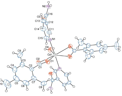
The asymmetric unit of the crystal structure of the mono-nuclear title complex contains half of a CdII cation (site symmetry 2), one 2,4,6-trimethylbenzoate (TMB) anion and one nicotinamide (NA) molecule together with half of a water molecule (point group symmetry 2), the TMB and NA ligands coordinating in bidentate and monodentate manners, respec-tively (Fig. 1).
The CdII cation is coordinated bidentately to the carboxylate O atoms (O1, O2, O1iand O2i) of two symmetry-related 2,4,6-trimethylbenzoate (TMB) anions and to the water O atom (O4) at distances of 2.297 (2), 2.527 (2) and 2.306 (3) A˚ , respectively, to form a distorted pentagonal
arrangement. The sum of the bond angles O1—Cd1—O1i [87.57 (11)], O1—Cd1—O2 [53.63 (7)], O1i
—Cd1—O2i [53.63 (7)], O2—Cd1—O4 [84.47 (5)] and O2i
—Cd1—O4 [84.47 (5)] in the basal plane around CdII
cation is 363.77
[symmetry code: (i) 1x,y,1
2z]. This confirms the presence of the CdIIcation with a small deviation from the basal plane. The distorted pentagonal–bipyramidal coordination sphere is completed by the two pyridine N atoms (N1 and N1i) of the two symmetry-related monodentate nicotinamide (NA) ligands at distances of 2.371 (3) A˚ in the axial positions (Fig. 1). The near equalities of the C1—O1 [1.249 (4) A˚ ] and C1— O2 [1.253 (3) A˚ ] bonds in the carboxylate groups indicate delocalized bonding arrangements, rather than localized single and double bonds. The O2—C1—O1 bond angle [121.7 (3)]
seems to be slightly decreased than that present in a free acid [122.2]. The O2—C1—O1 bond angle may be compared with
the corresponding values of 123.5 (2) and 120.4 (2) in (II),
125.2 (5) in (III), 119.2 (3) and 123.8 (2) in (IV), 123.6 (3)
and 119.4 (3)in (V) and 123.86 (13) and 118.49 (14)in (VI),
where the benzoate ions are coordinated to the metal atoms only monodentately in (III), and both monodentately and bidentately in (II), (IV), (V) and (VI). The Cd1 atom lies 0.0192 (1) A˚ above of the planar (O1/O2/C1) carboxylate group. The O1—Cd1—O2 angle is 53.63 (7). The
corre-sponding O—M—O angles are 58.79 (6)in (II), 59.02 (8)in
(IV), 58.45 (9) in (V) and 60.70 (4) in (VI). In the TMB
anion, the carboxylate group is twisted away from the attached benzene ring, A (C2–C7), ring by 60.94 (18), while the
benzene and pyridine rings [pyridine =B(N1/C11–C15)], are oriented at a dihedral angle of 50.32 (11). The
four-membered ring D (Cd1/O1/O2/C1) is nearly planar with a maximum deviation of 0.0029 (30) A˚ (for C1) from the mean plane, and it is oriented at dihedral angles of 60.98 (11) and 81.91 (7), with respect to theAandBrings.
research communications
Acta Cryst.(2018). E74, 246–251 Ho¨keleket al. [Cd(C
[image:2.610.319.567.495.690.2]10H11O2)2(C6H6N2O)2(H2O)]
247
Figure 1The molecular structure of the title complex with the atom-numbering scheme. Unlabelled atoms are related to labelled atoms by the symmetry operation (1x,y,1
2z). Displacement ellipsoids are drawn at the 50%
3. Supramolecular features
In the crystal, the molecules are linkedviaintermolecular N— HNA ONA, N—HNA OC, O—HW ONA and C— HTMB OC(NA = nicotinamide, C = carboxylate, W = water and TMB = 2,4,6-trimethylbenzoate) hydrogen bonds (Table 1) withR2
2(12), R 3 3(8), R
3 3(14), R
3 3(16), R
3 3(20), R
3 3(22),
R4 4(22),R
5 5(16),R
6
6(16) andR 6
6(18) ring motifs (Fig. 2), forming a three-dimensional architecture. Hydrogen-bonding and van der Waals contacts are the dominant interactions in the crystal packing. No significant – or C—H interactions are observed.
4. Hirshfeld surface analysis
Visulization and exploration of intermolecular close contacts of a structure is invaluable, and this can be achieved using Hirshfeld surface (HS) analysis (Hirshfeld, 1977; Spackman & Jayatilaka, 2009). An HS analysis was carried out by using
CrystalExplorer17.5 (Turner et al., 2017) to investigate the locations of atom atom short contacts with the potential to form hydrogen bonds and the quantitative ratios of these
interactions and the -stacking interactions in the crystal structure of the title complex.
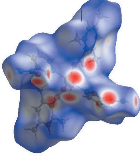
In the HS plotted over dnorm (Fig. 3), the white surface indicates contacts with distances equal to the sum of van der Waals radii, and the red and blue colours indicate distances shorter (in close contact) or longer (distinct contact) than the van der Waals radii, respectively (Venkatesanet al., 2016). The bright-red spots appearing near NA-O3, TMB-O1 and O2, and hydrogen atoms H2A, H2B, H41 and H8Cindicate their role as the respective donors and acceptors in the dominant O— H O, N—H O and C—H O hydrogen bonds; they also appear as blue and red regions corresponding to positive and negative potentials on the HS mapped over electrostatic potential (Spackman et al., 2008; Jayatilaka et al., 2005) as shown in Fig. 4. The blue regions indicate the positive elec-trostatic potential (hydrogen-bond donors), while the red regions indicate the negative electrostatic potential (hydrogen-bond acceptors). The shape-index of the HS is a tool to visualize the–stacking by the presence of adjacent red and blue triangles; if there are no adjacent red and/or blue
248
Ho¨keleket al. [Cd(C [image:3.610.317.562.446.718.2]10H11O2)2(C6H6N2O)2(H2O)] Acta Cryst.(2018). E74, 246–251
research communications
Figure 2
Part of the crystal structure. O—HW ONA, N—HNA OC and N—
HNA ONA (W = water, C = carboxylate and NA = nicotinamide)
hydrogen bonds, enclosingR2 2(12),R
3 3(8),R
3 3(14),R
3 3(16),R
3 3(20),R
3 3(22),
R4
4(22),R55(16),R66(16) andR66(18) ring motifs are shown as dashed lines.
C-bound H atoms have been omitted for clarity.
Table 1
Hydrogen-bond geometry (A˚ ,).
D—H A D—H H A D A D—H A
N2—H2A O3vi 0.89 (3) 2.26 (4) 3.047 (4) 147 (3)
N2—H2B O2vii 0.81 (3) 2.03 (3) 2.830 (4) 168 (4) O4—H41 O3iii 0.80 (3) 1.92 (3) 2.714 (3) 170 (3)
C8—H8C O1viii 0.96 2.55 3.468 (5) 161
Symmetry codes: (iii)xþ1;yþ2;zþ1; (vi)x;yþ2;zþ1
2; (vii)x;y;zþ1; (viii)
x;yþ1;z1 2.
Figure 3
View of the three-dimensional Hirshfeld surface of the title complex plotted overdnormin the range0.6741 to 1.6440 a.u.
Table 2
Selected interatomic distances (A˚ ).
O1 H8Ci 2.55 N2 H13 2.75
O2 H2Bii 2.03 (3) C6 H14ii 2.80
O3 H41iii 1.92 (3) C16 H41iii 2.85 (3)
O3 H2Aiv 2.26 (4) H8A H8Av 2.54
Symmetry codes: (i)x;yþ1;zþ1
2; (ii)x;y;z1; (iii)xþ1;yþ2;zþ1; (iv)
x;yþ2;z1
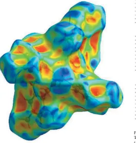
[image:3.610.45.298.482.689.2]triangles, then there are no – interactions. Fig. 5 clearly suggests that there are no–interactions in (I).
The overall two-dimensional fingerprint plot, Fig. 6a, and those delineated into H H, H C/C H, H O/O H, H N/N H, C C and O C/C O contacts (McKinnonet al., 2007) are illustrated in Fig. 6b–g, respectively, together with their relative contributions to the Hirshfeld surface. The most important interaction is H H, contributing 56.9% to the overall crystal packing, which is reflected in Fig. 6b as widely scattered points of high density due to the large hydrogen content of the molecule. The single spike in the centre atde=di= 1.2 A˚ in Fig. 6bis due to a short interatomic H H contact (Table 2). In the absence of C—H inter-actions in the crystal, the pair of characteristic wings resulting in the fingerprint plot delineated into H C/C H contacts, with 21.3% contribution to the HS, Fig. 6c; the pair of thin edges atde+di1.67 A˚ result from short interatomic H C/ C H contacts (Table 2). In the fingerprint plot delineated into H O/O H contacts, Fig. 6d, the 19.0% contribution to
research communications
Acta Cryst.(2018). E74, 246–251 Ho¨keleket al. [Cd(C
[image:4.610.48.293.75.343.2]10H11O2)2(C6H6N2O)2(H2O)]
249
Figure 4View of the three-dimensional Hirshfeld surface of the title complex plotted over electrostatic potential energy in the range0.1379 to 0.1988 a.u. using the STO-3G basis set at the Hartree–Fock level of theory. The N—H O, O—H O and C—H O hydrogen-bond donors and acceptors are viewed as blue and red regions around the atoms corresponding to positive and negative potentials, respectively.
Figure 5
[image:4.610.315.565.287.681.2] [image:4.610.50.540.326.712.2]Hirshfeld surface of the title complex plotted over shape-index.
Figure 6
The full two-dimensional fingerprint plots for the title complex, showing (a) all interactions, and delineated into (b) H H, (c) H C/C H, (d)
H O/O H, (e) H N/N H, (f) C C and (g) O C/C O
interactions. Thedianddevalues are the closest internal and external
[image:4.610.53.318.439.718.2]the HS arises from intermolecular O—H O hydrogen bonding and is viewed as a pair of spikes with the tip atde+di
1.74 A˚ . The short H O/O H contacts are masked by strong O—H O hydrogen bonding in this plot. The H N/ N H contacts in the structure, with a 1.9% contribution to the HS, has a symmetrical distribution of points, Fig. 6e, with the tips atde+di2.96 A˚ arising from the short interatomic H N/N H contact listed in Table 2. The Hirshfeld surface representations with the function dnorm plotted onto the surface are shown for the H H, H C/C H, H O/O H and H N/N H interactions in Fig. 7a–d, respectively.
The Hirshfeld surface analysis confirms the importance of H-atom contacts in establishing the packing. The large number of H H, H C/C H and H O/O H interactions suggest that van der Waals interactions and hydrogen bonding play the major roles in the crystal packing (Hathwar et al., 2015).
5. Synthesis and crystallization
The title compound was prepared by the reaction of 3CdSO48H2O (0.64 g, 2.5 mmol) in water (50 ml) and nico-tinamide (0.61 g, 5 mmol) in water (25 ml) with sodium 2,4,6-trimethylbenzoate (0.93 g, 5 mmol) in water (150 ml) at room temperature. The mixture was filtered and set aside to crys-tallize at ambient temperature for six weeks, giving colourless single crystals (yield: 1.42 g, 85%). Combustion analysis: found; C, 57.07, H, 5.67, N, 7.92%. Calculated: C32H36CdN4O7 C, 57.42; H, 5.43, N, 8.34%. FT–IR: 3390, 3122, 2921, 1669, 1619, 1539, 1445, 1399, 1113, 1038, 847, 731, 641 cm1.
6. Refinement
Crystal data, data collection and structure refinement details are summarized in Table 3. The H atoms of the NH2group and of the water molecule were located in difference-Fourier maps and refined freely. The C-bound H atoms were positioned geometrically with C—H = 0.93 and 0.96 A˚ for aromatic and methyl H atoms, respectively, and constrained to ride on their parent atoms, withUiso(H) =kUeq(C), wherek= 1.5 for methyl H-atoms and k = 1.2 for aromatic H-atoms.
Acknowledgements
The authors acknowledge the Aksaray University, Science and Technology Application and Research Center, Aksaray, Turkey, for the use of the Bruker SMART BREEZE CCD diffractometer (purchased under grant No. 2010K120480 of the State Planning Organization).
References
Adiwidjaja, G., Rossmanith, E. & Ku¨ppers, H. (1978).Acta Cryst.
B34, 3079–3083.
Antolini, L., Battaglia, L. P., Corradi, A. B., Marcotrigiano, G., Menabue, L., Pellacani, G. C. & Saladini, M. (1982).Inorg. Chem. 21, 1391–1395.
Bigoli, F., Braibanti, A., Pellinghelli, M. A. & Tiripicchio, A. (1972).
Acta Cryst.B28, 962–966.
Bruker (2012). APEX2, SAINT and SADABS. Bruker AXS Inc. Madison, Wisconsin, USA.
250
Ho¨keleket al. [Cd(C [image:5.610.46.291.74.309.2]10H11O2)2(C6H6N2O)2(H2O)] Acta Cryst.(2018). E74, 246–251
research communications
Figure 7
The Hirshfeld surface representations with the function dnorm plotted
onto the surface for (a) H H, (b) H C/C H, (c) H O/O H and
(d) H N/N H interactions.
Table 3
Experimental details.
Crystal data
Chemical formula [Cd(C10H11O2)2(C6H6N2O)2
-(H2O)]
Mr 701.05
Crystal system, space group Orthorhombic,Pbcn
Temperature (K) 296
a,b,c(A˚ ) 23.6876 (5), 15.6711 (4), 9.0682 (2)
V(A˚3) 3366.21 (13)
Z 4
Radiation type MoK
(mm1) 0.70
Crystal size (mm) 0.450.280.21
Data collection
Diffractometer Bruker SMART BREEZE CCD
Absorption correction Multi-scan (SADABS; Bruker, 2012)
Tmin,Tmax 0.784, 0.867
No. of measured, independent and observed [I> 2(I)] reflections
68263, 4213, 3681
Rint 0.028
(sin/)max(A˚1) 0.669
Refinement
R[F2> 2(F2)],wR(F2),S 0.047, 0.099, 1.32
No. of reflections 4213
No. of parameters 215
H-atom treatment H atoms treated by a mixture of
independent and constrained refinement
max,min(e A˚3) 0.32,0.47
[image:5.610.303.559.74.378.2]Catterick (nee´ Drew), J., Hursthouse, M. B., New, D. B. & Thornton, P. (1974).J. Chem. Soc. Chem. Commun.pp. 843–844.
Chen, H.-J. & Chen, X.-M. (2002).Inorg. Chim. Acta,329, 13–21. Farrugia, L. J. (2012).J. Appl. Cryst.45, 849–854.
Hathwar, V. R., Sist, M., Jørgensen, M. R. V., Mamakhel, A. H., Wang, X., Hoffmann, C. M., Sugimoto, K., Overgaard, J. & Iversen, B. B. (2015).IUCrJ,2, 563–574.
Hirshfeld, H. L. (1977).Theor. Chim. Acta,44, 129–138.
Ho¨kelek, T., C¸ aylak, N. & Necefog˘lu, H. (2008). Acta Cryst. E64, m505–m506.
Ho¨kelek, T., Dal, H., Tercan, B., Aybirdi, O¨ . & Necefog˘lu, H. (2009a).
Acta Cryst.E65, m1365–m1366.
Ho¨kelek, T., Dal, H., Tercan, B., Aybirdi, O¨ . & Necefog˘lu, H. (2009b).
Acta Cryst.E65, m1037–m1038.
Ho¨kelek, T., Dal, H., Tercan, B., Aybirdi, O¨ . & Necefog˘lu, H. (2009c).
Acta Cryst.E65, m627–m628.
Jayatilaka, D., Grimwood, D. J., Lee, A., Lemay, A., Russel, A. J., Taylor, C., Wolff, S. K., Cassam-Chenai, P. & Whitton, A. (2005).
TONTO. Available at: http://hirshfeldsurface.net/
Krishnamachari, K. A. V. R. (1974).Am. J. Clin. Nutr.27, 108– 111.
McKinnon, J. J., Jayatilaka, D. & Spackman, M. A. (2007).Chem.
Commun.pp. 3814–3816.
Nadzhafov, G. N., Shnulin, A. N. & Mamedov, Kh. S. (1981).Zh.
Strukt. Khim.22, 124–128.
Necefog˘lu, H., Ho¨kelek, T., Ersanlı, C. C. & Erdo¨nmez, A. (2002).
Acta Cryst.E58, m758–m761.
Sheldrick, G. M. (2008).Acta Cryst.A64, 112–122.
Shnulin, A. N., Nadzhafov, G. N., Amiraslanov, I. R., Usubaliev, B. T. & Mamedov, Kh. S. (1981).Koord. Khim.7, 1409–1416.
Spackman, M. A. & Jayatilaka, D. (2009).CrystEngComm,11, 19–32. Spackman, M. A., McKinnon, J. J. & Jayatilaka, D. (2008).Cryst. Eng.
Comm.10, 377–388.
Spek, A. L. (2009).Acta Cryst.D65, 148–155.
Turner, M. J., McKinnon, J. J., Wolff, S. K., Grimwood, D. J., Spackman, P. R., Jayatilaka, D. & Spackman, M. A. (2017).
CrystalExplorer17. The University of Western Australia.
Venkatesan, P., Thamotharan, S., Ilangovan, A., Liang, H. & Sundius, T. (2016).Spectrochim. Acta Part A,153, 625–636.
Wright, W. B. & King, G. S. D. (1954).Acta Cryst.7, 283–288. You, K.-S., Arnold, L. J. Jr, Allison, W. S. & Kaplan, N. O. (1978).
Trends Biochem. Sci.3, 265–268.
research communications
Acta Cryst.(2018). E74, 246–251 Ho¨keleket al. [Cd(C
supporting information
sup-1
Acta Cryst. (2018). E74, 246-251
supporting information
Acta Cryst. (2018). E74, 246-251 [https://doi.org/10.1107/S2056989018001494]
Crystal structure and Hirshfeld surface analysis of
aquabis(nicotinamide-κ
N
1)bis(2,4,6-trimethylbenzoato-
κ
2O
,
O
′
)cadmium(II)
Tuncer H
ö
kelek, Safiye
Ö
zkaya and Hacali Necefo
ğ
lu
Computing details
Data collection: APEX2 (Bruker, 2012); cell refinement: SAINT (Bruker, 2012); data reduction: SAINT (Bruker, 2012); program(s) used to solve structure: SHELXS97 (Sheldrick, 2008); program(s) used to refine structure: SHELXL97
(Sheldrick, 2008); molecular graphics: ORTEP-3 for Windows (Farrugia, 2012); software used to prepare material for publication: WinGX (Farrugia, 2012) and PLATON (Spek, 2009).
Aquabis(nicotinamide-κN1)bis(2,4,6-trimethylbenzoato-κ2O,O′)cadmium(II)
Crystal data
[Cd(C10H11O2)2(C6H6N2O)2(H2O)]
Mr = 701.05
Orthorhombic, Pbcn
Hall symbol: -P 2n 2ab
a = 23.6876 (5) Å
b = 15.6711 (4) Å
c = 9.0682 (2) Å
V = 3366.21 (13) Å3
Z = 4
F(000) = 1440
Dx = 1.383 Mg m−3
Mo Kα radiation, λ = 0.71073 Å Cell parameters from 9766 reflections
θ = 2.6–28.4°
µ = 0.70 mm−1
T = 296 K Block, colorless 0.45 × 0.28 × 0.21 mm
Data collection
Bruker SMART BREEZE CCD diffractometer
Radiation source: fine-focus sealed tube Graphite monochromator
φ and ω scans
Absorption correction: multi-scan (SADABS; Bruker, 2012)
Tmin = 0.784, Tmax = 0.867
68263 measured reflections 4213 independent reflections 3681 reflections with I > 2σ(I)
Rint = 0.028
θmax = 28.4°, θmin = 1.6°
h = −31→31
k = −20→20
l = −11→12
Refinement
Refinement on F2
Least-squares matrix: full
R[F2 > 2σ(F2)] = 0.047
wR(F2) = 0.099
S = 1.32 4213 reflections 215 parameters 0 restraints
Primary atom site location: structure-invariant direct methods
Secondary atom site location: difference Fourier map
Hydrogen site location: inferred from neighbouring sites
supporting information
sup-2
Acta Cryst. (2018). E74, 246-251
w = 1/[σ2(F
o2) + (0.0171P)2 + 5.5549P]
where P = (Fo2 + 2Fc2)/3
(Δ/σ)max = 0.001
Δρmax = 0.32 e Å−3
Δρmin = −0.47 e Å−3
Special details
Geometry. All esds (except the esd in the dihedral angle between two l.s. planes) are estimated using the full covariance matrix. The cell esds are taken into account individually in the estimation of esds in distances, angles and torsion angles; correlations between esds in cell parameters are only used when they are defined by crystal symmetry. An approximate (isotropic) treatment of cell esds is used for estimating esds involving l.s. planes.
Refinement. Refinement of F2 against ALL reflections. The weighted R-factor wR and goodness of fit S are based on F2,
conventional R-factors R are based on F, with F set to zero for negative F2. The threshold expression of F2 > 2sigma(F2) is
used only for calculating R-factors(gt) etc. and is not relevant to the choice of reflections for refinement. R-factors based on F2 are statistically about twice as large as those based on F, and R- factors based on ALL data will be even larger.
Fractional atomic coordinates and isotropic or equivalent isotropic displacement parameters (Å2)
x y z Uiso*/Ueq
supporting information
sup-3
Acta Cryst. (2018). E74, 246-251
H11 0.4655 0.8732 0.5176 0.043* C12 0.40932 (12) 0.83775 (17) 0.6698 (3) 0.0314 (6) C13 0.37296 (15) 0.7725 (2) 0.7083 (4) 0.0445 (8) H13 0.3509 0.7761 0.7929 0.053* C14 0.37015 (16) 0.7016 (2) 0.6178 (4) 0.0529 (9) H14 0.3455 0.6572 0.6398 0.064* C15 0.40397 (16) 0.6973 (2) 0.4953 (4) 0.0470 (8) H15 0.4017 0.6493 0.4353 0.056* C16 0.41406 (12) 0.91928 (18) 0.7553 (3) 0.0357 (6)
Atomic displacement parameters (Å2)
U11 U22 U33 U12 U13 U23
Cd1 0.03990 (16) 0.02586 (14) 0.03497 (16) 0.000 −0.00070 (14) 0.000 O1 0.0606 (14) 0.0357 (11) 0.0452 (13) −0.0048 (10) −0.0166 (11) 0.0054 (10) O2 0.0552 (14) 0.0349 (11) 0.0504 (14) −0.0075 (10) −0.0097 (12) 0.0037 (10) O3 0.0675 (15) 0.0323 (11) 0.0413 (12) −0.0071 (10) −0.0008 (12) 0.0041 (10) O4 0.0467 (19) 0.0269 (14) 0.071 (2) 0.000 −0.0015 (19) 0.000 N1 0.0449 (14) 0.0373 (13) 0.0356 (14) −0.0081 (11) 0.0017 (11) −0.0062 (11) N2 0.0645 (19) 0.0310 (13) 0.0346 (14) 0.0058 (13) −0.0028 (13) −0.0019 (12) C1 0.0408 (15) 0.0326 (14) 0.0299 (14) −0.0032 (12) 0.0014 (12) −0.0019 (12) C2 0.0418 (16) 0.0375 (15) 0.0369 (16) −0.0074 (13) −0.0042 (13) 0.0058 (13) C3 0.0457 (19) 0.073 (3) 0.057 (2) −0.0045 (18) 0.0016 (17) −0.003 (2) C4 0.042 (2) 0.121 (4) 0.081 (3) −0.018 (2) −0.003 (2) 0.005 (3) C5 0.072 (3) 0.101 (4) 0.067 (3) −0.043 (3) −0.016 (2) 0.001 (3) C6 0.085 (3) 0.056 (2) 0.058 (2) −0.030 (2) −0.011 (2) −0.0060 (19) C7 0.061 (2) 0.0349 (16) 0.0465 (19) −0.0115 (15) −0.0078 (16) 0.0007 (14) C8 0.074 (3) 0.057 (2) 0.083 (3) 0.010 (2) −0.005 (2) −0.032 (2) C9 0.055 (3) 0.130 (5) 0.113 (5) 0.017 (3) 0.009 (3) −0.042 (4) C10 0.103 (4) 0.189 (7) 0.137 (6) −0.085 (5) −0.034 (4) −0.022 (5) C11 0.0391 (15) 0.0323 (14) 0.0368 (15) −0.0100 (12) 0.0041 (13) 0.0004 (12) C12 0.0359 (14) 0.0310 (13) 0.0274 (13) −0.0027 (11) −0.0015 (11) 0.0032 (11) C13 0.0507 (19) 0.0490 (18) 0.0340 (16) −0.0122 (15) 0.0065 (14) 0.0034 (14) C14 0.068 (2) 0.0449 (18) 0.0457 (19) −0.0277 (17) 0.0084 (17) 0.0038 (15) C15 0.067 (2) 0.0339 (15) 0.0403 (17) −0.0140 (15) 0.0006 (16) −0.0033 (14) C16 0.0361 (14) 0.0356 (14) 0.0353 (14) 0.0009 (11) −0.0012 (13) 0.0013 (13)
Geometric parameters (Å, º)
Cd1—O1 2.297 (2) C4—H4 0.9300 Cd1—O1i 2.297 (2) C5—C10 1.536 (6)
Cd1—O2 2.527 (2) C6—C5 1.371 (7) Cd1—O2i 2.527 (2) C6—H6 0.9300
Cd1—O4 2.306 (3) C7—C6 1.392 (5) Cd1—N1 2.371 (3) C7—C8 1.503 (5) Cd1—N1i 2.371 (3) C8—H8A 0.9600
supporting information
sup-4
Acta Cryst. (2018). E74, 246-251
O1—C1 1.249 (4) C11—H11 0.9300 O2—C1 1.253 (3) C12—C11 1.380 (4) O3—C16 1.236 (4) C12—C13 1.382 (4) O4—H41 0.80 (3) C12—C16 1.499 (4) N1—C11 1.331 (4) C13—C14 1.382 (5) N1—C15 1.344 (4) C13—H13 0.9300 N2—C16 1.321 (4) C14—H14 0.9300 N2—H2A 0.90 (4) C15—C14 1.371 (5) N2—H2B 0.81 (4) C15—H15 0.9300 C1—C2 1.499 (4) C9—H9A 0.9600 C2—C3 1.388 (5) C9—H9B 0.9600 C2—C7 1.394 (5) C9—H9C 0.9600 C3—C4 1.397 (6) C10—H10A 0.9600 C3—C9 1.512 (6) C10—H10B 0.9600 C4—C5 1.370 (7) C10—H10C 0.9600
O1···H8Cii 2.55 N2···H13 2.75
O2···H2Biii 2.03 (3) C6···H14iii 2.80
O3···H41iv 1.92 (3) C16···H41iv 2.85 (3)
O3···H2Av 2.26 (4) H8A···H8Avi 2.54
O1—Cd1—O1i 87.57 (11) C7—C2—C1 118.9 (3)
O1—Cd1—O2 53.63 (7) C2—C3—C4 117.9 (4) O1i—Cd1—O2 137.06 (8) C2—C3—C9 121.5 (4)
O1—Cd1—O2i 137.06 (8) C4—C3—C9 120.6 (4)
O1i—Cd1—O2i 53.63 (7) C3—C4—H4 118.8
O1—Cd1—O4 136.22 (6) C5—C4—C3 122.3 (4) O1i—Cd1—O4 136.22 (6) C5—C4—H4 118.8
O1—Cd1—N1 85.85 (9) C4—C5—C6 118.6 (4) O1i—Cd1—N1 101.67 (9) C4—C5—C10 119.7 (5)
O1—Cd1—N1i 101.67 (9) C6—C5—C10 121.8 (5)
O1i—Cd1—N1i 85.85 (9) C5—C6—C7 121.8 (4)
O1—Cd1—C1 26.66 (8) C5—C6—H6 119.1 O1i—Cd1—C1 112.62 (9) C7—C6—H6 119.1
O1—Cd1—C1i 112.62 (9) C2—C7—C8 121.7 (3)
O1i—Cd1—C1i 26.66 (8) C6—C7—C2 118.5 (4)
O2—Cd1—O2i 168.94 (10) C6—C7—C8 119.7 (4)
O2—Cd1—C1 26.97 (7) C7—C8—H8A 109.5 O2i—Cd1—C1 163.57 (8) C7—C8—H8B 109.5
O2—Cd1—C1i 163.57 (8) C7—C8—H8C 109.5
O2i—Cd1—C1i 26.97 (7) H8A—C8—H8B 109.5
O4—Cd1—O2 84.47 (5) H8A—C8—H8C 109.5 O4—Cd1—O2i 84.47 (5) H8B—C8—H8C 109.5
O4—Cd1—N1 84.84 (6) C3—C9—H9A 109.5 O4—Cd1—N1i 84.84 (6) C3—C9—H9B 109.5
O4—Cd1—C1 110.64 (6) C3—C9—H9C 109.5 O4—Cd1—C1i 110.64 (6) H9A—C9—H9B 109.5
supporting information
sup-5
Acta Cryst. (2018). E74, 246-251
N1i—Cd1—O2 85.20 (9) H9B—C9—H9C 109.5
N1—Cd1—O2i 85.20 (9) C5—C10—H10A 109.5
N1i—Cd1—O2i 93.81 (9) C5—C10—H10B 109.5
N1—Cd1—N1i 169.68 (12) C5—C10—H10C 109.5
N1—Cd1—C1 89.67 (9) H10A—C10—H10B 109.5 N1i—Cd1—C1 93.97 (9) H10A—C10—H10C 109.5
N1—Cd1—C1i 93.97 (9) H10B—C10—H10C 109.5
N1i—Cd1—C1i 89.67 (9) N1—C11—C12 123.5 (3)
C1—Cd1—C1i 138.71 (12) N1—C11—H11 118.3
C1—O1—Cd1 97.78 (18) C12—C11—H11 118.3 C1—O2—Cd1 86.90 (18) C11—C12—C13 118.6 (3) Cd1—O4—H41 127 (2) C11—C12—C16 118.3 (3) C11—N1—Cd1 121.6 (2) C13—C12—C16 123.1 (3) C11—N1—C15 117.5 (3) C12—C13—C14 118.3 (3) C15—N1—Cd1 120.9 (2) C12—C13—H13 120.8 C16—N2—H2A 121 (2) C14—C13—H13 120.8 C16—N2—H2B 120 (3) C13—C14—H14 120.3 H2A—N2—H2B 119 (4) C15—C14—C13 119.5 (3) O1—C1—Cd1 55.57 (15) C15—C14—H14 120.3 O1—C1—O2 121.7 (3) N1—C15—C14 122.6 (3) O1—C1—C2 117.6 (3) N1—C15—H15 118.7 O2—C1—Cd1 66.13 (17) C14—C15—H15 118.7 O2—C1—C2 120.7 (3) O3—C16—N2 124.0 (3) C2—C1—Cd1 173.1 (2) O3—C16—C12 119.1 (3) C3—C2—C1 120.2 (3) N2—C16—C12 116.9 (3) C3—C2—C7 121.0 (3)
O1i—Cd1—O1—C1 −160.5 (2) N1i—Cd1—C1—O2 −71.26 (19)
O2—Cd1—O1—C1 −0.25 (18) C1i—Cd1—C1—O1 14.27 (18)
O2i—Cd1—O1—C1 175.96 (17) C1i—Cd1—C1—O2 −165.28 (19)
O4—Cd1—O1—C1 19.5 (2) Cd1—O1—C1—O2 0.5 (3) N1—Cd1—O1—C1 97.6 (2) Cd1—O1—C1—C2 −178.5 (2) N1i—Cd1—O1—C1 −75.3 (2) Cd1—O2—C1—O1 −0.4 (3)
C1i—Cd1—O1—C1 −169.85 (13) Cd1—O2—C1—C2 178.6 (3)
O1—Cd1—O2—C1 0.25 (17) Cd1—N1—C11—C12 −177.2 (2) O1i—Cd1—O2—C1 29.9 (2) C15—N1—C11—C12 2.4 (5)
O2i—Cd1—O2—C1 −166.18 (18) Cd1—N1—C15—C14 177.8 (3)
O4—Cd1—O2—C1 −166.18 (18) C11—N1—C15—C14 −1.8 (5) N1—Cd1—O2—C1 −81.75 (19) O1—C1—C2—C3 118.2 (4) N1i—Cd1—O2—C1 108.55 (19) O1—C1—C2—C7 −60.7 (4)
C1i—Cd1—O2—C1 36.3 (5) O2—C1—C2—C3 −60.9 (4)
O1—Cd1—N1—C11 −162.7 (3) O2—C1—C2—C7 120.2 (3) O1i—Cd1—N1—C11 110.6 (2) C1—C2—C3—C4 −178.2 (4)
O1—Cd1—N1—C15 17.7 (3) C1—C2—C3—C9 0.1 (6) O1i—Cd1—N1—C15 −68.9 (3) C7—C2—C3—C4 0.7 (6)
O2—Cd1—N1—C11 −109.6 (2) C7—C2—C3—C9 179.0 (4) O2i—Cd1—N1—C11 59.3 (2) C3—C2—C7—C6 −0.3 (5)
supporting information
sup-6
Acta Cryst. (2018). E74, 246-251
O2i—Cd1—N1—C15 −120.2 (3) C3—C2—C7—C8 176.8 (4)
O4—Cd1—N1—C11 −25.6 (2) C1—C2—C7—C8 −4.3 (5) O4—Cd1—N1—C15 154.9 (3) C2—C3—C4—C5 −0.5 (7) N1i—Cd1—N1—C11 −25.6 (2) C9—C3—C4—C5 −178.8 (5)
N1i—Cd1—N1—C15 154.9 (3) C3—C4—C5—C6 −0.2 (8)
C1—Cd1—N1—C11 −136.3 (2) C3—C4—C5—C10 −180.0 (5) C1i—Cd1—N1—C11 84.8 (3) C7—C6—C5—C4 0.6 (7)
C1—Cd1—N1—C15 44.1 (3) C7—C6—C5—C10 −179.6 (5) C1i—Cd1—N1—C15 −94.7 (3) C2—C7—C6—C5 −0.4 (6)
O1i—Cd1—C1—O1 21.1 (3) C8—C7—C6—C5 −177.6 (4)
O1—Cd1—C1—O2 −179.6 (3) C13—C12—C11—N1 −1.1 (5) O1i—Cd1—C1—O2 −158.40 (18) C16—C12—C11—N1 −179.1 (3)
O2—Cd1—C1—O1 179.6 (3) C11—C12—C13—C14 −0.8 (5) O2i—Cd1—C1—O1 −9.8 (4) C16—C12—C13—C14 177.1 (3)
O2i—Cd1—C1—O2 170.68 (14) C11—C12—C16—O3 36.2 (4)
O4—Cd1—C1—O1 −165.73 (18) C11—C12—C16—N2 −143.9 (3) O4—Cd1—C1—O2 14.72 (19) C13—C12—C16—O3 −141.7 (3) N1—Cd1—C1—O1 −81.4 (2) C13—C12—C16—N2 38.2 (4) N1i—Cd1—C1—O1 108.3 (2) C12—C13—C14—C15 1.3 (6)
N1—Cd1—C1—O2 99.07 (19) N1—C15—C14—C13 0.0 (6)
Symmetry codes: (i) −x+1, y, −z+1/2; (ii) x, −y+1, z+1/2; (iii) x, y, z−1; (iv) −x+1, −y+2, −z+1; (v) x, −y+2, z−1/2; (vi) −x+1, −y+1, −z.
Hydrogen-bond geometry (Å, º)
D—H···A D—H H···A D···A D—H···A
N2—H2A···O3vii 0.89 (3) 2.26 (4) 3.047 (4) 147 (3)
N2—H2B···O2viii 0.81 (3) 2.03 (3) 2.830 (4) 168 (4)
O4—H41···O3iv 0.80 (3) 1.92 (3) 2.714 (3) 170 (3)
C8—H8C···O1ix 0.96 2.55 3.468 (5) 161