N
000-[(
E
)-4-Methoxybenzylidene]-2-(5-methoxy-2-methyl-1
H
-indol-3-yl)-acetohydrazide
Mehmet Akkurt,aJoel T. Mague,bShaaban K. Mohamed,c,d Antar A. Abelhamideand Mustafa R. Albayatif*
aDepartment of Physics, Faculty of Sciences, Erciyes University, 38039 Kayseri, Turkey,bDepartment of Chemistry, Tulane University, New Orleans, LA 70118, USA,cChemistry and Environmental Division, Manchester Metropolitan University, Manchester M1 5GD, England,dChemistry Department, Faculty of Science, Mini University, 61519 El-Minia, Egypt,eDepartment of Chemistry, Faculty of Science, Sohag University, 82524 Sohag, Egypt, andfKirkuk University, College of Science, Department of Chemistry, Kirkuk, Iraq
Correspondence e-mail: shaabankamel@yahoo.com
Received 30 September 2013; accepted 2 October 2013
Key indicators: single-crystal X-ray study;T= 100 K; mean(C–C) = 0.002 A˚; Rfactor = 0.036;wRfactor = 0.090; data-to-parameter ratio = 12.7.
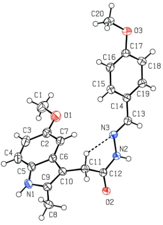
The conformation adopted by the title compound, C20H21
-N3O3, in the crystal is ‘J’-shaped and appears to be at least
partially directed by a weak intramolecular C—H N hydrogen bond. In the crystal, molecules are linked by N— H O hydrogen bonds, forming dimers with R22(8) motifs.
Furthermore, these dimers connect to each other via C— H O and N—H O hydrogen bonds to form a three-dimensional network.
Related literature
For general medical applications of non-steriodal anti-inflammatory drugs (NSAIDs), see: Richyet al.(2004). For the undesirable side effects of such drugs, see: Allisonet al.(1992); McMahon (2001); Rochaet al.(2001); Halenet al.(2009). For a similar structure, see: Mague et al. (2013). For hydrogen-bond motifs, see: Bernsteinet al.(1995).
Experimental
Crystal data
C20H21N3O3 Mr= 351.40
Triclinic,P1 a= 7.1894 (2) A˚ b= 10.4055 (3) A˚ c= 12.4403 (4) A˚
= 107.983 (2)
= 92.451 (2)
= 97.882 (2) V= 873.24 (5) A˚3 Z= 2
CuKradiation
= 0.74 mm1 T= 100 K
0.140.120.08 mm
Data collection
Bruker D8 VENTURE PHOTON 100 CMOS diffractometer Absorption correction: multi-scan
(SADABS; Bruker, 2013) Tmin= 0.85,Tmax= 0.94
8928 measured reflections 3121 independent reflections 2579 reflections withI> 2(I) Rint= 0.029
Refinement
R[F2> 2(F2)] = 0.036 wR(F2) = 0.090 S= 1.04 3121 reflections 246 parameters
H atoms treated by a mixture of independent and constrained refinement
max= 0.19 e A˚ 3
min=0.18 e A˚ 3
Table 1
Hydrogen-bond geometry (A˚ ,).
D—H A D—H H A D A D—H A
N1—H1 O2i
0.88 (2) 2.04 (2) 2.9212 (17) 174.0 (18) N2—H2 O2ii
0.920 (18) 1.988 (18) 2.9025 (16) 171.9 (15) C4—H4 O3iii 0.95 2.50 3.410 (2) 161 C11—H11B N3 0.99 2.36 2.8373 (19) 109 C20—H20A O1iv
0.98 2.49 3.215 (2) 131
Symmetry codes: (i) xþ2;yþ1;zþ2; (ii) xþ2;yþ2;zþ2; (iii)
xþ1;y1;z; (iv)x1;y;z.
Data collection:APEX2(Bruker, 2013); cell refinement:SAINT (Bruker, 2013); data reduction:SAINT; program(s) used to solve structure: SHELXTL (Sheldrick, 2008); program(s) used to refine structure: SHELXL2013 (Sheldrick, 2008); molecular graphics: ORTEP-3 for Windows(Farrugia, 2012); software used to prepare material for publication: WinGX (Farrugia, 2012) and PLATON (Spek, 2009).
The authors are deeply grateful to Tulane University, Erciyes University and Manchester Metropolitan University for supporting this study.
Supplementary data and figures for this paper are available from the IUCr electronic archives (Reference: SJ5357).
References
Allison, M. C., Howatson, A. G., Torrance, C. J., Lee, F. D. & Russell, R. I. (1992).N. Engl. J. Med.327, 749–754.
Bernstein, J., Davis, R. E., Shimoni, L. & Chang, N.-L. (1995).Angew. Chem. Int. Ed. Engl.34, 1555–1573.
Bruker (2013).APEX2,SAINTandSADABS. Bruker AXS Inc., Madison, Wisconsin, USA.
Farrugia, L. J. (2012).J. Appl. Cryst.45, 849–854.
Halen, P. K., Prashant, R., Murumkar, P. R., Giridhar, R. & Mange Ram Yadav, M. R. (2009).Mini Rev. Med. Chem.9, 124–139.
Mague, J. T., Akkurt, M., Mohamed, S. K., El-Remaily, M. A. A. & Albayati, M. R. (2013).Acta Cryst. E69, o1614.
organic compounds
o1660
Akkurtet al. doi:10.1107/S1600536813027050 Acta Cryst.(2013). E69, o1660–o1661Acta Crystallographica Section E Structure Reports
Online
Richy, F., Bruyere, O., Ethgen, O., Rabenda, V., Bouvenot, G., Audran, M., Herrero-Beaumont, G., Moore, A., Eliakim, R., Haim, M. & Reginster, J.-Y. (2004).Ann. Rheum. Dis.63, 759–766.
Burg, M. B. (2001).Proc. Natl Acad. Sci. USA,98, 5317–5322. Sheldrick, G. M. (2008).Acta Cryst.A64, 112–122.
supporting information
sup-1
Acta Cryst. (2013). E69, o1660–o1661
supporting information
Acta Cryst. (2013). E69, o1660–o1661 [doi:10.1107/S1600536813027050]
N
′
-[(E)-4-Methoxybenzylidene]-2-(5-methoxy-2-methyl-1H-indol-3-yl)acetohydrazide
Mehmet Akkurt, Joel T. Mague, Shaaban K. Mohamed, Antar A. Abelhamid and Mustafa R.
Albayati
S1. Comment
Indomethacin like other non-steroidal anti-inflammatory drugs (NSAIDs) is widely used in treatment of pain, fever, and
inflammation (Richy et al., 2004). Prolonged administration of such drugs is commonly associated with several undesired
side-effects. The most common of these are gastrointestinal hemorrhage, ulceration, and decreased renal function (Allison
et al., 1992; McMahon 2001; Rocha et al., 2001). The existence of a free carboxylic acid group in the parent drug has
been considred to be the major factor in establishing superficial stomach erosion, particularly in the corpus region of the
stomach (Halen et al., 2009). Thus, it was considered essential to mask or to remove this functional group in order to
produce a safer and more tolerant prodrug profile. Following this reasoning, we report here the synthesis and crystal
structure of the title compound.
The "J" shaped conformation of the title molecule (I) is shown in Fig. 1. The bond lengths and bond angles of (I)
compare well with those in related compounds (Mague et al., 2013).
In the crystal, the molecules form inversion dimers with R22(8) motifs (Bernstein et al., 1995) through N—H···O
hydrogen bonds (Fig. 2, Table 1). In addition, the dimers are linked by C—H···O and N—H···O hydrogen bonds (Table
1), forming a three-dimensional network.
S2. Experimental
A mixture of 233 mg (1 mmol) 2-(5-methoxy-2-methyl-1H-indol-3-yl)acetohydrazide and 136 mg (1 mmol) of
4-meth-oxybenzaldehyde in 30 ml ethanol containing few drops of glacial acetic acid was refluxed for 5 h. The reaction mixture
was allowed to cool to room temperature and the excess solvent was evaporated under vacuum. The residual solid was
collected, washed with cold ethanol and recrystallized from ethanol. Colourless blocks of X-ray quality were obtained.
M.p. 453–455 K.
S3. Refinement
The H atoms of the amino group were found in the difference Fourier maps, and were refined freely. C-bound H atoms
were placed geometrically and refined using a riding model with C—H = 0.95 - 0.99 Å, and with Uiso(H) = 1.2 or
Figure 1
supporting information
sup-3
[image:5.610.131.484.69.416.2]Acta Cryst. (2013). E69, o1660–o1661 Figure 2
Packing viewed along a showing the hydrogen bonds as dotted lines.
N′-[(E)-4-Methoxybenzylidene]-2-(5-methoxy-2-methyl-1H-indol-3-yl)-acetohydrazide
Crystal data
C20H21N3O3 Mr = 351.40
Triclinic, P1 Hall symbol: -P 1
a = 7.1894 (2) Å
b = 10.4055 (3) Å
c = 12.4403 (4) Å
α = 107.983 (2)°
β = 92.451 (2)°
γ = 97.882 (2)°
V = 873.24 (5) Å3
Z = 2
F(000) = 372
Dx = 1.336 Mg m−3
Cu Kα radiation, λ = 1.54178 Å Cell parameters from 6305 reflections
θ = 3.8–68.2°
µ = 0.74 mm−1 T = 100 K Block, colourless 0.14 × 0.12 × 0.08 mm
Data collection
Bruker D8 VENTURE PHOTON 100 CMOS diffractometer
Radiation source: INCOATEC IµS micro–focus source
Mirror monochromator
Detector resolution: 10.4167 pixels mm-1
ω scans
Absorption correction: multi-scan (SADABS; Bruker, 2013)
Tmin = 0.85, Tmax = 0.94
Rint = 0.029
θmax = 68.2°, θmin = 3.8°
k = −11→12
l = −14→12
Refinement
Refinement on F2
Least-squares matrix: full
R[F2 > 2σ(F2)] = 0.036 wR(F2) = 0.090 S = 1.04 3121 reflections 246 parameters 0 restraints
H atoms treated by a mixture of independent and constrained refinement
w = 1/[Σ2(F
o2) + (0.0398P)2 + 0.2493P]
where P = (Fo2 + 2Fc2)/3
(Δ/σ)max = 0.001
Δρmax = 0.19 e Å−3
Δρmin = −0.18 e Å−3
Special details
Geometry. Bond distances, angles etc. have been calculated using the rounded fractional coordinates. All su's are estimated from the variances of the (full) variance-covariance matrix. The cell e.s.d.'s are taken into account in the estimation of distances, angles and torsion angles
Refinement. Refinement on F2 for ALL reflections except those flagged by the user for potential systematic errors.
Weighted R-factors wR and all goodnesses of fit S are based on F2, conventional R-factors R are based on F, with F set to
zero for negative F2. The observed criterion of F2 > σ(F2) is used only for calculating -R-factor-obs etc. and is not
relevant to the choice of reflections for refinement. R-factors based on F2 are statistically about twice as large as those
based on F, and R-factors based on ALL data will be even larger.
Fractional atomic coordinates and isotropic or equivalent isotropic displacement parameters (Å2)
x y z Uiso*/Ueq
supporting information
sup-5
Acta Cryst. (2013). E69, o1660–o1661
C19 0.3475 (2) 1.05862 (15) 0.72627 (12) 0.0305 (5) C20 −0.2801 (2) 0.84238 (17) 0.57471 (14) 0.0380 (5) H1 0.855 (3) 0.295 (2) 0.9360 (16) 0.052 (5)*
H1A 0.86220 0.41770 0.42890 0.0720*
H1B 0.71490 0.49270 0.38010 0.0720*
H1C 0.64190 0.35780 0.41200 0.0720*
H2 0.847 (2) 0.9799 (18) 0.9426 (14) 0.037 (4)*
H3 0.79600 0.28050 0.54110 0.0470*
H4 0.82900 0.20030 0.69610 0.0460*
H7 0.66820 0.63830 0.74730 0.0370*
H8A 0.66310 0.43210 1.13970 0.0570*
H8B 0.79780 0.57680 1.17140 0.0570*
H8C 0.88670 0.43840 1.13960 0.0570*
H11A 0.67420 0.71790 1.09300 0.0340* H11B 0.55260 0.71370 0.98090 0.0340*
H13 0.64810 1.04650 0.84880 0.0330*
H15 0.23020 0.79590 0.81260 0.0350*
H16 −0.05350 0.78380 0.70910 0.0360*
H18 0.16500 1.12090 0.63150 0.0390*
H19 0.45010 1.12800 0.72920 0.0370*
H20A −0.23210 0.75960 0.53170 0.0570* H20B −0.38450 0.85740 0.52910 0.0570* H20C −0.32500 0.83130 0.64510 0.0570*
Atomic displacement parameters (Å2)
U11 U22 U33 U12 U13 U23
C17 0.0296 (8) 0.0303 (8) 0.0264 (7) 0.0094 (6) −0.0022 (6) 0.0042 (6) C18 0.0362 (8) 0.0313 (8) 0.0316 (8) 0.0066 (6) −0.0019 (6) 0.0136 (7) C19 0.0298 (8) 0.0301 (8) 0.0316 (8) 0.0026 (6) −0.0004 (6) 0.0111 (6) C20 0.0293 (8) 0.0443 (9) 0.0367 (8) 0.0029 (7) −0.0040 (6) 0.0098 (7)
Geometric parameters (Å, º)
O1—C1 1.422 (2) C14—C15 1.393 (2)
O1—C2 1.3776 (19) C15—C16 1.388 (2)
O2—C12 1.2422 (17) C16—C17 1.390 (2)
O3—C17 1.3653 (18) C17—C18 1.389 (2)
O3—C20 1.425 (2) C18—C19 1.377 (2)
N1—C5 1.386 (2) C1—H1A 0.9800
N1—C9 1.383 (2) C1—H1B 0.9800
N2—N3 1.3818 (16) C1—H1C 0.9800
N2—C12 1.3475 (19) C3—H3 0.9500
N3—C13 1.2810 (19) C4—H4 0.9500
N1—H1 0.88 (2) C7—H7 0.9500
N2—H2 0.920 (18) C8—H8A 0.9800
C2—C3 1.406 (2) C8—H8B 0.9800
C2—C7 1.382 (2) C8—H8C 0.9800
C3—C4 1.384 (2) C11—H11A 0.9900
C4—C5 1.383 (2) C11—H11B 0.9900
C5—C6 1.415 (2) C13—H13 0.9500
C6—C10 1.434 (2) C15—H15 0.9500
C6—C7 1.400 (2) C16—H16 0.9500
C8—C9 1.490 (2) C18—H18 0.9500
C9—C10 1.369 (2) C19—H19 0.9500
C10—C11 1.501 (2) C20—H20A 0.9800
C11—C12 1.514 (2) C20—H20B 0.9800
C13—C14 1.460 (2) C20—H20C 0.9800
C14—C19 1.398 (2)
C1—O1—C2 118.05 (13) C14—C19—C18 121.03 (14) C17—O3—C20 117.85 (13) O1—C1—H1A 109.00
C5—N1—C9 109.09 (13) O1—C1—H1B 109.00
N3—N2—C12 122.52 (12) O1—C1—H1C 109.00 N2—N3—C13 114.94 (12) H1A—C1—H1B 109.00
C9—N1—H1 120.9 (12) H1A—C1—H1C 109.00
C5—N1—H1 126.0 (13) H1B—C1—H1C 109.00
N3—N2—H2 118.7 (10) C2—C3—H3 120.00
C12—N2—H2 118.8 (10) C4—C3—H3 120.00
O1—C2—C3 123.67 (14) C3—C4—H4 121.00
O1—C2—C7 115.23 (14) C5—C4—H4 121.00
C3—C2—C7 121.10 (15) C2—C7—H7 120.00
C2—C3—C4 120.20 (16) C6—C7—H7 120.00
supporting information
sup-7
Acta Cryst. (2013). E69, o1660–o1661
C4—C5—C6 121.74 (15) C9—C8—H8B 109.00
N1—C5—C6 107.17 (14) C9—C8—H8C 110.00
N1—C5—C4 131.07 (15) H8A—C8—H8B 109.00 C5—C6—C10 107.06 (13) H8A—C8—H8C 109.00 C7—C6—C10 134.17 (14) H8B—C8—H8C 109.00 C5—C6—C7 118.73 (14) C10—C11—H11A 109.00 C2—C7—C6 119.35 (15) C10—C11—H11B 109.00 N1—C9—C8 120.08 (14) C12—C11—H11A 109.00 N1—C9—C10 109.40 (14) C12—C11—H11B 109.00 C8—C9—C10 130.47 (15) H11A—C11—H11B 108.00 C6—C10—C11 126.49 (13) N3—C13—H13 119.00 C9—C10—C11 126.29 (13) C14—C13—H13 119.00 C6—C10—C9 107.22 (13) C14—C15—H15 119.00 C10—C11—C12 113.58 (12) C16—C15—H15 119.00 N2—C12—C11 119.29 (12) C15—C16—H16 120.00 O2—C12—N2 118.72 (13) C17—C16—H16 120.00 O2—C12—C11 121.97 (13) C17—C18—H18 120.00 N3—C13—C14 121.97 (13) C19—C18—H18 120.00 C13—C14—C19 119.12 (13) C14—C19—H19 119.00 C15—C14—C19 118.23 (13) C18—C19—H19 119.00 C13—C14—C15 122.65 (13) O3—C20—H20A 110.00 C14—C15—C16 121.06 (14) O3—C20—H20B 109.00 C15—C16—C17 119.73 (14) O3—C20—H20C 109.00 O3—C17—C16 124.68 (14) H20A—C20—H20B 109.00 C16—C17—C18 119.73 (13) H20A—C20—H20C 109.00 O3—C17—C18 115.59 (14) H20B—C20—H20C 109.00 C17—C18—C19 120.21 (15)
C4—C5—C6—C10 −177.19 (14) O3—C17—C18—C19 179.57 (13) N1—C5—C6—C10 1.37 (16) C16—C17—C18—C19 0.4 (2) C7—C6—C10—C11 3.1 (3) C17—C18—C19—C14 −1.2 (2) C5—C6—C10—C11 −179.56 (13)
Hydrogen-bond geometry (Å, º)
D—H···A D—H H···A D···A D—H···A
N1—H1···O2i 0.88 (2) 2.04 (2) 2.9212 (17) 174.0 (18)
N2—H2···O2ii 0.920 (18) 1.988 (18) 2.9025 (16) 171.9 (15)
C4—H4···O3iii 0.95 2.50 3.410 (2) 161
C11—H11B···N3 0.99 2.36 2.8373 (19) 109 C20—H20A···O1iv 0.98 2.49 3.215 (2) 131