Isovaline monohydrate
Ray J. Butcher,a* Greg Brewer,bAaron S. Burtoncand Jason P. Dworkind
a
Department of Chemistry, Howard University, 525 College Street NW, Washington, DC 20059, USA,bDepartment of Chemistry, Catholic University of America, Washington, DC 20064, USA,cNASA Johnson Space Center, Astromaterial and
Exploration Science Directorate, Houston, TX 77058, USA, anddSolar System
Exploration Division, NASA Goddard Space Flight Center, Greenbelt, MD 20771, USA
Correspondence e-mail: [email protected]
Received 23 October 2013; accepted 20 November 2013
Key indicators: single-crystal X-ray study;T= 123 K; mean(C–C) = 0.005 A˚; Rfactor = 0.056;wRfactor = 0.162; data-to-parameter ratio = 13.2.
The title compound, C5H11NO2H2O, is an isomer of the -amino acid valine that crystallizes from water in its zwitterion form as a monohydrate. It is not one of the 20 proteinogenic amino acids that are used in living systems and differs from the natural amino acids in that it has no-H atom. The compound exhibits hydrogen bonding between the water molecule and the carboxylate O atoms and an amine H atom. In addition, there are intermolecular hydrogen-bonding interactions between the carboxylate O atoms and amine H atoms. In the crystal, these extensive N—H O and O—H O hydrogen bonds lead to the formation of a three-dimensional network.
Related literature
The structure of the title compound or its salts have not been reported to the CCDC but there are reports of homoleptic coordination complexes of zinc(II) with isovaline, see: Stras-deltet al.(2001). For literature related to eighty amino acids that have been detected in meteorites or comets, see: Glavin & Dworkin (2009); Burton et al.(2012). For the role that crys-tallization plays in chiral separation, see: Blackmond & Klussmann (2007); Blackmondet al.(2008). For the role of the H atom on the-C atom in enhancing the rate of racemiza-tion, see: Yamada et al. (1983). For the mechanism of race-mization of amino acids lacking an-H atom, see: Pizzarello & Groy (2011). For the role that crystallization can play in the enrichment of l-isovaline, see: Glavin & Dworkin (2009);
Bada (2009); Bonner et al. (1979). For normal bond lengths and angles, see: Orpen (1993).
Experimental
Crystal data
C5H11NO2H2O
Mr= 135.16
Orthorhombic,P212121
a= 5.9089 (5) A˚ b= 10.4444 (10) A˚ c= 11.9274 (11) A˚
V= 736.10 (12) A˚3 Z= 4
CuKradiation
= 0.84 mm1
T= 123 K
0.480.080.06 mm
Data collection
Agilent Xcalibur (Ruby, Gemini) diffractometer
Absorption correction: multi-scan (CrysAlis PRO; Agilent, 2012) Tmin= 0.383,Tmax= 1.000
1662 measured reflections 1204 independent reflections 1072 reflections withI> 2(I) Rint= 0.072
Refinement
R[F2> 2(F2)] = 0.056 wR(F2) = 0.162
S= 1.11 1204 reflections 91 parameters 3 restraints
H atoms treated by a mixture of independent and constrained refinement
max= 0.37 e A˚
3 min=0.27 e A˚
3
Table 1
Hydrogen-bond geometry (A˚ ,).
D—H A D—H H A D A D—H A
O1W—H1W1 O1i
0.83 (2) 2.05 (2) 2.811 (3) 152 (4) O1W—H1W2 O2ii
0.83 (2) 1.96 (2) 2.787 (3) 171 (5) N1—H1A O2i
0.91 1.84 2.745 (3) 177 N1—H1C O1W 0.91 2.09 2.792 (4) 133 N1—H1B O1iii
0.91 1.98 2.832 (3) 156
Symmetry codes: (i)xþ1;y;z; (ii)xþ1
2;yþ1;zþ 1 2; (iii)xþ
1 2;yþ
1 2;zþ1.
Data collection: CrysAlis PRO(Agilent, 2012); cell refinement: CrysAlis PRO; data reduction: CrysAlis PRO; program(s) used to solve structure: SHELXS97(Sheldrick, 2008); program(s) used to refine structure:SHELXL97(Sheldrick, 2008); molecular graphics: SHELXTL(Sheldrick, 2008); software used to prepare material for publication:SHELXTL.
RJB wishes to acknowledge the NSF–MRI program (grant CHE-0619278) for funds to purchase the diffractometer. GB wishes to acknowledge support of this work from NASA (NNX10AK71A)
Supplementary data and figures for this paper are available from the IUCr electronic archives (Reference: JJ2178).
organic compounds
Acta Cryst.(2013). E69, o1829–o1830 doi:10.1107/S1600536813031620 Butcheret al.
o1829
Acta Crystallographica Section E Structure Reports Online
Agilent (2012). CrysAlis PRO. Agilent Technologies UK Ltd, Yarnton, England.
Bada, J. L. (2009).Proc. Natl Acad. Sci.106, E85.
Blackmond, D. G. & Klussmann, M. (2007).Chem. Commun.pp. 3990–3996. Blackmond, D., Viedma, C., Ortiz, J., Torres, T. & Izuma, T. (2008).J. Am.
Chem. Soc.130, 15274–15275.
Bonner, W. A., Blair, N. E., Lemmon, R. M., Flores, J. J. & Pollock, G. E. (1979).Geochim. Cosmochim. Acta,43, 1841–1846.
Chem. Soc. Rev.41, 5459–5472.
Glavin, D. P. & Dworkin, J. P. (2009).Proc. Natl Acad. Sci.106, 5487–5492. Orpen, G. A. (1993).Chem. Soc. Rev.22, 191–197.
Pizzarello, S. & Groy, T. L. (2011).Geochim. Cosmochim. Acta,75, 645–656. Sheldrick, G. M. (2008).Acta Cryst.A64, 112–122.
Strasdelt, H., Busching, I., Behrends, S., Saak, W. & Barklage, W. (2001). Chem. Eur. J.7, 1133–1137.
supporting information
sup-1
Acta Cryst. (2013). E69, o1829–o1830
supporting information
Acta Cryst. (2013). E69, o1829–o1830 [doi:10.1107/S1600536813031620]
Isovaline monohydrate
Ray J. Butcher, Greg Brewer, Aaron S. Burton and Jason P. Dworkin
S1. Comment
The alpha amino acids are essential for life as they are the building blocks of all proteins and enzymes. Nature uses
almost exclusively the L form of the nineteen naturally occurring chiral amino acids. Glycine is achiral. However, it is
known that there are over eighty amino acids that have been detected in meteorites or comets (Glavin & Dworkin 2009;
Burton et al., 2012). One of these extraterrestrial non proteinogenic amino acids is isovaline. The majority of these amino
acids show little or no enrichment of one enantiomer over the other. An intriguing question is the process that lead to the
separation and enrichment of the L enantiomer over the D. There are several possible explanations for this including the
role that crystallization plays (Blackmond & Klussmann, 2007). Only two of the twenty amino acids used biologically
crystallize in a chiral space group, which allows for spontaneous separation of enantiomers, at the level of the crystal,
from a racemic solution (Blackmond et al., 2008). Isovaline, a non proteinogenic amino acid also allows for this
separation of enantiomers at the level of the crystal as it crystallizes in the chiral space group, P212121.
Another important aspect in the prebiotic chemistry of the amino acids is the role of racemization. All of the nineteen
naturally occurring chiral amino acids have a hydrogen atom on the alpha carbon atom, which enhances the rate of
racemization (Yamada et al., 1983). However, little is known about the mechanism of racemization of amino acids
lacking an alpha hydrogen atom (Pizzarello & Groy, 2011). The structure of isovaline given here can be used to examine
the role that crystallization can play in the enrichment of L isovaline (Glavin & Dworkin 2009; Bada, 2009), and as a
starting point in mechanistic studies of racemization mechanisms of isovaline (Bonner et al., 1979).
In the structure of the title compound the amino acid is in the usual zwitterionic form. While the structure of the title
compound or its salts have not been reported there are reports of homoleptic coordination complexes of isovaline with
zinc(II) (Strasdelt et al. (2001). However, there are several important differences between these structures and the title
compound. The metal complexes all crystallized in non-chiral space groups and, of course, when coordinated to a metal,
the isovaline will not be in a zwitterionic form but, apart from the COO- group, all the the bond lengths and angles are in
the normal range for such compounds (Orpen, 1993). There is extensive N—H···O and O—H···O hydrogen bonding
linking the zwitterions into a 3-D array.
S2. Experimental
A sample of the title compound was obtained from Acros Organics. Crystals of the title compound were grown from the
slow evaporation of a racemic solution of the amino acid in water.
S3. Refinement
H atoms were placed in geometrically idealized positions and constrained to ride on their parent atoms with a C—H
distances of 0.98 and 0.99 Å and an N—H distance of 0.91 \%A Uiso(H) = 1.2Ueq(C) and 0.96 Å for NH3 [Uiso(H) =
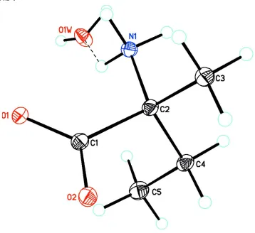
Figure 1
Diagram of the title compound showing atom labeling. Atomic displacement parameters are at the 30% probablity level.
supporting information
sup-3
[image:5.610.130.480.73.416.2]Acta Cryst. (2013). E69, o1829–o1830
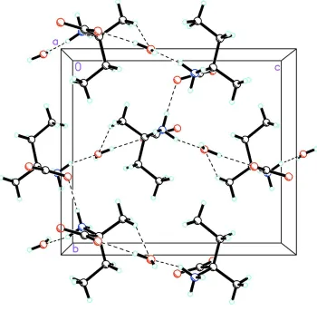
Figure 2
Packing diagram of the title compound viewed along the a axis showing tthe extensive N—H···O and O—H···O hydrogen
bonds as dashed lines.
2-Azaniumyl-2-methylbutanoate monohydrate
Crystal data
C5H11NO2·H2O Mr = 135.16
Orthorhombic, P212121 Hall symbol: P 2ac 2ab a = 5.9089 (5) Å b = 10.4444 (10) Å c = 11.9274 (11) Å V = 736.10 (12) Å3 Z = 4
F(000) = 296 Dx = 1.220 Mg m−3
Cu Kα radiation, λ = 1.54184 Å Cell parameters from 679 reflections θ = 3.7–75.3°
µ = 0.84 mm−1 T = 123 K Needle, colorless 0.48 × 0.08 × 0.06 mm
Data collection
Agilent Xcalibur (Ruby, Gemini) diffractometer
Radiation source: Enhance (Cu) X-ray Source Graphite monochromator
Detector resolution: 10.5081 pixels mm-1 ω scans
θmax = 75.4°, θmin = 5.6° h = −7→4
l = −14→14
Refinement
Refinement on F2 Least-squares matrix: full R[F2 > 2σ(F2)] = 0.056 wR(F2) = 0.162 S = 1.11 1204 reflections 91 parameters 3 restraints
Primary atom site location: structure-invariant direct methods
Secondary atom site location: difference Fourier map
Hydrogen site location: inferred from neighbouring sites
H atoms treated by a mixture of independent and constrained refinement
w = 1/[σ2(F
o2) + (0.093P)2 + 0.2964P] where P = (Fo2 + 2Fc2)/3
(Δ/σ)max < 0.001 Δρmax = 0.37 e Å−3 Δρmin = −0.27 e Å−3
Special details
Geometry. All e.s.d.'s (except the e.s.d. in the dihedral angle between two l.s. planes) are estimated using the full
covariance matrix. The cell e.s.d.'s are taken into account individually in the estimation of e.s.d.'s in distances, angles and torsion angles; correlations between e.s.d.'s in cell parameters are only used when they are defined by crystal symmetry. An approximate (isotropic) treatment of cell e.s.d.'s is used for estimating e.s.d.'s involving l.s. planes.
Refinement. Refinement of F2 against ALL reflections. The weighted R-factor wR and goodness of fit S are based on F2,
conventional R-factors R are based on F, with F set to zero for negative F2. The threshold expression of F2 > σ(F2) is used only for calculating R-factors(gt) etc. and is not relevant to the choice of reflections for refinement. R-factors based on F2 are statistically about twice as large as those based on F, and R- factors based on ALL data will be even larger.
Fractional atomic coordinates and isotropic or equivalent isotropic displacement parameters (Å2)
x y z Uiso*/Ueq
O1 0.0058 (4) 0.37096 (19) 0.50201 (18) 0.0316 (5)
O2 −0.1536 (4) 0.4257 (3) 0.33811 (19) 0.0385 (6)
O1W 0.6455 (4) 0.4874 (3) 0.6172 (2) 0.0467 (7)
H1W1 0.776 (5) 0.465 (5) 0.601 (3) 0.070*
H1W2 0.661 (8) 0.517 (5) 0.682 (2) 0.070*
N1 0.4267 (4) 0.3850 (2) 0.4302 (2) 0.0297 (6)
H1A 0.5663 0.3957 0.3994 0.045*
H1B 0.4074 0.3014 0.4493 0.045*
H1C 0.4141 0.4348 0.4925 0.045*
C1 0.0133 (6) 0.4049 (2) 0.4006 (3) 0.0302 (6)
C2 0.2504 (5) 0.4229 (3) 0.3472 (2) 0.0294 (6)
C3 0.2766 (6) 0.3382 (3) 0.2440 (3) 0.0367 (7)
H3A 0.2503 0.2486 0.2648 0.055*
H3B 0.4301 0.3474 0.2139 0.055*
H3C 0.1664 0.3640 0.1869 0.055*
C4 0.2832 (6) 0.5657 (3) 0.3178 (3) 0.0344 (7)
H4A 0.4384 0.5779 0.2885 0.041*
H4B 0.1762 0.5888 0.2572 0.041*
C5 0.2477 (7) 0.6556 (3) 0.4151 (3) 0.0451 (9)
H5A 0.2565 0.7442 0.3886 0.068*
supporting information
sup-5
Acta Cryst. (2013). E69, o1829–o1830
H5C 0.0984 0.6401 0.4482 0.068*
Atomic displacement parameters (Å2)
U11 U22 U33 U12 U13 U23
O1 0.0326 (11) 0.0293 (9) 0.0329 (10) 0.0009 (8) 0.0031 (9) 0.0061 (9)
O2 0.0268 (11) 0.0510 (13) 0.0376 (11) 0.0010 (11) 0.0007 (9) 0.0110 (11)
O1W 0.0379 (13) 0.0627 (17) 0.0396 (13) 0.0087 (13) −0.0008 (11) −0.0148 (12)
N1 0.0268 (12) 0.0270 (11) 0.0352 (14) −0.0004 (9) 0.0016 (11) 0.0015 (11)
C1 0.0324 (15) 0.0214 (12) 0.0367 (15) −0.0017 (11) 0.0027 (12) 0.0020 (11)
C2 0.0279 (15) 0.0275 (13) 0.0328 (14) −0.0009 (12) 0.0009 (11) 0.0051 (13)
C3 0.0371 (18) 0.0392 (16) 0.0338 (16) 0.0040 (15) −0.0005 (13) 0.0010 (14)
C4 0.0320 (15) 0.0317 (15) 0.0394 (15) −0.0038 (13) −0.0001 (13) 0.0085 (14)
C5 0.055 (2) 0.0260 (14) 0.054 (2) −0.0030 (14) 0.0026 (18) 0.0020 (15)
Geometric parameters (Å, º)
O1—C1 1.261 (4) C2—C4 1.545 (4)
O2—C1 1.255 (4) C3—H3A 0.9800
O1W—H1W1 0.830 (19) C3—H3B 0.9800
O1W—H1W2 0.832 (19) C3—H3C 0.9800
N1—C2 1.490 (4) C4—C5 1.507 (5)
N1—H1A 0.9100 C4—H4A 0.9900
N1—H1B 0.9100 C4—H4B 0.9900
N1—H1C 0.9100 C5—H5A 0.9800
C1—C2 1.550 (4) C5—H5B 0.9800
C2—C3 1.524 (4) C5—H5C 0.9800
H1W1—O1W—H1W2 102 (3) C2—C3—H3B 109.5
C2—N1—H1A 109.5 H3A—C3—H3B 109.5
C2—N1—H1B 109.5 C2—C3—H3C 109.5
H1A—N1—H1B 109.5 H3A—C3—H3C 109.5
C2—N1—H1C 109.5 H3B—C3—H3C 109.5
H1A—N1—H1C 109.5 C5—C4—C2 114.2 (3)
H1B—N1—H1C 109.5 C5—C4—H4A 108.7
O2—C1—O1 126.2 (3) C2—C4—H4A 108.7
O2—C1—C2 116.4 (3) C5—C4—H4B 108.7
O1—C1—C2 117.4 (3) C2—C4—H4B 108.7
N1—C2—C3 108.1 (3) H4A—C4—H4B 107.6
N1—C2—C4 108.6 (3) C4—C5—H5A 109.5
C3—C2—C4 111.3 (3) C4—C5—H5B 109.5
N1—C2—C1 109.1 (2) H5A—C5—H5B 109.5
C3—C2—C1 110.7 (3) C4—C5—H5C 109.5
C4—C2—C1 108.9 (2) H5A—C5—H5C 109.5
C2—C3—H3A 109.5 H5B—C5—H5C 109.5
O2—C1—C2—N1 −175.8 (3) O1—C1—C2—C4 −114.2 (3)
O1—C1—C2—C3 123.0 (3) C1—C2—C4—C5 54.7 (4)
O2—C1—C2—C4 65.8 (3)
Hydrogen-bond geometry (Å, º)
D—H···A D—H H···A D···A D—H···A
O1W—H1W1···O1i 0.83 (2) 2.05 (2) 2.811 (3) 152 (4)
O1W—H1W2···O2ii 0.83 (2) 1.96 (2) 2.787 (3) 171 (5)
N1—H1A···O2i 0.91 1.84 2.745 (3) 177
N1—H1C···O1W 0.91 2.09 2.792 (4) 133
N1—H1B···O1iii 0.91 1.98 2.832 (3) 156