©
201
7 M
a
cmillan Publis
hers Limited,
p
ar
t o
f S
p
ri
n
g
er
N
at
u
re
.
All rights reserved.
IntroDuctIon
Adult bile ducts consist of highly functional biliary epithelial cells1, which regulate bile homeostasis and modulate inflamma-tory responses. These cells are also known as cholangiocytes and represent the main cell type affected in cholangiopathies2,3—a diverse group of liver disorders including diseases such as primary biliary cirrhosis and primary sclerosing cholangitis. Despite the growing importance of these diseases, research in biliary physiol-ogy and the development of new therapeutics has been hampered by the lack of robust platforms for disease modeling and high-throughput drug screening3. Although animal models exist, their capacity for fully reproducing human pathophysiology is lim-ited4,5, and access to primary biliary tissue remains problematic, prohibiting large-scale experiments. Here, we describe a protocol for the generation of large quantities of CLCs from human hPSCs, which can be applied to model cholangiopathies in vitro and to validate the effects of therapeutic compounds6.
Development of the protocol
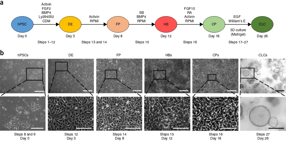
The protocol for the generation of CLCs6 was developed by reca-pitulating key stages of native bile duct development (Fig. 1a). Cholangiocytes originate from HBs, a bipotent population of embryonic liver progenitor cells7 that can also differentiate into hepatocytes. Hepatoblasts surrounding the portal vein give rise to a monolayer of immature cholangiocyte progenitor cells (the ductal plate)7, which undergoes a process of 3D remodeling and maturation, resulting in functional bile ducts.
The generation of bipotent HBs was based on our established methodology for producing hPSC-derived hepatocyte-like cells8. To achieve biliary commitment of HBs, we used physiological
cues reported to control biliary specification, such as activin A (a member of the TGF-β superfamily)7,9 and fibroblast growth factor (FGF) 10 (ref. 10). Screening a variety of growth factors, we also identified a requirement for retinoic acid6. The combined activation of these signaling pathways was sufficient to promote differentiation of HBs into CPs expressing early biliary markers, including KRT19 and SOX9 (ref. 6).
Maturation of native cholangiocytes happens in synchrony with 3D re-arrangement of the ductal plate into tubular structures7. Most of the functional properties of the biliary epithelium are associated with absorption and secretion processes, which require a polarized epithelium forming a lumen and therefore cannot be accurately reproduced by cells organized in monolayer11,12. Consequently, for the final stage of our protocol promoting CP maturation to CLCs, we developed a 3D culture system based on previous studies using Matrigel and epidermal growth factor (EGF)13,14 that promotes spontaneous differentiation of HBs into cystic structures express-ing early biliary markers such as KRT19 (refs. 13,14). Prolonged culture of CPs under these conditions resulted in CLC organoids with a central lumen demonstrating characteristic functional prop-erties such as γ-glutamyl transferase (GGT) activity6.
Applications
The mechanisms controlling development of the human biliary tree remain poorly understood. Indeed, developmental studies in humans are limited by minimal access to fetal tissue, whereas animal models fail to fully recapitulate the development of the human biliary tree or the phenotype of developmental disorders5. Our in vitro system could address some of these challenges, as it
Directed differentiation of human induced
pluripotent stem cells into functional
cholangiocyte-like cells
Fotios Sampaziotis1–3, Miguel Cardoso de Brito1,6, Imbisaat Geti1,6, Alessandro Bertero1,6, Nicholas RF Hannan4,7 & Ludovic Vallier1,2,5,7
1Wellcome Trust-Medical Research Council Stem Cell Institute, Cambridge Stem Cell Institute, Anne McLaren Laboratory, Department of Surgery, University of Cambridge, Cambridge, UK. 2Department of Surgery, University of Cambridge and NIHR Cambridge Biomedical Research Centre, Cambridge, UK. 3Department of Hepatology, Cambridge University Hospitals NHS Foundation Trust, Cambridge, UK. 4Center for Biomolecular Sciences, University of Nottingham, UK. 5Wellcome Trust Sanger Institute, Hinxton, UK. 6These authors contributed equally to this work. 7These authors jointly directed this work. Correspondence should be addressed to L.V. (lv225@cam.ac.uk).
Published online 23 March 2017; doi:10.1038/nprot.2017.011
©
201
7 M
a
cmillan Publis
hers Limited,
p
ar
t o
f S
p
ri
n
g
er
N
at
u
re
.
All rights reserved.
relies on a stepwise differentiation protocol that closely mimics embryonic bile duct development. Therefore, substantial numbers of cells corresponding to different embryological stages can be easily generated, enabling mechanistic large-scale studies in biliary specification or developmental disorders. Accordingly, we applied this methodology to interrogate the role of TGF-β and Notch signaling in biliary tubulogenesis, and reproduce the Alagille syn-drome phenotype in vitro6. The same principle could be used in future studies to explore a broad spectrum of pathways that could be involved in bile duct development and pathogenesis.
CLCs also recapitulate many physiological functions of cholan-giocytes in vitro, as well as their defects in the context of disease when using hPSCs derived from patients with cholangiopa-thies6. Consequently, CLCs could present an optimal platform for modeling biliary disease, validating therapeutic compounds and screening for novel treatment agents. We have already dem-onstrated proof of principle for the feasibility of this application by reproducing the effects of the drugs verapamil and octreotide in our culture system6 and using patient-specific hiPSCs to iden-tify a new application for the experimental compound VX809 in the management of cystic fibrosis cholangiopathy6. Importantly, the capacity of our system for generating substantial numbers of CLCs6, combined with its compatibility with large-scale experi-mental formats (24- and 48-well plates)6, could set the foundation for the use of patient-derived CLCs to develop high-throughput drug screening platforms for cholangiopathies in the future.
Comparison with other methods
Primary cholangiocyte isolation has been reported15–17. However, such methodologies are technically challenging, support only
short-term growth with limited expansion and generate limited numbers of cells, all of which are not compatible with large-scale experiments15–17. Furthermore, primary cholangiocytes cultured in monolayer systems have not been shown to maintain their functional properties15–17.
Two other protocols have been described for generating biliary epithelium from hPSCs18,19. The method by Dianat et al. results in cells with a transcriptional signature6,19 compatible with a subpop-ulation of cholangiocytes located in the canals of Hering, known as small cholangiocytes20. Therefore, this approach is optimized for studies on small cholangiocytes and complements our protocol, which is aimed toward the production of large cholangiocytes. The method by Ogawa et al. generates cholangiocyte organoids expressing mature markers and demonstrating biliary functional-ity; however, biliary specification is based on a coculture system with mouse OP9 cells12. Although a mixed culture system may recapitulate more closely the native niche of HBs/cholangiocytes, it is technically more challenging and presents several limitations. Indeed, OP9 cells are derived from bone marrow and are known to promote hematopoietic differentiation of ESCs by secreting factors such as macrophage colony-stimulating factor (M-CSF). This poses substantial limitations for mechanistic studies in biliary specification and early biliary development, as unknown secreted factors could interfere with experimental outcomes. Furthermore, the heterogeneity of the cell population in a coculture system renders –omics studies, such as genome-wide analyses, more chal-lenging. Consequently, the platform by Ogawa et al. may be better suited for studies in which accurate reproduction of a complex cellular niche is critical, whereas our system is more optimized for mechanistic studies in biliary development and therapeutics. Activin
FGF2 BMP4 Ly294002
CDM
a
b
FGF10 RA Activin
RPMI
EGF William’s E
3D culture (Matrigel) SB
BMP4 RPMI Activin
RPMI hPSC
Day 0
Steps 1–12
hPSCs DE FP HBs CPs CLCs
Steps 8 and 9 Day 0
Steps 12 Day 3
Steps 14 Day 8
Steps 15 Day 12
Steps 16 Day 16
Steps 27 Day 26 Steps 13 and 14 Steps 15 Steps 16 Steps 17–27
Day 3 Day 8 Day 12 Day 16 Day 26
[image:2.594.49.544.60.304.2]DE FP HB CP CLC
©
201
7 M
a
cmillan Publis
hers Limited,
p
ar
t o
f S
p
ri
n
g
er
N
at
u
re
.
All rights reserved.
Limitations
There are two main limitations to our platform. Our system relies on a complex extracellular matrix (Matrigel). The composition of Matrigel is not fully defined, and variation in the growth factor and protein contents of each batch could affect the efficiency of the final stage of our protocol. Furthermore, the use of Matrigel could render the translation of our platform to good manu-facturing practice conditions challenging and prevent in vivo applications toward cell-based therapy and regenerative medicine. Another important consideration is the maturity of the gener-ated cells. CLC organoids express both early and mature biliary markers and maintain some fetal characteristics corresponding more accurately to a stage between fetal and fully mature bile ducts. Consequently, before modeling adult biliary disorders, CLCs should be tested for the presence of the relevant mature markers and functionality.
Experimental design
Our method describes the generation of hPSC-derived CLC orga-noids over a period of 26 d. Biliary differentiation is achieved through five key stages of recapitulating bile duct development (Fig. 1). Our protocol starts with the plating of hPSCs on day 0 (d0), and we refer to the first day of differentiation as day 1. The first stage (days 1–3) results in the generation of definitive endoderm (DE) cells. These cells correspond to the common pro-genitor from which the liver, lung, pancreas, alimentary tract and thyroid arise. Subsequently, DE cells are differentiated into FP cells (stage 2, days 4–8), which correspond to precursors of the liver, pancreas, lung and thyroid lineages found in the anterior portion of the embryonic alimentary canal. In the third stage (days 9–12), FP cells are differentiated into HBs, bipotent pro-genitors of hepatocytes and cholangiocytes, which can give rise to both. The fourth stage (days 13–16) results in biliary commit-ment of HBs and the generation of CPs, which represent early cholangiocytes forming the ductal plate in vivo. In the final stage of our method (days 17–26), CPs form functional CLC organoids in 3D culture conditions.
Starting population considerations. We have demonstrated that this protocol is reproducible with four different hPSC lines6 and embryonic stem (ES) cells (Fig. 2). Variability in differen-tiation capacity is a common issue with hPSC lines, which may reflect the efficiency and timing of our protocol. Therefore, some minor optimization steps may be required for each hPSC line, as described in the following sections.
Preparation of hPSCs. To achieve high differentiation efficiency, the generation of a near-homogeneous DE population is critical.
For that, hPSCs must exhibit optimal morphology and minimal background differentiation. They should first be allowed to grow to near confluence (70–80%), and then they should be broken into small clumps and plated at high density, as described in the sections below (Fig. 1b; Steps 1–9). Clump size and density have a critical role in this step. Very small clumps or single cells are not viable after the first day of differentiation, whereas large clumps differentiate only partially, maintaining the expression of pluripo-tency markers at their center. Low densities prevent the cells from reaching near confluence by the end of the first stage. This can have a negative impact on paracrine signaling, cell migration and cell-to-cell contact, which are critical factors for efficient forma-tion of the foregut epithelium. A minimum of 24 h should be allowed for the hPSCs to adhere to the plate before starting dif-ferentiation; however, this period can be extended to a maximum of 48 h if the clump size is thought to be too small.
Generation of DE and FP. Definitive endoderm differentiation is characterized by morphological changes, epithelial–mesenchymal transition (Fig. 1b), substantial proliferation of the cells and increased death of cells that fail to differentiate. By the end of d3, the cells should be approaching confluence (Fig. 1b) and express-ing Sox17 and EOMES homogeneously (>90%, Figs. 3 and 4). Cell proliferation continues during the FP stage, and by the end of d8 the cells should be forming a confluent epithelium with cells exhibiting a characteristic rhomboidal morphology (Fig. 1b). The generation of a near-homogeneous population of FP cells is critical for the efficiency of later stages. Therefore, we recommend that differentiations be optimized to generate cell populations with >95% purity for endoderm and foregut markers (Figs. 3
and 4) such as GATA4 and FOXA2. In particular, for resistant hPSC lines with substantial contamination from partially dif-ferentiated cells, we recommend splitting the cells at the foregut stage (day 6). For lines with lower proliferation rates and good differentiation efficiency, this step is optional. If the cells are split at this stage, it is very important that they be dissociated to single cells and replated at a density allowing the formation of a fully confluent epithelium by day 8.
Generation of bipotent HBs. Cell proliferation begins to reduce at this stage, although cells should continue to proliferate at a lower rate. We have noticed variability in the proliferation rates of different hPSC lines. Differentiation of FP to HBs should result in a near-homogeneous population (>95%) expressing hepatoblast markers (CK19, AFP) (Figs. 3 and 4), which is important for the efficiency of subsequent steps. A high cell density of FPs forming a monolayer of relatively small cells is critical to the success of this stage. For resistant hPSC lines, this stage could be prolonged by 24 h to improve differentiation efficiency. However, substantial prolongation of HB differentiation carries the risk of committing a large proportion of cells to the hepatic lineage, which results in increased hepatoblast/hepatocyte contamination at the next stage and reduced biliary lineage commitment.
Generation of CPs. Cell proliferation should increase compared with the previous stage, and by the fourth day of cholangiocyte progenitor differentiation substantial overgrowth should be seen (100% confluence and/or areas of cells forming multiple layers). Differentiation of HBs to CPs is heterogeneous, resulting
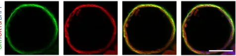
CK7
/
CK19
/
[image:3.594.49.287.62.119.2]DAPI
©
201
7 M
a
cmillan Publis
hers Limited,
p
ar
t o
f S
p
ri
n
g
er
N
at
u
re
.
All rights reserved.
in a mixed population of CPs (75%), HBs (15%) and cells at intermediate stages (Fig. 4). Consequently, although hepatic markers such as AFP can still be detected, early biliary mark-ers such as CK19 and SOX9 should also be expressed almost homogeneously (Figs. 2 and 3). Poor efficiency at this stage (<75% SOX9+ cells) could result in incomplete maturation of CLC organoids in the next step. The quality and duration of the HB stage is critical to limiting HB contamination and ensuring biliary commitment.
Therefore, for resistant hPSC lines, we recommend optimizing the duration and efficiency of HB differentiation as described in the previous section (Generation of bipotent HBs) and in the Troubleshooting section.
Generation of CLC organoids. For the final stage of our protocol, CPs are dissociated into small clumps and transferred to 3D culture conditions. Density and clump size are the factors most critical to the success of this step. Very high densities do not allow adequate
hPSCs
104
1.98 93.5
1.02 0.71 0.95
97.7 Q1
0.74
Q4
3.95 0.059Q3 Q2
95.3 4.00 × 10
–3
0.34 2.61
97.1 0.25
5.78 17.4
76.6 5.65
17.6 0.49
76.3 0.64
3.46
104 103
103 102
102 101
101 100
100
104
104 103
103 102
102 101
101 100
100
104
104 103
103 102
102 101
101 100
100
104
104 103
103 102
102 101
101 100
100
104
104 103
103 102
102 101
101 100
100
104
104 103
103 102
102 101
101 100
100
Log APC
Log FITC
DE FP HBs CPs CLCs
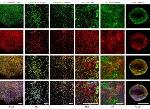
[image:4.594.51.546.57.418.2]OCT4/NANOG EOMES/SOX17 GATA4/FOXA2 CK19/AFP CK19/SOX9 CK7/SOX9
Figure 4| Flow cytometry analyses demonstrating the expression of characteristic markers at key stages of CLC differentiation. CLC organoids were harvested as described in Procedure Steps 40–47. Cells were dissociated into single cells following incubation with TrypLE for 5 min at 37 °C and fixed with 4% (wt/vol) PFA for 20 min at 4 °C. The cells were stained for IF as previously described6, using the antibodies provided in table 1. A standard gating strategy was used22 (demonstrated in supplementary Fig. 2). A minimum of 2 × 10⁴ gated events were used for analysis. Postsort fractions are indicated in the quadrants of each graph. The average efficiency of differentiation from hPSCs to CLCs across three lines (CK7+/Sox9+ organoids) was 77% (s.d. = 6.5%)6.
OCT4/NANOG/DAPI
hPSCs DE FP HBs CPs CLCs
[image:4.594.72.541.578.676.2]EOMES/SOX17/DAPI GATA4/FOXA2/DAPI CK19/AFP/DAPI CK19/SOX9/DAPI CK7/SOX9/DAPI
© 201 7 M a cmillan Publis hers Limited, p ar t o f S p ri n g er N at u re .
All rights reserved.
space for the cells to expand and reorganize into organoids with a central lumen. Instead, proliferating clumps of cells merge together into large aggregates. Single cells or very small clumps may not be viable, whereas large clumps gradually migrate and attach to
the bottom of the plate, forming a monolayer. Consequently, the efficiency of this phase depends on careful manipulation of the quantity of cells, Matrigel and media. Minor adjustments to density and clump size may be required for different hPSC lines (see Troubleshooting and Procedure sections, Steps 17–26). For resistant lines such as H9, we recommend adding forskolin (optional step), which promotes intraluminal fluid secretion and facilitates the formation of organoids with a lumen.
Of note, this step starts with a heterogeneous population of cells including HBs and CPs, and thus some hepatic contamination is expected. However, cells expressing hepatic markers such as AFP fail to form organoids and usually gravitate to the bottom of the plate. By contrast, by the end of this stage CLC organoids should express biliary (CK19, CK7, SOX9; Figs. 3 and 4), but not hepatic (AFP), markers and demonstrate functional properties character-istic of biliary epithelium, such as GGT and alkaline phosphatase (ALP) activity (Fig. 5). Importantly, we have noticed differences in differentiation efficiency with different batches of Matrigel. For resistant hPSC lines, Matrigel should be screened for batches that support organoid formation and cholangiocyte functionality.
Controls
Intrahepatic cholangiocytes are not commercially available. Therefore, we recommend the use of fresh bile duct tissue obtained from liver donors, or frozen isolated common bile duct cholangiocytes commercially available (Celprogen) as a positive control for the expression of biliary markers.
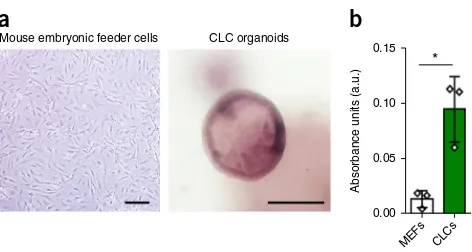
Mouse embryonic feeder cells
a
b
CLC organoids 0.15 * 0.10 0.05 0.00 MEFs CLCs [image:5.594.49.285.59.185.2]Absorbance units (a.u.)
Figure 5| Functional properties of CLC organoids. (a) CLC organoids demonstrating characteristic ALP staining. Mouse embryonic feeder cells are used as a negative control. Scale bars, 100 µm. (b) GGT activity of CLC organoids measured in absorbance units (a.u.); n = 3; mouse embryonic feeder cells (MEFs) are used as a negative control. Error bars, s.d.; individual data points are demonstrated; *P < 0.05, two-tailed Student’s t-test; F-test was used to compare variances, P = 0.1218 (no significant difference in variance). GGT and ALP activity were assessed using commercially available kits (MaxDiscovery gamma-Glutamyl Transferase (GGT) Enzymatic Assay Kit and BCIP/NBT Color Development Substrate, respectively) according to the manufacturer’s instructions.
MaterIals REAGENTS
crItIcal All the reagents listed are reconstituted and stored per the manufacturer’s instructions unless specifically stated.
hPSCs (All hPSC lines were derived by the Cambridge Biomedical Research Campus (BRC) hIPSC core facility (ethics ref. no. 08/H0311/201 for Hertfordshire Regional Ethics Committee (REC) and 09/H0304/77 for National Research Ethics Service (NRES) Committee East of England, Cambridge East)) !cautIon HPSC derivation should always occur in compliance with appropriate national laws and institutional regulations. Informed consent must be obtained from human subjects !cautIon The cell lines used in your research should be regularly checked to ensure that they are authentic and that they are not infected with mycoplasma.
Gelatin (Sigma, cat. no. G1890)
Water for embryo transfer (Sigma, cat. no. W1503) Advanced DMEM/F12 (Life Technologies, cat. no. 12634028) Penicillin–streptomycin (Life Technologies, cat. no. 15140122) l-Glutamine (Life Technologies, cat. no. 25030024)
β-Mercaptoethanol (Sigma, cat. no. M6250) !cautIonβ-Mercaptoethanol is toxic if ingested or inhaled, or following prolonged skin exposure. Wear protective clothing and use a fume hood.
FBS (Life Technologies, cat. no. 10500064) crItIcal Because of batch-to-batch variability in serum, serum batches should be screened for their capacity to maintain pluripotency for a minimum of two passages. Key features of pluripotent cell growth include characteristic colony morphology, differentiation potential to all three germ layers and expression of pluripotency markers such as NANOG, POU5F1 and SOX2.
Ham’s F-12 Nutrient Mix, GlutaMAX Supplement (Life Technologies, cat. no. 31765068)
Iscove’s Modified Dulbecco’s Medium (IMDM; Life Technologies, cat. no. 21980065)
Chemically defined lipid concentrate (Life Technologies, cat. no. 11905031) Monothioglycerol (Sigma, cat. no. M6145)
Transferrin (30 mg/ml; Roche, cat. no. 652202) Insulin (10 mg/ml; Roche, cat. no. 1376497) • • • • • • • • • • • • •
Poly(vinyl alcohol) (PVA) 87–90% hydrolyzed (Sigma, cat. no. P8136) KnockOut Serum Replacement (KOSR; Life Technologies, cat. no. 10828028) Collagenase IV (Life Technologies, cat. no. 17104019)
Dispase (Invitrogen, cat. no. 17105041)
DMEM/F-12 (Life Technologies, cat. no. 11330032) RPMI 1640 + GlutaMAX (Gibco, cat. no. 61870)
B-27 supplement containing insulin (Gibco, cat. no. 17504-044) crItIcal Because of batch-to-batch variability in B27, batches should be screened for their capacity to support HB and CP differentiation in a minimum of two different differentiation experiments. HB and CP differentiation should be assessed based on appropriate markers in flow cytometry analyses. These include >95% expression of CK19 and AFP for HBs and >75% expression of Sox9 and CK19 for CPs (Fig. 4).
MEM nonessential amino acids (MEM-NEAA; Gibco, cat. no. 1140) Dulbecco’s PBS (DPBS; Life Technologies, cat. no. 14190)
Cell dissociation buffer, enzyme-free, PBS (Gibco, cat. no. 13151014) William’s E Medium, no phenol red (Invitrogen, cat. no. A12176-01) Dexamethasone (R&D Systems, cat. no. 1126/100)
DMSO (Sigma, cat. no. D2650)
ITS+ Universal Cell Culture Supplement Premix, 20 ml, 2-liter equivalent
(Corning, cat. no. 354352)
Nicotinamide (Sigma, cat. no. N0636) d-Glucose (Invitrogen, cat. no. 15023021) Sodium bicarbonate powder (Sigma, cat. no. S5761)
2-Phospho-l-ascorbic acid trisodium salt (Sigma, cat. no. 49752) HEPES solution (Sigma, cat. no. H0887-20ML)
Sodium pyruvate (Invitrogen, cat. no. 11360-070)
Recombinant human activin A (R&D Systems, cat. no. 338-AC) Recombinant human BMP-4 (R&D Systems, cat. no. 314-BP) Recombinant human FGF basic, 146 amino acids (R&D Systems, cat. no. 233-FB)
LY294002 (Promega, cat. no. V1201) CHIR99021 (Tocris, cat. no. 4423) SB431542 (Tocris Bioscience, cat. no. 1614)
Recombinant Human Keratinocyte Growth Factor-2 (FGF10; Source Bioscience, cat. no. ABC144)
©
201
7 M
a
cmillan Publis
hers Limited,
p
ar
t o
f S
p
ri
n
g
er
N
at
u
re
.
All rights reserved.
Retinoic acid (Sigma, cat. no. R2625)
Y27632 (ROCK inhibitor; Selleck, cat. no. S1049)
Matrigel (BD Biosciences, cat. no. 356237) crItIcal Because of batch-to-batch variability in Matrigel, batches should be screened for their capac-ity to support organoid formation and maturation in a minimum of two different differentiation experiments. Organoids should be clearly identified following 5 d of culture in Matrigel, although small ring structures can be seen as early as 48–72 h. CLC maturation should be assessed based on appropriate marker expression in flow cytometry analyses and functional assays. These include >75% expression of Sox9 and CK7, and ALP and GGT activity (Figs. 3–5).
Recombinant Human EGF Protein (R&D Systems, cat. no. 236-EG) Cell recovery solution (SLS, cat. no. 354253)
Donkey serum (AbD Serotec, cat. no. c06sb) Triton-X100 solution (Sigma, cat. no. X100-500ML)
Paraformaldehyde 16% (wt/vol) (PFA; Alfa Aesar, cat. no. 30525-89-4) GenElute Mammalian Total RNA Miniprep Kit (Sigma, cat. no. RTN-350) TrypLE Express Enzyme (1×), no phenol red (Gibco, cat. no. 12604021) Cytokeratin 7 antibody (RCK105; Abcam, cat. no. ab9021; Table 1) Cytokeratin 7 antibody (Abcam, cat. no. ab68459; Table 1) Cytokeratin 19 antibody (Abcam, cat. no. ab7754; Table 1) SOX9 H-90 antibody (Santa Cruz, cat. no. sc-20095; Table 1) TBX3 (A-20) antibody (Santa Cruz, cat. no. sc-17871; Table 1) HNF4 (H-171) antibody (Santa Cruz, cat. no. sc-8987; Table 1) Alpha fetoprotein (AFP) antibody (DAKO, cat. no. A0008; Table 1) Sox17 antibody (R&D, cat. no. AF1924; Table 1)
TBR2/EOMES antibody (Abcam, cat. no. ab23345; Table 1) GATA4 (G-4) antibody (Santa Cruz, cat. no. sc-25310; Table 1) HNF3b/FoxA2 antibody (R&D, cat. no. AF2400; Table 1) Oct-3/4 (H-134) antibody (Santa Cruz, cat. no. sc-9081; Table 1) Anti-human NANOG antibody (R&D, cat. no. AF1997; Table 1) MaxDiscovery gamma-Glutamyl Transferase (GGT) Enzymatic Assay Kit (Bioo Scientific, 5601-01)
BCIP/NBT Color Development Substrate (Promega, S3771) Trigene (Distel concentrate; Starlab, cat. no. TM309) EQUIPMENT
CO2 incubator (Sanyo, cat. no. MCO-18AC)
• • •
• • • • • • • • • • • • • • • • • • • • •
• •
•
Centrifuge (Eppendorf, cat. no. 5804)
Counting chamber (Superior Marienfeld, cat. no. 0640410)
Disposable serological pipettes, 5, 10 and 25 ml (Corning, cat. nos. 4487, 4488 and 4489)
Graduated filter tips, 1,000 µl, 200 µl, 20 µl, 10 µl (Starlab, cat. nos. S1122-1830, S1120-8810, S1120-1810, S1120-3810) Centrifuge tubes, 15 ml and 50 ml (Corning, cat. nos. 430791 and 430291)
500-ml Vacuum Filter/Storage Bottle System, 0.22-µm pore (Corning, cat. no. 431097)
100-mm TC-Treated Culture Dish (Corning, cat. no. 430167)
Costar 12-Well Clear TC-Treated Multiple-Well Plates (Corning, cat. no. 3513) Costar 24-Well Clear TC-Treated Multiple-Well Plates (Corning, cat. no. 3526) Plate heater (TAP Biosystem, cat. no. 016-0R10)
Inverted microscope (Olympus, cat. no. CKX41) REAGENT SETUP
Gelatin for coating tissue culture plates (500 ml) Dissolve 0.5 g of gelatin in 500 ml of water for embryo transfer. Heat the mixture at 56 °C until the gelatin has fully dissolved (~30 min). crItIcal Sterilize gelatin solution using a vacuum filter/storage bottle system. Store it at room temperature (18–25 °C) for up to 1 month.
Serum-containing medium for coating tissue culture plates (500 ml) Add 50 ml of FBS, 5 ml of glutamine, 5 ml of penicillin–streptomycin (pen/strep) and 3.5 µl of β-mercaptoethanol to 450 ml of Advanced DMEM/F12. crItIcal Sterilize serum-containing medium using a vacuum filter/ storage bottle system. Mix the medium well before filtration. Store it at 4 °C for up to 1 month.
Chemically defined medium—PVA (CDM–PVA) medium for maintenance of hPSCs Combine 0.5 g of PVA, 250 ml of F12 + GlutaMAX, 250 ml of IMDM, 5 ml of concentrated lipids, 20 µl of thioglycerol, 350 µl of insulin, 250 µl of transferrin and 5 ml of pen/strep. Store the solution at 4 °C for up to 1 month. Dissolve PVA in IMDM by adding 0.5 g of PVA to 50 ml of IMDM and mixing overnight at 4 °C (e.g., using a 50-ml Falcon tube on a roller). crItIcal Sterilize CDM–PVA medium using a vacuum filter/storage bottle system. Mix the medium well before filtration. Warm it to 37 °C before use.
• • •
•
•
•
• • • • •
taBle 1 | Antibody list.
target
antigen supplier
catalog number
cell
type analyses
Fluorophore
type clone Buffer concentration Dilution
SOX17 R&D AF1924 Endoderm IF Unconjugated Polyclonal Donkey serum (DS)
200 µg/ml 1:100
GATA4 Santa Cruz sc-25310 FP FC Unconjugated G-4 DS 200 µg/ml 1:100
HNF4A Santa Cruz sc-8987 HB IF Unconjugated H-171 DS 200 µg/ml 1:100
AFP DAKO A-0008 HB IF/FC Unconjugated Polyclonal DS 1.4 g/liter 1:100
TBX3 Santa Cruz sc-17871 HB IF Unconjugated A-20 DS 200 µg/ml 1:100
SOX9 Santa Cruz sc-20095 CP/CLCs IF/FC Unconjugated H-90 DS 200 µg/ml 1:100
CK7 Abcam ab68459 CLC IF/FC Unconjugated EPR1619Y DS 0.111 mg/ml 1:100
CK7 Abcam ab9021 CLC IF/FC Unconjugated RCK105 DS 1 mg/ml 1:100
NANOG R&D AF1997 hPSCs IF/FC Unconjugated Polyclonal DS 200 µg/ml 1:100
Oct3-4 Santa Cruz sc-9081 hPSCs IF/FC Unconjugated H-134 DS 200 µg/ml 1:100
CK19 Abcam ab7754 CLC IF/FC Unconjugated A53-B/A2 DS 1 mg/ml 1:100
TBR2/EOMES Abcam ab23345 DE IF/FC Unconjugated Polyclonal DS 200 µg/ml 1:100
FOXA2 R&D AF2400 FP FC Unconjugated Polyclonal DS 200 µg/ml 1:100
©
201
7 M
a
cmillan Publis
hers Limited,
p
ar
t o
f S
p
ri
n
g
er
N
at
u
re
.
All rights reserved.
Collagenase, 500 ml Dissolve 500 mg of collagenase IV in 400 ml of Advanced DMEM/F12 combined with 100 ml of KOSR, 5 ml of l-glutamine and 3.5 µl of β-mercaptoethanol. crItIcal Sterilize collagenase using a vacuum filter/storage bottle system. Mix it well before filtration. Store collagenase at 4 °C for up to 1 month. Warm it to 37 °C before use. Dispase, 500 ml Dissolve 500 mg of Dispase in 500 ml of DMEM/F-12. crItIcal Sterilize dispase using a vacuum filter/storage bottle system. Mix it well before filtration. Store the solution at 4 °C for up to 1 month. Warm it to 37 °C before use.
1:1 Collagenase/dispase solution for dissociation of hPSCs Warm collagenase and dispase to 37 °C. Mix 1 volume of collagenase with 1 volume of dispase immediately before use. The volumes used are dependent on the number and type of plates used. For each 10-cm plate, mix 3 ml of collagenase with 3 ml of dispase.
RPMI/B-27 differentiation medium for the differentiation of FP, HBs and CPs (500 ml) Add 10 ml of B-27, 5 ml of NEAA and 5 ml of pen/strep to 500 ml of RPMI-1640. crItIcal We have noticed variation between different batches of B-27. B-27 batches should be screened for the capacity to support FP, HB and CP differentiation. Appropriate markers for the efficiency of each stage are provided in the Experimental design section. Store the medium at 4 °C for a maximum of 3 weeks. Warm it to 37 °C before use.
Nicotinamide 0.4 M stock solution Dissolve 24.4 g of nicotinamide powder in 500 ml of embryo transfer water. crItIcal Sterilize nicotinamide stock solution using a vacuum filter/storage bottle system. Mix it well before filtration. Store the solution at 4 °C for up to 3 months.
Sodium bicarbonate 1 M stock solution preparation Dissolve 42 g of sodium bicarbonate powder in 500 ml of embryo transfer water. crItIcal Sterilize sodium bicarbonate stock solution using a vacuum filter/storage bottle system. Mix it well before filtration. Store the solution at 4 °C for up to 3 months.
Ascorbic acid trisodium salt 100 mM stock solution preparation Dissolve 16.1 g of ascorbic acid trisodium salt powder in 500 ml of embryo transfer water. crItIcal Sterilize ascorbic acid trisodium salt stock solution using a vacuum filter/storage bottle system. Mix the solution well before filtration. Store it at 4 °C for up to 3 months. Protect it from light.
d-Glucose 1 M stock solution preparation Dissolve 90.1 g of d-glucose
powder in 500 ml of embryo transfer water. Warm the mixture to 50 °C to facilitate dissolution. crItIcal Sterilize d-glucose stock solution using a vacuum filter/storage bottle system. Mix the solution well before filtration. Store it at 4 °C for up to 3 months.
Dexamethasone 10 mM stock solution Dissolve 100 mg of dexamethasone in 25.4797 ml of DMSO. Prepare 50- to 100-µl aliquots. Store them at −80 °C for up to 12 months.
Supplemented William’s E medium for the maturation of CPs to CLCs in 3D culture Combine 443 ml of William’s E (WE) medium with 12.5 ml of nicotinamide stock solution, 8.5 ml of sodium bicarbonate stock solution,
1 ml of ascorbic acid trisodium salt stock solution, 7 ml of glucose stock solution, 3.15 ml of sodium pyruvate, 10 ml of HEPES solution, 5 ml of ITS+ premix, 5 µl of dexamethasone (R&D Systems), 5.3 ml of glutamine
and 5 ml of pen/strep. crItIcal Sterilize supplemented WE medium using a vacuum filter/storage bottle system. Mix the medium well before filtration. Store it at 4 °C for up to 1 month. Warm it to 37 °C before use.
Matrigel preparation 10-ml Matrigel vials should be thawed slowly in a refrigerator placed at 4 °C overnight. Thawed Matrigel should be mixed well and then divided into 1-ml aliquots. Aliquotting of Matrigel should always be performed in a tissue culture hood to avoid bacterial contamination. Matrigel should be kept constantly on ice to avoid solidification. All equipment coming into contact with Matrigel should be precooled to 4 °C. This includes pipette tips and media for diluting Matrigel. Tubes for aliquotting should be kept on ice. Store Matrigel aliquots at −20 °C or −80 °C for up to 3 months. crItIcal Each aliquot should undergo a maximum of two freeze–thaw cycles. This can be achieved by adjusting aliquot volumes accordingly. 50% (vol/vol) Matrigel solution preparation Add 1 volume of supplemented WE medium to 1 volume of Matrigel and mix thoroughly. To calculate the volume of supplemented WE medium and Matrigel that need to be mixed, please use the following formula:
Volume of suppleme ed WEnt =[(number of24-plate wells)×50l]/2
The number of wells is multiplied by 50 µl, which corresponds to the volume of each dome (see Step 24) and is divided by 2 to reflect the Matrigel–media ratio (50% or 1:1). crItIcal The supplemented WE medium should be precooled to 4 °C. crItIcal Both Matrigel and the supplemented WE medium should be kept on ice during and after the preparation of the 50% (vol/vol) solution to avoid solidification.
EQUIPMENT SETUP
Gelatin/serum-coated tissue culture plates Add enough gelatin solution to fully cover the surface of the plate. Indicative volumes are 6 ml for a 10-cm plate and 1 ml for each well of a 12-well plate. Coat for a minimum of 30 min at room temperature, and then aspirate the gelatin and replace it with enough volume of serum-containing medium to fully cover the surface of the plate. Indicative volumes are 6 ml for a 10-cm plate and 1 ml for each well of a 12-well plate. Store plate in an incubator at 37 °C for up to 1 week. crItIcal Allow a minimum of 24 h at 37 °C before using the plate.
Plate heater setup Clean the plate heater with trigene and 70% (vol/vol) ethanol and place it in a tissue culture hood. Set the temperature to 37 °C and place a 24-well plate on the heating surface. crItIcal Allow a minimum of 30 min for the plate to warm up, before plating Matrigel with cells. If you are using multiple plates, these can be prewarmed in an incubator for a minimum of 30 min, with each plate placed on the plate heater immediately before plating.
proceDure
passaging of hpscs ● tIMInG 1 d
1| Ensure that hPSC colonies are growing and maintaining their characteristic morphology21. We recommend using lines
that have been stable in culture for at least ten passages. Change the medium daily using CDM–PVA supplemented with activin (10 ng/ml) and FGF (12 ng/ml). Proceed to the next step when the cells are 70–80% confluent.
2| Aspirate the medium and wash the plate with Ca2+/Mg2+-free PBS. The volume of PBS depends on the type of plate
used. Indicative minimum volumes are 6 ml for a 10-cm plate, 1–2 ml for a well of a six-well plate and 0.5 ml for a well of a 12-well plate.
3| Aspirate the PBS and add the appropriate volume of 1:1 collagenase/dispase solution. Refer to Step 2 for indicative volumes. Incubate at 37 °C for 30–60 min until the majority of the colonies (>90%) have detached.
©
201
7 M
a
cmillan Publis
hers Limited,
p
ar
t o
f S
p
ri
n
g
er
N
at
u
re
.
All rights reserved.
5| Allow 1–2 min for the colonies to settle to a loose pellet. Aspirate the supernatant and add 6 ml of CDM–PVA. Repeat this step twice for a total of three washes with CDM–PVA.
6| Aspirate the supernatant and resuspend the pellet in 1 ml of CDM–PVA supplemented with activin (10 ng/ml) and bFGF (12 ng/ml).
crItIcal step Using a 1,000-µl pipette, gently break the colonies into small clumps. Clump size can affect differentiation
efficiency. Aim for clumps of 50–100 cells (Fig. 1b).
7| Prepare new plates by washing gelatin-coated plate with PBS, as described in Step 2. Aspirate the PBS and add the appropriate volume of CDM–PVA supplemented with activin (10 ng/ml) and bFGF (12 ng/ml), as described in Step 2.
8| Add 100 µl of the cell suspension (from Step 6) to each 10-cm dish.
crItIcal step hPSCs should be plated at a density that will allow them to reach 80% confluence in 6–8 d for maintenance
plates and 3–6 d for differentiation (Fig. 1b). This is usually achieved by using a 1:6–1:10 split ratio. Adjust the volume of cell suspension added to each plate based on your split ratio. Optimal split ratios vary and need to be adjusted for each individual hPSC line depending on its growth parameters. A typical plating density for our lines is 200,000 cells per 10-cm plate for maintenance and 500,000–1,000,000 cells for differentiation.
9| Incubate the cells at 37 °C overnight.
Differentiation of hpscs into De ● tIMInG 3 d
10| Day 1. Ensure that hPSCs have fully attached after plating. Aspirate the medium and add freshly prepared CDM–PVA supplemented with activin A (100 ng/ml), bFGF (80 ng/ml), BMP-4 (10 ng/ml), LY294002 (10 µM) and CHIR99021 (3 µM). Incubate the cells at 37 °C overnight.
?trouBlesHootInG
11| Day 2. Replace the medium with freshly prepared CDM–PVA supplemented with activin A (100 ng/ml), bFGF (80 ng/ml), BMP-4 (10 ng/ml) and LY294002 (10 µM). Incubate the cells at 37 °C overnight.
12| Day 3. Replace the medium with freshly prepared RPMI/B27 medium supplemented with activin A (100 ng/ml) and bFGF (80 ng/ml). Incubate the cells at 37 °C overnight. The typical morphology of the cells at the end of this stage is dem-onstrated in Figure 1b. A proportion of the cells can be further characterized with flow cytometry and immunofluorescence (IF) for the expression of endoderm markers such as Sox17, anticipating >90% positive cells (Figs. 3 and 4).
?trouBlesHootInG
Differentiation of De into Fp cells ● tIMInG 5 d
13| Day 4–6. Replace the medium daily with freshly prepared RPMI/B27 medium supplemented with activin A (50 ng/ml).
14| Day 7. Assess the homogeneity and morphology of the cells. The typical morphology of the cells on day 7 is shown in Figure 1b. If the cells exhibit optimal morphology with minimal contamination from undifferentiated or partially differentiated cells, then complete FP differentiation without splitting the cells (option A). For populations with suboptimal morphological characteristics and substantial contamination with poorly differentiated cells, or if the cells are overgrown, proceed to split the cells (option B).
(a) completion of Fp differentiation without splitting ● tIMInG 2 d
(i) Day 7 and 8. Replace the medium daily with freshly prepared RPMI/B27 medium supplemented with activin A (50 ng/ml). ? trouBlesHootInG
(B) splitting of cells and completion of Fp differentiation ● tIMInG 2 d (i) Day 7. Prepare new plates as described in Step 7.
(ii) Wash the cells once with PBS, as described in Step 2. Add the appropriate volume of cell dissociation buffer as described in Step 2 and incubate at 37 °C for 20 min until the cells have detached. Tap the plate to facilitate detachment.
(iii) Transfer the cells to a 15-ml tube. Gently aspirate and resuspend the cell solution using a 5-ml serological pipette, to facilitate dissociation to single cells.
(iv) Wash the plate that contained the cells with 1 volume of RPMI/B27 medium, and transfer the wash to the 15-ml tube. (v) Centrifuge at 444g for 3 min at room temperature. Aspirate the supernatant and resuspend the cells in 6 ml of
RPMI/B27 medium.
©
201
7 M
a
cmillan Publis
hers Limited,
p
ar
t o
f S
p
ri
n
g
er
N
at
u
re
.
All rights reserved.
(vii) Centrifuge the mixture at 444g for 3 min at room temperature. Aspirate the supernatant and resuspend the cells in an appropriate volume of freshly prepared RPMI/B27 medium supplemented with activin A (50 ng/ml) and Rho kinase inhibitor Y-27632 (10 µm) for a final concentration of 1 × 10⁶ cells/ml.
crItIcal step Y-27632 should always be freshly added and kept in the culture for a minimum of 24 h to improve
cell survival.
(viii) Add the appropriate volume of cell suspension to the new plates to provide a coverage of 150,000 cells/cm2. Ensure
that this is more than the minimum volume indicated in Step 2, and supplement with freshly prepared RPMI/B27 medium supplemented with activin A (50 ng/ml) if required.
(ix) Incubate the cells at 37 °C overnight.
crItIcal step The density of the cells following the split may affect the efficiency of the later steps of
differentiation. It is critical to plate the cells at an appropriately high density so that the cells are almost confluent (90%) following the split. In some cases, not all the cells attach; therefore, it is critical to look at the plates and, if necessary, increase the cell number plated to achieve the right confluence. Very high densities promoting growth of cells in overlapping layers also have a negative impact on differentiation efficiency and should be avoided. (x) Day 8. Replace the medium with freshly prepared RPMI/B27 medium supplemented with activin A (50 ng/ml).
Incubate the cells at 37 °C overnight. Further characterize a proportion of the cells with IF and flow cytometry analyses for the expression of foregut markers such as GATA4 (Figs. 3 and 4), anticipating >90% positive cells.
crItIcal step The typical morphology of the cells at the end of this stage can be seen in Figure 1b.
? trouBlesHootInG
Differentiation of Fp cells into HBs ● tIMInG 4 d
15| Day 9–12. Replace the medium daily with freshly prepared RPMI/B27 medium supplemented with SB-431542 (10 µM) and BMP-4 (50 ng/ml). Monitor hepatoblast differentiation through the expression of HNF4A, AFP and TBX3 by IF and flow cytometry analyses.
crItIcal step The typical morphology of the cells is demonstrated in Figure 1b. Optimal hepatoblast differentiation
is necessary for efficient differentiation of later stages. AFP expression should be observed in >95% of the cells by day 12 (Figs. 3 and 4).
?trouBlesHootInG
Differentiation of HBs into cps ● tIMInG 4 d
16| Day 13–16. Replace the medium daily with freshly prepared RPMI/B27 medium supplemented with FGF10 (50 ng/ml), activin A (50 ng/ml) and retinoic acid (3 µM). Monitor CP differentiation through the expression of Sox9, which should be observed in >75% of the cells by day 16 (Fig. 4).
crItIcal step The typical morphology of the cells is demonstrated in Figure 1b. Optimal CP differentiation is necessary
for efficient differentiation of later stages. ?trouBlesHootInG
passaging of cps and transfer to 3D culture conditions ● tIMInG 1–2 h
crItIcal Before starting this step, ensure that the Matrigel and related equipment are prepared as described in the
Rea-gent Setup and that the plate heater and the required number of plates are prepared as described in the Equipment Setup. 17| Day 17. Wash the cells once with PBS and add the appropriate volume of cell dissociation buffer as described in Step 2. Incubate the cells at 37 °C for 20 min.
18| Tap the plate to facilitate detachment. The cells should detach as a monolayer or in large clumps. If no detachment can be identified after 20 min, proceed to mechanical dissociation with a pipette using a combination of horizontal, perpendicular and circular movements. We used a 1,000-µl pipette for harvesting cells from 1 well of a 12-well plate.
19| Transfer the cells to a 15-ml tube. Gently aspirate and resuspend the cell solution 2–3 times, using a 1,000-µl pipette, to facilitate dissociation to small clumps.
crItIcal step Clump size is critical to the efficiency of the following differentiation step and the formation of organoids.
Aim for clumps of 10–50 cells. Very small clumps and single cells exhibit poor survival, whereas large clumps gravitate to the bottom of the plate and fail to form organoids. Optimization of clump size may be required between different lines.
?trouBlesHootInG
©
201
7 M
a
cmillan Publis
hers Limited,
p
ar
t o
f S
p
ri
n
g
er
N
at
u
re
.
All rights reserved.
21| Centrifuge the mixture at 444g for 3 min at room temperature. Aspirate the supernatant.
22| Resuspend the cells in the appropriate volume of freshly prepared 50% (vol/vol) Matrigel supplemented with EGF (20 ng/ml) and the Rho kinase inhibitor Y-27632 (10 µm). Mix thoroughly.
crItIcal step Cholangiocyte progenitors should be plated at a density that will allow the emerging CLC organoids to
reach 80% confluence in 10 d. This is usually achieved by using a 1:6–1:10 split ratio (1 well of a 12-well plate split into 10 wells of a 24-well plate). Optimal split ratios vary and must be adjusted for each individual hPSC line depending on its growth parameters and differentiation efficiency. A typical plating density for our lines is 1–2 × 10⁵ cells.
crItIcal step The 50% (vol/vol) Matrigel cell suspension should be kept on ice at all times to avoid solidification.
23| Mix the 50% (vol/vol) Matrigel cell suspension thoroughly while keeping on ice.
crItIcal step Ensure that 24-well plates have been placed on a plate heater or an incubator at least 30 min before
plating, as described in the Equipment Setup section. Plating of the 50% (vol/vol) Matrigel cell suspension should happen with the plate on the plate heater.
24| To form a Matrigel dome in one well of a 24-well plate, hold the tip of the 1,000-µl pipette close to the surface of a well and start pipetting 50 µl of the 50% (vol/vol) Matrigel cell suspension until a small droplet forms. Lower the pipette tip so that the droplet touches the warm plate surface, and gently pipette the remainder of the 50 µl.
crItIcal step Ensure that the droplet does not touch the walls of the well, which could lead to collapse of the Matrigel dome.
25| Allow 1–2 min for the 50% (vol/vol) Matrigel cell suspension to solidify. This can be assessed by gently tilting the plate. Turn the plate upside down and incubate at 37 °C for 30 min.
26| Add enough supplemented WE with EGF (20 ng/ml) and Rho kinase inhibitor Y-27632 (10 µm) to cover the Matrigel domes. For 1 well of a 24-well plate, we use 1 ml of medium.
crItIcal step Y-27632 should always be freshly added and kept in the culture for a minimum of 24 h to improve
cell survival.
?trouBlesHootInG
Differentiation of cps into clc organoids ● tIMInG 10 d
27| Day 17–26. Replace the medium every 2 d with freshly prepared supplemented WE medium with EGF (20 ng/ml). Organoids should start forming following 2–4 d of culture. CLC differentiation can be monitored through the expression of CK7, which should be observed in >75% of the cells by day 26 (Fig. 4); positive ALP staining (Fig. 5a); and GGT activity (Fig. 5b) of CLC organoids.
crItIcal step The typical morphology of the cells is demonstrated in Figure 1b and supplementary Figure 1.
?trouBlesHootInG
characterization of clc organoids
28| Follow option A to characterize CLC organoids by IF, or follow option B to extract CLCs from Matrigel for further analyses.
(a) Immunofluorescence ● tIMInG 2 d
crItIcal step A Matrigel dilution of 50% (vol/vol) or less should be used for the generation of CLC organoids for staining
to allow adequate antibody penetration. (i) Day 1. Aspirate the culture medium.
(ii) Add 1 ml of 4% (wt/vol) PFA per well of a 24-well plate and incubate the plate for 20 min at room temperature to fix the CLC organoids in Matrigel.
(iii) Aspirate the PFA.
(iv) Wash twice with PBS (10 min per wash).
(v) Permeabilize the organoids and block them for 1 h with a 10% (vol/vol) donkey serum and 0.1% (vol/vol) Triton-X100 solution in PBS at room temperature.
(vi) Stain the organoids overnight at 4 °C with primary antibody diluted in a solution of 1% (vol/vol) donkey serum and 0.1% (vol/vol) TritonX-100 in PBS.
(vii) Day 2. Wash the organoids three times with PBS (45 min per wash).
©
201
7 M
a
cmillan Publis
hers Limited,
p
ar
t o
f S
p
ri
n
g
er
N
at
u
re
.
All rights reserved.
(x) Add a solution of Hoechst 33258 1:10,000 (vol/vol) in PBS for 10 min at room temperature. (xi) Wash the organoids three times with PBS (45 min per wash).
(xii) Image using a confocal microscope. All IF images (Figs. 2 and 3) were acquired using a Zeiss LSM 700 confocal microscope. Imagej 1.48k software (Wayne Rasband, NIHR, USA, http://imagej.nih.gov/ij) was used for image processing such as merging of different channels.
? trouBlesHootInG
(B) extraction of clcs from Matrigel for further analyses ● tIMInG 40 min (i) Aspirate the medium.
(ii) Add 500µl per well of a 24-well plate cell recovery solution.
(iii) Mechanically dissociate the Matrigel and CLC organoids by scraping with the tip of a P1000 pipette and transfer the mixture to a 15-ml Falcon tube.
(iv) Incubate the resulting suspension of fragments of Matrigel/CLC organoids in cell recovery solution for 30 min at 4 °C. (v) Centrifuge at 444g, for 4 min at room temperature.
(vi) Aspirate the supernatant.
(vii) Wash the organoids twice with supplemented WE medium.
(viii) Harvest and lyse CLC organoids for RNA extraction using any commercially available kit (we used the GenElute Mammalian Total RNA Miniprep Kit) or dissociate into single cells for flow cytometry following incubation with TrypLE for 5 min at 37 °C.
?trouBlesHootInG
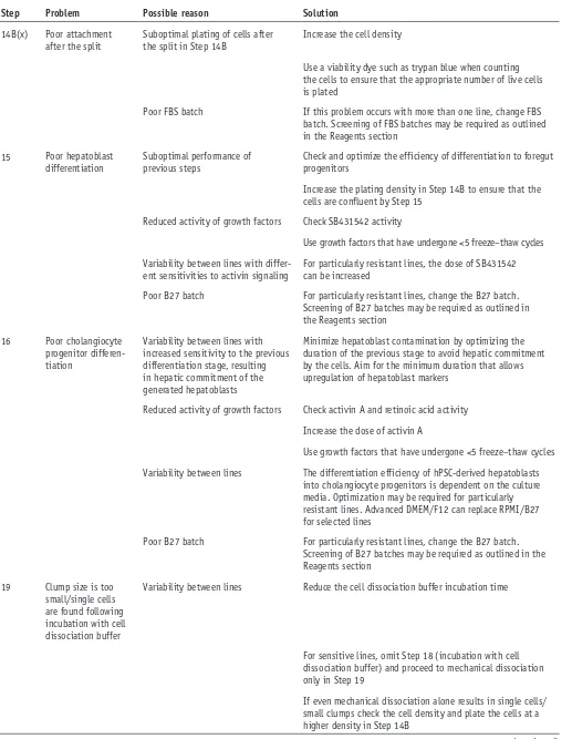
[image:11.594.42.559.337.733.2]Troubleshooting advice can be found in table 2.
taBle 2 | Troubleshooting table.
step problem possible reason solution
10 Poor attachment
of hPSCs Longer attachment time is required Repeat Step 9, incubating the cells for 1 more day before proceeding to Step 10
Colony size is too small Break colonies into slightly bigger clumps that gravitate to the bottom of the plate more easily, facilitating attachment
Variability between hPSC lines Add the Rho kinase inhibitor Y-27632 to the medium during passaging
Poor FBS batch If this problem occurs with more than one line, change the FBS batch. Screening of FBS batches may be required as outlined in the Reagents section
12 Poor endoderm
differ-entiation efficiency Suboptimal plating of hPSCs for differentiation Decrease clump size and increase plating density
Variability between lines with different sensitivity to activin or Wnt signaling
For particularly resistant lines, optimize the dose of activin A in Steps 10–12 and that of CHIR in Step 10, by monitoring the impact of increased doses on the efficiency of endoderm differentiation
14A(i), 14B(x)
Poor FP differentia-tion efficiency
Variability between lines For persistent contamination with poorly differentiated cells, split the cells as described in Step 14B
Suboptimal plating of cells following split in Step 14B
For poor differentiation efficiency following a split, optimize cell density, ensuring that the cells are confluent by the following day
Reduced activity of growth factors Check activin-A activity
Use growth factors that have undergone <5 freeze–thaw cycles
Poor B27 batch For particularly resistant lines, change B27 batch. Screening of B27 batches may be required as outlined in the Reagents section
©
201
7 M
a
cmillan Publis
hers Limited,
p
ar
t o
f S
p
ri
n
g
er
N
at
u
re
.
[image:12.594.46.563.75.742.2]All rights reserved.
taBle 2 | Troubleshooting table (continued).
step problem possible reason solution
14B(x) Poor attachment after the split
Suboptimal plating of cells after the split in Step 14B
Increase the cell density
Use a viability dye such as trypan blue when counting the cells to ensure that the appropriate number of live cells is plated
Poor FBS batch If this problem occurs with more than one line, change FBS batch. Screening of FBS batches may be required as outlined in the Reagents section
15 Poor hepatoblast differentiation
Suboptimal performance of previous steps
Check and optimize the efficiency of differentiation to foregut progenitors
Increase the plating density in Step 14B to ensure that the cells are confluent by Step 15
Reduced activity of growth factors Check SB431542 activity
Use growth factors that have undergone <5 freeze–thaw cycles
Variability between lines with
differ-ent sensitivities to activin signaling For particularly resistant lines, the dose of SB431542 can be increased
Poor B27 batch For particularly resistant lines, change the B27 batch. Screening of B27 batches may be required as outlined in the Reagents section
16 Poor cholangiocyte progenitor differen-tiation
Variability between lines with increased sensitivity to the previous differentiation stage, resulting in hepatic commitment of the generated hepatoblasts
Minimize hepatoblast contamination by optimizing the duration of the previous stage to avoid hepatic commitment by the cells. Aim for the minimum duration that allows upregulation of hepatoblast markers
Reduced activity of growth factors Check activin A and retinoic acid activity
Increase the dose of activin A
Use growth factors that have undergone <5 freeze–thaw cycles
Variability between lines The differentiation efficiency of hPSC-derived hepatoblasts into cholangiocyte progenitors is dependent on the culture media. Optimization may be required for particularly resistant lines. Advanced DMEM/F12 can replace RPMI/B27 for selected lines
Poor B27 batch For particularly resistant lines, change the B27 batch. Screening of B27 batches may be required as outlined in the Reagents section
19 Clump size is too small/single cells are found following incubation with cell dissociation buffer
Variability between lines Reduce the cell dissociation buffer incubation time
For sensitive lines, omit Step 18 (incubation with cell dissociation buffer) and proceed to mechanical dissociation only in Step 19
If even mechanical dissociation alone results in single cells/ small clumps check the cell density and plate the cells at a higher density in Step 14B
©
201
7 M
a
cmillan Publis
hers Limited,
p
ar
t o
f S
p
ri
n
g
er
N
at
u
re
.
All rights reserved.
● tIMInG
Steps 1–9, passaging of hPSCs: 1 d
Steps 10–12, differentiation of hPSCs into DE: 3 d Steps 13 and 14, differentiation of DE into FP cells: 5 d Step 15, differentiation of FP cells into HBs: 4 d Step 16, differentiation of HBs into CPs: 4 d
Steps 17–26, passaging of CPs and transfer to 3D culture conditions: 1–2 h Step 27, differentiation of CPs into CLC organoids: 10 d
Step 28A, IF staining of CLC organoids: 2 d
Step 28B, extraction of cells from Matrigel for further analyses: 40 min
antIcIpateD results
We describe a methodology for the differentiation of hPSCs into functional CLC organoids in 26 d. The early stages of our protocol (DE, FP, HB) result in >90% cells expressing endoderm and then FP markers (Fig. 4). However, biliary specification of HBs results in 75% CK19+/SOX9+ CPs, which mature into a population of 75% CK7+ CLCs during the final step of our
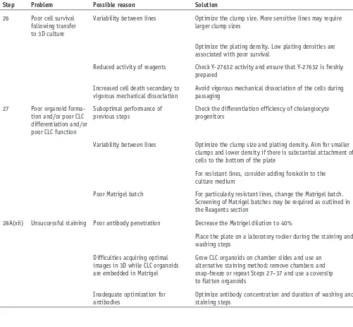
[image:13.594.43.544.82.545.2]differentiation (Fig. 4). The resulting CLC organoids should express biliary markers such as CK19 and CK7 in IF analyses taBle 2 | Troubleshooting table (continued).
step problem possible reason solution
26 Poor cell survival following transfer to 3D culture
Variability between lines Optimize the clump size. More sensitive lines may require larger clump sizes
Optimize the plating density. Low plating densities are associated with poor survival
Reduced activity of reagents Check Y-27632 activity and ensure that Y-27632 is freshly prepared
Increased cell death secondary to vigorous mechanical dissociation
Avoid vigorous mechanical dissociation of the cells during passaging
27 Poor organoid forma-tion and/or poor CLC differentiation and/or poor CLC function
Suboptimal performance of previous steps
Check the differentiation efficiency of cholangiocyte progenitors
Variability between lines Optimize the clump size and plating density. Aim for smaller clumps and lower density if there is substantial attachment of cells to the bottom of the plate
For resistant lines, consider adding forskolin to the culture medium
Poor Matrigel batch For particularly resistant lines, change the Matrigel batch. Screening of Matrigel batches may be required as outlined in the Reagents section
28A(xii) Unsuccessful staining Poor antibody penetration Decrease the Matrigel dilution to 40%
Place the plate on a laboratory rocker during the staining and washing steps
Difficulties acquiring optimal images in 3D while CLC organoids are embedded in Matrigel
Grow CLC organoids on chamber slides and use an alternative staining method; remove chambers and snap-freeze or repeat Steps 27–37 and use a coverslip to flatten organoids
Inadequate optimization for antibodies
©
201
7 M
a
cmillan Publis
hers Limited,
p
ar
t o
f S
p
ri
n
g
er
N
at
u
re
.
All rights reserved.
(Figs. 2 and 3). Hepatic markers (AFP, albumin) can still be detected in these stages because of the presence of a contaminating population of hepatic-lineage cells, but these should be identified only in clumps of cells without a lumen or attached to the bottom of the plate. Furthermore, CLC organoids can be validated further for additional cholangiocyte markers such as CFTR, AE2 and secretin receptor6, and they should demonstrate functional properties such as luminal
accumulation of rhodamine-123, and GGT and ALP activity (Fig. 5). The methods used to characterize CLC organoids (flow cytometry, IF, rhodamine-123 accumulation, GGT activity and ALP staining) have been described elsewhere6.
Our platform promotes substantial cell expansion. Using three different hPSC lines, we observed an average yield of >50 × 106 CLCs per 1 × 10⁶ hPSCs. Proliferation should be particularly evident during the generation of CLC organoids.
1 × 10⁵ CPs should give rise to 50–100 CLC organoids with diameters ranging between 100 and 1,000 µm. However,
variations in terms of the expansion potential and the differentiation efficiency of our protocol can occur. This can be attributed to inherent differences between hPSC lines and batch-to-batch variability for some of the reagents, including Matrigel. For reproducible results, the use of fresh medium and well-preserved, small-molecule, recombinant protein and Matrigel stocks is essential.
Note: Any Supplementary Information and Source Data files are available in the
online version of the paper.
acknoWleDGMents This work was funded by ERC starting grant Relieve IMDs (L.V., N.R.F.H.), the Cambridge Hospitals National Institute for Health Research Biomedical Research Center (L.V., N.R.F.H., F.S.), the Evelyn trust (N.R.F.H.) and the EU Fp7 grant TissuGEN (M.C.d.B.). F.S. was supported by an Addenbrooke’s Charitable Trust Clinical Research Training Fellowship and a joint MRC-Sparks Clinical Research Training Fellowship.
The authors thank the Cambridge BRC hIPSCs core facility for the derivation of the Cystic Fibrosis hIPSC line, P. Materek (Wellcome Trust-Medical Research Council Stem Cell Institute, Cambridge Stem Cell Institute, Anne McLaren Laboratory, Department of Surgery, University of Cambridge) for the provision of cells used as negative controls, D. Ortmann for his input into the design of the figures and P.-A. Tsagkaraki for her help with the generation of the manuscript figures and statistical analyses.
autHor contrIButIons F.S.: design and concept of study, execution of experiments and data acquisition, development of protocols and validation, collection of data, production of figures, manuscript writing and editing, and final approval of the manuscript. M.C.d.B., I.G. and A.B.: execution of experiments, collection and provision of data. N.R.F.H.: design and concept of study, editing and final approval of the manuscript. L.V.: design and concept of the study, editing and final approval of the manuscript. M.C.d.B., I.G. and A.B. contributed equally to this work. L.V. and N.R.F.H. jointly directed this work, contributing equally.
coMpetInG FInancIal Interests The authors declare competing financial interests: details are available in the online version of the paper.
Reprints and permissionsinformation is available online at http://www.nature. com/reprints/index.html.
1. O’Hara, S.P., Tabibian, J.H., Splinter, P.L. & Larusso, N.F. The dynamic biliary epithelia: molecules, pathways, and disease. J. Hepatol.58, 575–582 (2013).
2. Park, S.M. The crucial role of cholangiocytes in cholangiopathies. Gut Liver 6, 295–304 (2012).
3. Lazaridis, K.N. The cholangiopathies Konstantinos. Mayo Clin. Proc.90, 791–800 (2015).
4. Pollheimer, M.J., Trauner, M. & Fickert, P. Will we ever model PSC? - ‘It’s hard to be a PSC model!’. Clin. Res. Hepatol. Gastroenterol.35, 792–804 (2011).
5. Lemaigre, F.P. Notch signaling in bile duct development: new insights raise new questions. Hepatology48, 358–360 (2008).
6. Sampaziotis, F. et al. Cholangiocytes derived from human induced pluripotent stem cells for disease modeling and drug validation.
Nat. Biotechnol.33, 845–852 (2015).
7. Si-Tayeb, K., Lemaigre, F.P. & Duncan, S.A. Organogenesis and development of the liver. Dev. Cell18, 175–189 (2010).
8. Hannan, N.R.F., Segeritz, C.-P., Touboul, T. & Vallier, L. Production of hepatocyte-like cells from human pluripotent stem cells. Nat. Protoc.8, 430–437 (2013).
9. Clotman, F. et al. Control of liver cell fate decision by a gradient of TGF beta signaling modulated by Onecut transcription factors. Genes Dev.19, 1849–1854 (2005).
10. Yanai, M. et al. FGF signaling segregates biliary cell-lineage from chick hepatoblasts cooperatively with BMP4 and ECM components in vitro.
Dev. Dyn.237, 1268–1283 (2008).
11. Tabibian, J.H., Masyuk, A.I., Masyuk, T.V., O’Hara, S.P. & LaRusso, N.F. Physiology of cholangiocytes. Compr. Physiol.3, 541–565 (2013). 12. Ogawa, M. et al. Directed differentiation of cholangiocytes from human
pluripotent stem cells. Nat. Biotechnol.33, 853–861 (2015).
13. Tanimizu, N., Miyajima, A. & Mostov, K.E. Liver progenitor cells develop cholangiocyte-type epithelial polarity in three-dimensional culture. Mol. Biol. Cell18, 1472–1479 (2007).
14. Zhao, D. et al. Derivation and characterization of hepatic progenitor cells from human embryonic stem cells. PLoS One4, e6468 (2009).
15. Tabibian, J.H. et al. Characterization of cultured cholangiocytes isolated from livers of patients with primary sclerosing cholangitis. Lab. Invest. 94, 1126–1133 (2014).
16. Grant, A.G. & Billing, B.H. The isolation and characterization of a bile ductule cell population from normal and bile-duct ligated rat livers.
Br. J. Exp. Pathol.58, 301–310 (1977).
17. Joplin, R., Strain, A.J. & Neuberger, J.M. Immuno-isolation and culture of biliary epithelial cells from normal human liver. In Vitro Cell. Dev. Biol. 25, 1189–1192 (1989).
18. Zaret, K.S. et al. Directed differentiation of cholangiocytes from human pluripotent stem cells. Eur. J. Cell Biol.33, 1–48 (2015).
19. Dianat, N. et al. Generation of functional cholangiocyte-like cells from human pluripotent stem cells and HepaRG cells. Hepatology60, 700–714 (2014). 20. Glaser, S. et al. Heterogeneity of the intrahepatic biliary epithelium.
World J. Gastroenterol.12, 3523–3536 (2006).
21. Kent, L. Culture and maintenance of human embryonic stem cells.
J. Vis. Exp. 2–5 (2009).