Methylation of
HOXA9
and
ISL1
Predicts
Patient Outcome in High-Grade Non-Invasive
Bladder Cancer
Mark O. Kitchen1,2*, Richard T. Bryan3, Kim E. Haworth1, Richard D. Emes4, Christopher Luscombe2, Lyndon Gommersall2, K. K. Cheng3, Maurice P. Zeegers5, Nicholas D. James6, Adam J. Devall3, Anthony A. Fryer1, William E. Farrell1
1Institute for Science and Technology in Medicine, Keele University, Stoke-on-Trent, United Kingdom, 2Urology Department, University Hospitals of North Midlands NHS Trust, Stoke-on-Trent, United Kingdom, 3School of Cancer Sciences, University of Birmingham, Birmingham, United Kingdom,4Advanced Data Analysis Centre, University of Nottingham, Nottingham, United Kingdom,5Department of Complex Genetics, NUTRIM School for Nutrition, Toxicology and Metabolism, Maastricht University Medical Centre, Maastricht, The Netherlands,6Cancer Research Unit, University of Warwick, Coventry, United Kingdom
*m.o.kitchen@keele.ac.uk
Abstract
Introduction
Inappropriate DNA methylation is frequently associated with human tumour development, and in specific cases, is associated with clinical outcomes. Previous reports of DNA methylation in low/intermediate grade non-muscle invasive bladder cancer (NMIBC) have suggested that specific patterns of DNA methylation may have a role as diagnostic or prog-nostic biomarkers. In view of the aggressive and clinically unpredictable nature of high-grade (HG) NMIBC, and the current shortage of the preferred treatment option (Bacillus: Calmette-Guerin), novel methylation analyses may similarly reveal biomarkers of disease outcome that could risk-stratify patients and guide clinical management at initial diagnosis.
Methods
Promoter-associated CpG island methylation was determined in primary tumour tissue of 36 initial presentation high-grade NMIBCs, 12 low/intermediate-grade NMIBCs and 3 nor-mal bladder controls. The genesHOXA9,ISL1,NKX6-2,SPAG6,ZIC1andZNF154were selected for investigation on the basis of previous reports and/or prognostic utility in low/ intermediate-grade NMIBC. Methylation was determined by Pyrosequencing of sodium-bisulphite converted DNA, and then correlated with gene expression using RT-qPCR. Meth-ylation was additionally correlated with tumour behaviour, including tumour recurrence and progression to muscle invasive bladder cancer or metastases.
Results
TheISL1genes’promoter-associated island was more frequently methylated in recurrent and progressive high-grade tumours than their non-recurrent counterparts (60.0%vs. 18.2%,
OPEN ACCESS
Citation:Kitchen MO, Bryan RT, Haworth KE, Emes RD, Luscombe C, Gommersall L, et al. (2015) Methylation ofHOXA9andISL1Predicts Patient Outcome in High-Grade Non-Invasive Bladder Cancer. PLoS ONE 10(9): e0137003. doi:10.1371/ journal.pone.0137003
Editor:Bing-Hua Jiang, Thomas Jefferson University, UNITED STATES
Received:May 14, 2015
Accepted:August 11, 2015
Published:September 2, 2015
Copyright:© 2015 Kitchen et al. This is an open access article distributed under the terms of the
Creative Commons Attribution License, which permits unrestricted use, distribution, and reproduction in any medium, provided the original author and source are credited.
Data Availability Statement:All relevant data are within the paper and its Supporting Information files.
p= 0.008).ISL1andHOXA9showed significantly higher mean methylation in recurrent and progressive tumours compared to non-recurrent tumours (43.3%vs. 20.9%,p= 0.016 and 34.5%vs17.6%,p= 0.017, respectively). ConcurrentISL1/HOXA9methylation in HG-NMIBC reliably predicted tumour recurrence and progression within one year (Positive Pre-dictive Value 91.7%), and was associated with disease-specific mortality (DSM).
Conclusions
In this study we report methylation differences and similarities between clinical sub-types of high-grade NMIBC. We report the potential ability of methylation biomarkers, at initial diag-nosis, to predict tumour recurrence and progression within one year of diagnosis. We found that specific biomarkers reliably predict disease outcome and therefore may help guide patient treatment despite the unpredictable clinical course and heterogeneity of high-grade NMIBC. Further investigation is required, including validation in a larger patient cohort, to confirm the clinical utility of methylation biomarkers in high-grade NMIBC.
Introduction
High-grade non-muscle invasive bladder cancer (HG-NMIBC) is a clinically important sub-type of bladder transitional cell carcinoma (TCC), accounting for 10–15% of all TCC at presen-tation[1].The unpredictable nature of HG-NMIBC with regard to recurrence and progression to invasive or metastatic disease, presents many challenges for successful management. With no robust methods for predicting outcomes (recurrence, progression, Bacillus:Calmette-Guerin (BCG) failure) at initial diagnosis, patients may be under-treated with intravesical therapy alone or over-treated with immediate cystectomy, both with attributed adverse patient out-comes[2,3].
Multiple studies have described the importance of epigenetic modifications in tumourigen-esis, most frequently apparent as inappropriate DNA methylation within gene promoter-associated CpG islands, and/or changes that lead to histone tail modification(s)[4,5].These modifications impact upon gene expression and promote tumourigenesis predominantly by silencing of tumour-suppressor and/or apoptotic pathway genes[5,6]. Such epigenetically-mediated gene silencing has been demonstrated in NMIBC and muscle-invasive bladder cancer (MIBC), and is reported to be associated with tumour recurrence, progression, invasion, and metastasis, but also early events in tumour development such as the‘field-defect’phenomenon [7,8]. Recent studies highlight the potential clinical utility of epigenetic biomarkers in bladder cancer, describing tumour, blood and urine DNA methylation markers of, for example, NMIBC recurrence or chemo-resistance in MIBC[9–11].
Promoter-associated CpG Island methylation of theHOXA9,ISL1,NKX6-2,SPAG6,ZIC1
andZNF154genes are frequent findings in bladder cancer. In these cases, methylation appears associated with aggressive tumour characteristics, and may independently predict disease recurrence, progression, or disease-specific mortality (DSM)[9,12,13]. However the majority of these reports examine heterogeneous cohorts, comprising predominantly low/intermediate-grade NMIBC; high-low/intermediate-grade NMIBC has not been considered discretely for disease and/or sub-type specific epigenetic modifications.
We assessed a unique cohort of patients with HG-NMIBC for inappropriate promoter-asso-ciated CpG Island methylation at initial presentation and removal of the primary tumour(s),
supported by funding from the Birmingham Experimental Cancer Medicine Centre. The funders had no role in study design, data collection and analysis, decision to publish, or preparation of the manuscript.
with respect to known prospectively collected one-year outcomes of no-recurrence, recurrence, and progression (to MIBC or metastatic disease), and relative to low/intermediate-grade NMIBC. Identifying inappropriate methylation‘at diagnosis’of HG-NMIBC permitted a pre-liminary identification of novel and potentially clinically useful prognostic biomarkers.
Materials and Methods
Human tissue samples
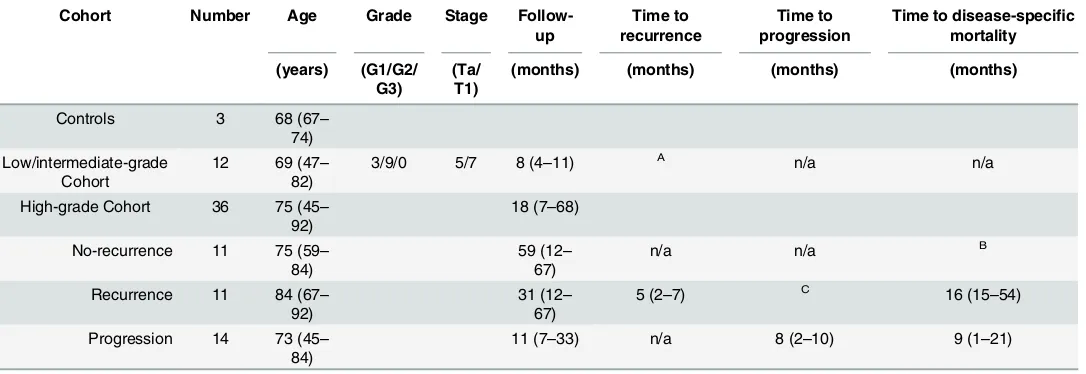
The primary tumour and normal bladder tissues used were provided by the Bladder Cancer Prognosis Programme (BCPP, Nottingham Research Ethics Committee: 05/Q2404/173)[14], the University of Birmingham Human Biomaterials Resource Centre (National Research Ethics Service (North West 5): 09/H1010/75), and the University Hospitals of North Midlands NHS Trust (National Research Ethics Service (South Central–Oxford C): 12/SC/0725). All samples were obtained after informed written consent and under the approval of appropriate national ethics review boards (reference numbers stated above). All samples were confirmed histologi-cally; repeat trans-urethral resection or cystectomy were performed, and/or intravesical ther-apy provided, where suggested by European Association of Urology Guidelines [15]. All primary human tissues (Table 1) were stored at -80°C prior to nucleic acid extraction.
DNA extraction and bisulphite modification
Genomic DNA was extracted from tumour and control tissues using a standard phenol-chloro-form extraction procedure[16] and subsequently bisulphite-modified as previously described [4]. Bisulphite conversion of DNA was confirmed in all cases by successful PCR using primers specific to bisulphite-converted DNA (primer sequences provided in supporting information
S1 Table). To increase the relative amount and stability of bisulphite-converted DNA,
whole-Table 1. Patient and Tumour characteristics.
Cohort Number Age Grade Stage Follow-up
Time to recurrence
Time to progression
Time to disease-specific mortality
(years) (G1/G2/ G3)
(Ta/ T1)
(months) (months) (months) (months)
Controls 3 68 (67–
74) Low/intermediate-grade
Cohort
12 69 (47– 82)
3/9/0 5/7 8 (4–11) A n/a n/a
High-grade Cohort 36 75 (45– 92)
18 (7–68)
No-recurrence 11 75 (59– 84)
59 (12– 67)
n/a n/a B
Recurrence 11 84 (67– 92)
31 (12– 67)
5 (2–7) C 16 (15–54)
Progression 14 73 (45– 84)
11 (7–33) n/a 8 (2–10) 9 (1–21)
Number of samples in each cohort, with median age, range in brackets, and median follow-up period, range in brackets, after initial tumour resection (diagnosis).
AOne patient suffered a 2mm uni-focal recurrence at 6 months.
BOne patient in the no-recurrence group suffered disease specific mortality at 16 months. COne patient in the recurrence one-year group suffered progression at 21 months.
[image:3.612.37.580.450.637.2]genome amplification (WGA) was performed as previously described[4] (described further in supporting informationS1 Text).
Pyrosequencing of bisulphite-converted DNA
CpG island sequences were identified from the UCSC Genome Browser (http://genome.ucsc. edu/), and imported into PyroMark Assay Design 2.0 Software for primer design (Qiagen, Manchester, UK). Dependent on the frequency and density of CpG dinucleotides within the sequence of interest, designed primers encompassed 4–7 consecutive CpGs in each gene (primer sequences inS1 Tableand details of genomic location inS2 Table). After PCR amplifi-cation of the target sequence, Pyrosequencing was performed using a PyroMark Q24 Pyrose-quencer using PyroMark Q24 Software 2.0 and PyroMark Gold Q24 Reagents (Qiagen), as previously described [17] (S1 Text).
Methylation was stringently defined in tumours as comprising a mean level of methylation across the CpGs surveyed of greater than four standard deviations (4SD) above the mean in normal controls [4]. The number of tumours methylated (by this definition) for any given gene describes thefrequencyof methylation, whereas the mean percentage methylation of all CpGs surveyed describes themean levelof methylation for a tumour in any particular gene.
Quantitative RT-PCR
Total RNA was extracted from control and tumour samples using a standard guanidinium thiocyanate-phenol-chloroform protocol[16], and complementary DNA synthesised as described previously[18].Thermal cycling using SYBR (III) Green was as previously described [19], with target genes normalised to glyceraldehyde-3-phosphate dehydrogenase (GAPDH) (S1 Table) as endogenous control (S1 Text). Relative quantification of transcript expression was performed using the 2-ΔΔcycle threshold (CT) method[20]. Reduced transcript expression in each tumour was regarded significant if lower than a 3-fold reduction relative to mean expression in control samples; the converse was true for increased transcript expression [18](S1 Text).
Informatics and statistics
Microsoft Excel 2010 and STATA (v8, Stata Corporation, TX) were used to perform Fisher’s exact, Students’t, and log-rank analyses, and sensitivity, specificity, and positive and negative predictive values of methylation with respect to clinical outcomes.
Results
Frequency of methylation within low/intermediate- and high-grade
NMIBC
We initially determined the methylation status of the six candidate genes in the high- and low/ intermediate-grade tumour cohorts by Pyrosequence analyses of bisulphite-converted DNA. Inappropriate methylation of promoter-associated CpG islands (S2 Table) was a frequent find-ing in both cohorts for theNKX6-2,SPAG6,ZIC1andZNF154genes, relative to normal blad-der (Table 2).
Table 2shows that the methylation frequency in low/intermediate-grade tumours was lower forISL1than for all other genes investigated (2/12 tumours; 16.7%), although by com-parison, methylation frequency increased within HG tumours (17/36; 45.0%,p= 0.091). Con-versely, frequency of methylation was lower in HG-NMIBCs forHOXA9(20/36; 55.6%) compared to low/intermediate-grade tumours (10/12; 83.3%,p= 0.167); (data summarised in
To further investigate potential reasons for the observed differences in the frequency of methylation between the high- and low/intermediate-grade tumour cohorts for specific genes, we determined gene-specific methylation frequencies relative to the clinical characteristics of the HG tumours, encompassing one-year clinical outcomes of no-recurrence, recurrence, and progression.
ForNKX6-2,SPAG6,ZIC1andZNF154, methylation frequencies in recurrence and progres-sion tumours were marginally greater than their no-recurrence counterparts, and were overall broadly similar to the frequencies apparent in low/intermediate-grade tumours (Fig 1).
[image:5.612.40.576.89.216.2]HOXA9demonstrated a similarly increased methylation frequency in recurrence and pro-gression tumours relative to their no-recurrence counterparts, however, as shown inFig 1, the methylation frequency in no-recurrence tumours was significantly lower compared to that observed in low/intermediate-grade tumours (p= 0.036). Although the trend of an increase in methylation frequency in recurrence and progression (relative to no-recurrence) tumours was
Table 2. Methylation frequency and mean level of methylation in low/intermediate- and high-grade tumour groups.
Methylation Frequency Mean Level of Methylation
Low/intermediate-grade High-grade Normal bladder control Low/intermediate-grade High-grade
Gene name Number (%) Number (%) pvalue (%) (%) (%) pvalue
HOXA9 10/12 (83.3) 20/36 (55.6) 0.167 6.1 44.0 29.3 0.057
ISL1 2/12 (16.7) 17/36 (47.2) 0.091 7.6 22.1 38.9 0.061
NKX6-2 10/12 (83.3) 32/36 (88.9) 0.631 5.4 38.9 50.2 0.126
SPAG6 10/12 (83.3) 32/36 (88.9) 0.631 8.2 38.5 52.1 0.097
ZIC1 11/12 (91.7) 31/36 (86.1) 0.999 17.3 62.2 58.6 0.545
ZNF154 10/12 (83.3) 32/36 (88.9) 0.631 7.6 44.1 53.1 0.318
Number and percentage of low/intermediate- and high-grade tumours that are methylated for each of the six candidate genes (left side of table) and the mean level of methylation in each cohort for each of the gene (right side of table). Methylation is defined using four standard deviations above the mean of the normal bladder controls as a cut-off, as described previously[4]. Differences in the number of tumours methylated comparing high- to low/intermediate-grade tumours were assessed using Fisher’s exact or Chi-squared tests (two-tailed), wherep<0.05 is considered significant. Differences in mean methylation were assessed by Students t test,p<0.05 again considered significant.
doi:10.1371/journal.pone.0137003.t002
Fig 1. Proportion of methylated tumours in low/intermediate- and high-grade tumour cohorts.From left to right, tumour cohort methylation frequency/proportion, as determined by Pyrosequencing within
HOXA9,ISL1,SPAG6,NKX6-2,ZIC1andZNF154, for the low/Intermediate-grade cohort (LG), no-recurrence (NR) and recurrence and progression tumours (R+P) respectively. The filled bars represent the proportion of methylated tumours relative to controls in each case. Differences between the frequencies of methylation are indicated as statistically significant by‘*’, wherep<0.05 as determined by Fisher’s exact or Chi-squared tests (two-tailed). Methylation was defined as described in the materials and methods.
[image:5.612.200.450.507.634.2]apparent for all genes investigated, this was distinctly more marked for theISL1gene. In this case, methylation frequency was significantly greater in recurrence and progression tumours relative to no-recurrence (15/25 (60.0%)vs2/11 (18.2%),p= 0.031), and also to their low/inter-mediate-grade counterparts (15/25 (60.0%)vs2/12 (16.7%),p= 0.017) (Fig 1).
Mean methylation levels within low/intermediate- and high-grade NMIBC
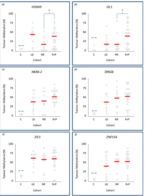
Our methylation analyses also showed considerable differences in, and between, the mean and range of methylation levels within individual tumours (Fig 2). Therefore, in addition to the analyses described above, we also assessed mean methylation levels within HG and low/inter-mediate-grade cohorts and within the HG tumour sub-types, to determine whether the level of methylation demonstrated any relationship with clinical characteristics.The mean level of methylation was greater, albeit not significantly, in HG relative to their low/intermediate-grade counterparts for theISL1,SPAG6,NKX6-2,and ZNF154genes
[image:6.612.201.437.304.622.2](Table 2). This increase approached significance forISL1(22.1% vs 36.5%,p= 0.061). Paradoxi-cally, the mean level of methylation forHOXA9was lower in HG compared to their low/interme-diate-grade counterparts, again approaching significance (29.3%vs 44.0%,p= 0.057) (Table 2).
Fig 2. Level of methylation within low/intermediate- and high-grade tumour cohorts.Panels (a) to (f) representingHOXA9,ISL1,NKX6-2,SPAG6,ZIC1andZNF154, respectively. Each panel displays individual tumour methylation values, as determined by Pyrosequencing, represented by grey circles within control (c), low/Intermediate-grade (LG), no-recurrence (NR) and recurrence and progression (R+P) tumour groups. The solid horizontal bars represent the overall mean methylation within each control or tumour group; differences between the means are indicated as statistically significant by‘*’, wherep<0.05 determined by Students-t testing. The double-headed arrow represents the cut-off point above which tumours are defined as methylated relative to normal bladder controls.
Using this approach, we also determined gene-specific methylation levels within HG tumours and relative to their clinical outcomes. Mean methylation levels withinISL1and
HOXA9were significantly higher in recurrence and progression tumours compared to their no-recurrence counterparts (43.3% vs. 20.9%,p= 0.016 and 34.5% vs. 17.6%,p= 0.017, respec-tively) (Fig 2). The mean methylation level increase from no-recurrence to recurrence and pro-gression tumours was not significant forNKX6-2,SPAG6,ZIC1andZNF154.
Correlation of methylation within clinical outcomes of high-grade NMIBC
We next determined the correlation between gene-specific methylation with clinical outcomes within the HG tumour cohort;HOXA9andISL1were assessed as the only genes demonstrating a significant difference in frequency or level of methylation.HOXA9promoter methylation demonstrated 72.7% specificity and an 84.2% positive predictive value (PPV) for tumour recur-rence and/or progression within one year of initial diagnosis, whilst methylation within theISL1promoter demonstrated specificity of 81.8% and PPV 87.5% for the same clinical out-comes, shown inTable 3. Moreover, concomitant methylation ofHOXA9andISL1at initial diagnosis predicted one-year recurrence and/or progression, with a PPV of 91.7%, whilst main-taining a specificity of 90.9%. To more rigorously assess association between these potential biomarkers and disease outcome, we employed logistical regression analysis.Table 3shows that when considered individually, methylation of eitherHOXA9orISL1attained statistical significance with tumour recurrence and/or progression (p= 0.050 andp= 0.047 respectively). However, in combination, while the biomarkers were less significantly associated with disease outcome (p= 0.067), they demonstrated a stronger odds ratio of tumour recurrence and/or progression than when either was considered separately (7.86 vs 4.74 and 5.73, respectively).
In addition to tumour behaviour we also considered promoter methylation as a predictor of disease-specific mortality: In this case,HOXA9promoter methylation demonstrated 57.1% specificity and a 70.6% negative predictive value (NPV) for disease-specific mortality, whilst
ISL1methylation suggested 57.1% and 60.0% for these outcome measures.
Methylation-associated changes in gene expression in high-grade
NMIBC
[image:7.612.34.578.89.195.2]Quantitative RT-PCR was used to evaluate associations between methylation and gene expres-sion in four of the six genes within a sub-set of 10–14 tumours, in comparison to controls.Fig 3shows that, relative to controls, 90.1% (29 of 32) methylated tumours display reduced tran-script expression, and 75.0% (24 of 32) show significantly reduced expression. Conversely,
Table 3. Methylation biomarker utility in high-grade NMIBC.
Outcome Potential Biomarker Odds Ratio 95% Confidence Interval pvalue
HOXA9 4.7 1.0–22.5 0.05
Recurrence or Progression ISL1 5.7 2.1–32.1 0.047
HOXA9 + ISL1 7.9 0.9–71.1 0.067
Outcome Potential Biomarker Sensitivity Specificity Positive Predictive Value Negative Predictive Value
HOXA9 64.00% 72.70% 84.20% 47.10%
Recurrence or Progression ISL1 56.00% 81.80% 87.50% 45.00%
HOXA9 + ISL1 44.00% 90.90% 91.70% 41.70%
Values for sensitivity, specificity, positive predictive value, negative predictive value, odds ratio (with 95%CI) and p-value of potential methylation biomarkers HOXA9 and ISL1 individually, and in combination, to predict high-grade NMIBC recurrence or progression at one year after initial diagnosis.
56.3% (9 of 16) unmethylated tumours displayed expression levels similar to, or in some cases higher than, that apparent in controls.
Discussion
The epigenomic landscape of bladder cancer is an area of growing research interest[21], specifi-cally in relation to identifying clinispecifi-cally viable biomarkers. Due to the current limitations in predicting the diverse clinical outcomes observed in HG-NMIBC, and the present BCG short-age, biomarkers that guide clinical decisions are of particular importance[22,23]. As described above, our analyses revealed frequent, and in some cases differential methylation, present at initial HG-NMIBC diagnosis, that appeared to correlate with tumour characteristics and clini-cal parameters.
The six genes selected for analyses are predominantly members of transcription factor fami-lies, primarily regulating gene-expression, enzyme-binding, and cell differentiation[24]. Their selection for analyses was on the basis of their frequent inappropriate methylation in bladder cancer [9,12,13,25]. In previous reports,HOXA9andZNF154methylation has been associated with tumour recurrence, andISL1methylation with tumour progression in predominantly low/intermediate-grade tumours[9,13].
[image:8.612.209.476.86.243.2]Our initial analyses revealed similar methylation frequencies in low/intermediate- and HG-NMIBC forNKX6-2,SPAG6,ZIC1andZNF154. In these cases, methylation frequencies were similar to those previously reported in the literature[9,12,13,25]. However, a difference in the frequency of methylation was apparent between the low/intermediate- and HG-NMIBC cohorts forHOXA9andISL1genes: although the methylation frequency of theHOXA9 pro-moter in low/intermediate-grade tumours was similar to that previously described in the litera-ture [12,13], it was found at markedly lower frequency in HG-NMIBC. Conversely, forISL1, methylation frequency in low/intermediate-grade tumours was lower than described previ-ously by others[13]; however, the methylation frequency increase that is apparent in the recur-rence and progression tumours is more consistent with these reports.
Fig 3. Association between high-grade NMIBC tumour methylation and gene expression.Quantitative RT-PCR analyses ofHOXA9,ISL1,SPAG6andZNF154transcript expression in individual high-grade tumours (HG). Expression is reported relative to the mean of three normal bladder controls (C, triangles), where the mean value is expressed as equal to 100%. Filled and unfilled circles denote methylated or unmethylated tumours, and represent the mean value from two independent experiments performed in triplicate. The horizontal bars within the control column represent the mean of the controls, a 3-fold reduction beneath this lies the double-headed arrow in each gene plot, representing the cut-off for significantly reduced expression in tumours relative to controls.
The differential methylation frequency inHOXA9andISL1may relate to several confound-ers, including the method in this study employed to define tumour methylation, and the rela-tively few low/intermediate-grade tumours for analyses. However, the concordance between our methylation data and those reported by others forNKX6-2,SPAG6,ZIC1andZNF154, confirms the robustness of our approach. On this basis, we reasoned that the differences in methylation frequency observed forHOXA9andISL1may be consequent to different clinical characteristics and/or outcomes within our HG tumour cohort; a phenomenon similarly described in, for example, breast, colon and pituitary tumours[4,26,27]. We therefore per-formed HG-NMIBC sub-type analyses and found that the methylation profiles of recurrence and progression tumours, although similar to each other, were mostly quite distinct from their no-recurrence counterparts; on this basis we grouped recurrence and progression tumours for analyses.
The methylation frequency in recurrence and progression tumours was consistently higher across all six genes compared to their no-recurrence counterparts; these differences were most pronounced forHOXA9andISL1and achieved statistical significance forISL1. Paradoxically, inHOXA9, no-recurrence, recurrence and progression tumours were less frequently methyl-ated than in their low/intermediate-grade tumour counterparts. Although the reasons for this are unclear, possible explanations may relate to the number, or heterogeneity, of tumours investigated, though we cannot discount the possibility that there are epigenetic differences in particular genes between low/intermediate- and high-grade tumours. Conversely, methylation frequency in recurrence and progression tumours inISL1was markedly greater than in their low/intermediate-grade counterparts. Similar explanations to those described forHOXA9
might account for these observations.
Through quantitative Pyrosequence analyses, we determined mean methylation across mul-tiple CpG sites showing that, further to methylation frequency differences, there exist differ-ences in methylation levels between tumour cohorts for particular genes. Specifically, differences in mean methylation levels between low/intermediate- and HG tumours, and also between the no-recurrence, and their recurrence and progression tumour counterparts, analo-gous to observations reported by others in breast and colon cancer sub-types[26,27]. For both
HOXA9andISL1, there was a significant increase in the mean level of methylation in the recur-rence and progression tumours compared to their no-recurrecur-rence counterparts.
The observed differences in methylation frequency and mean methylation level between these clinically divergent subgroups, suggests that the frequency and/or mean level of methyla-tion increases with tumour aggressiveness. Similar trends have been reported in other tumour types[26,27] and this is thought to represent accumulation of epigenetic aberrations over time, similar to the accumulation of genetic mutations and genomic instability apparent during tumour progression[28].
Since our findings support previous associations ofHOXA9andISL1methylation with tumour characteristics and behaviour[9,13], we appraised potential clinical correlates of
HOXA9andISL1methylation, including their prognostic potential. In our HG-NMIBC cohort,HOXA9orISL1methylation at initial diagnosis reliably predicted tumour recurrence or progression within one year. In this context, concurrent methylation of bothHOXA9and
ISL1improved the positive predictive value to 91.7%, comparing favourably with other postu-lated biomarkers of recurrent/progressive disease in NMIBC [13,29]. Moreover, logistic regres-sion appeared to confirm that these findings were not consequent to methylation in a single gene. Furthermore, and as previously described[9,13], concurrent methylation inHOXA9and
inappropriate methylation and HG-NMIBC disease outcome will require validation in larger, independent tumour cohorts.
Finally, to reveal potential functional relevance of methylation within high-grade disease, we assessed the relationship between methylation and gene expression; across the four genes studied, we found an association of abnormal methylation with reduced transcript expression, consistent with findings from other groups[9,13]. These findings were not significant, possibly relating to number of tumours studied, or the passenger/driver phenomenon, whereby epige-netic marks may be present but not causative of altered gene expression[30].
These findings are the first to report similarities and differences of gene-associated methyla-tion in HG-NMIBC relative to low/intermediate-grade tumours. Furthermore, we have shown that specific methylation patterns at initial diagnosis predict one-year HG-NMIBC clinical out-comes, pointing to the exciting potential for methylation as a prognostic biomarker in this clin-ically unpredictable disease. Further investigations exploiting genome-wide array analyses are required to further characterise epigenetic similarities and differences between low/intermedi-ate-and HG-NMIBC, and to reveal biomarkers that may serve as therapeutic targets or guide clinical management, such as the timing, dose and length of BCG therapy, or timing of cystectomy.
Supporting Information
S1 Table. Primer table.List of bisulphite-converted PCR primers, Pyrosequencing (sequenc-ing) primers, and RT-qPCR primers.
(DOCX)
S2 Table. CpG island information.Table listing the genomic location of the promoter-associ-ated CpG island regions assessed for each of the six genes.
(DOCX)
S3 Table. Raw data file.Excel spreadsheet containing mean values for Pyrosequencing and RT-qPCR data.
(XLSX)
S1 Text. Supplemental materials and methods.Further description of primary tissue samples, and further description of DNA extraction, sodium bisulphite-conversion, Pyrosequencing, RNA extraction and RT-qPCR procedures.
(DOCX)
Acknowledgments
We would like to thank all the West Midlands Consultant Urologists and their units involved with BCPP, as well as the BCPP research nurses and Margaret Grant, Deborah Bird, Jennifer Barnwell, Duncan Nekeman and Eline van Roekel.
Author Contributions
References
1. Boustead GB, Fowler S, Swamy R, Kocklebergh R, Hounsome L, et al. (2014) Stage, grade and patho-logical characteristics of bladder cancer in the UK: British Association of Uropatho-logical Surgeons (BAUS) urological tumour registry. BJU Int 113: 924–930. doi:10.1111/bju.12468PMID:24447825
2. Raj GV, Herr H, Serio AM, Donat SM, Bochner BH, et al. (2007) Treatment paradigm shift may improve survival of patients with high risk superficial bladder cancer. J Urol 177: 1283–1286; discussion 1286. PMID:17382713
3. De Berardinis E, Busetto GM, Antonini G, Giovannone R, Gentile V (2011) T1G3 high-risk NMIBC (non-muscle invasive bladder cancer): conservative treatment versus immediate cystectomy. Int Urol Nephrol 43: 1047–1057. doi:10.1007/s11255-011-9941-xPMID:21442469
4. Duong CV, Emes RD, Wessely F, Yacqub-Usman K, Clayton RN, et al. (2012) Quantitative, genome-wide analysis of the DNA methylome in sporadic pituitary adenomas. Endocr Relat Cancer 19: 805– 816. doi:10.1530/ERC-12-0251PMID:23045325
5. Dawson MA, Kouzarides T (2012) Cancer epigenetics: from mechanism to therapy. Cell 150: 12–27. doi:10.1016/j.cell.2012.06.013PMID:22770212
6. Sharma S, Kelly TK, Jones PA (2010) Epigenetics in cancer. Carcinogenesis 31: 27–36. doi:10.1093/ carcin/bgp220PMID:19752007
7. Wolff EM, Chihara Y, Pan F, Weisenberger DJ, Siegmund KD, et al. (2010) Unique DNA methylation patterns distinguish noninvasive and invasive urothelial cancers and establish an epigenetic field defect in premalignant tissue. Cancer Res 70: 8169–8178. doi:10.1158/0008-5472.CAN-10-1335PMID: 20841482
8. Catto JW, Azzouzi AR, Rehman I, Feeley KM, Cross SS, et al. (2005) Promoter hypermethylation is associated with tumor location, stage, and subsequent progression in transitional cell carcinoma. J Clin Oncol 23: 2903–2910. PMID:15753461
9. Reinert T, Borre M, Christiansen A, Hermann GG, Orntoft TF, et al. (2012) Diagnosis of bladder cancer recurrence based on urinary levels of EOMES, HOXA9, POU4F2, TWIST1, VIM, and ZNF154 hyper-methylation. PLoS One 7: e46297. doi:10.1371/journal.pone.0046297PMID:23056278
10. Yu J, Zhu T, Wang Z, Zhang H, Qian Z, et al. (2007) A novel set of DNA methylation markers in urine sediments for sensitive/specific detection of bladder cancer. Clin Cancer Res 13: 7296–7304. PMID: 18094410
11. Tada Y, Yokomizo A, Shiota M, Tsunoda T, Plass C, et al. (2011) Aberrant DNA methylation of T-cell leukemia, homeobox 3 modulates cisplatin sensitivity in bladder cancer. Int J Oncol 39: 727–733. doi: 10.3892/ijo.2011.1049PMID:21617853
12. Reinert T, Modin C, Castano FM, Lamy P, Wojdacz TK, et al. (2011) Comprehensive genome methyla-tion analysis in bladder cancer: identificamethyla-tion and validamethyla-tion of novel methylated genes and applicamethyla-tion of these as urinary tumor markers. Clin Cancer Res 17: 5582–5592. doi: 10.1158/1078-0432.CCR-10-2659PMID:21788354
13. Kim YJ, Yoon HY, Kim JS, Kang HW, Min BD, et al. (2013) HOXA9, ISL1 and ALDH1A3 methylation patterns as prognostic markers for nonmuscle invasive bladder cancer: array-based DNA methylation and expression profiling. Int J Cancer 133: 1135–1142. doi:10.1002/ijc.28121PMID:23436614
14. Zeegers MP, Bryan RT, Langford C, Billingham L, Murray P, et al. (2010) The West Midlands Bladder Cancer Prognosis Programme: rationale and design. BJU Int 105: 784–788. doi:10.1111/j.1464-410X. 2009.08849.xPMID:19751260
15. Babjuk M, Burger M, Zigeuner R, Shariat SF, van Rhijn BW, et al. (2013) EAU guidelines on non-mus-cle-invasive urothelial carcinoma of the bladder: update 2013. Eur Urol 64: 639–653. doi:10.1016/j. eururo.2013.06.003PMID:23827737
16. Chomczynski P, Sacchi N (1987) Single-step method of RNA isolation by acid guanidinium thiocya-nate-phenol-chloroform extraction. Anal Biochem 162: 156–159. PMID:2440339
17. Fryer AA, Emes RD, Ismail KM, Haworth KE, Mein C, et al. (2011) Quantitative, high-resolution epige-netic profiling of CpG loci identifies associations with cord blood plasma homocysteine and birth weight in humans. Epigenetics 6: 86–94. doi:10.4161/epi.6.1.13392PMID:20864804
18. Dudley KJ, Revill K, Whitby P, Clayton RN, Farrell WE (2008) Genome-wide analysis in a murine Dnmt1 knockdown model identifies epigenetically silenced genes in primary human pituitary tumors. Mol Cancer Res 6: 1567–1574. doi:10.1158/1541-7786.MCR-08-0234PMID:18922972
19. Al-Azzawi H, Yacqub-Usman K, Richardson A, Hofland LJ, Clayton RN, et al. (2011) Reversal of endogenous dopamine receptor silencing in pituitary cells augments receptor-mediated apoptosis. Endocrinology 152: 364–373. doi:10.1210/en.2010-0886PMID:21177832
21. Netto GJ (2012) Molecular biomarkers in urothelial carcinoma of the bladder: are we there yet? Nat Rev Urol 9: 41–51.
22. Gontero P, Sylvester R, Pisano F, Joniau S, Vander Eeckt K, et al. (2014) Prognostic Factors and Risk Groups in T1G3 Non-Muscle-invasive Bladder Cancer Patients Initially Treated with Bacillus Calmette-Guerin: Results of a Retrospective Multicenter Study of 2451 Patients. Eur Urol.
23. Vedder MM, Marquez M, de Bekker-Grob EW, Calle ML, Dyrskjot L, et al. (2014) Risk prediction scores for recurrence and progression of non-muscle invasive bladder cancer: an international validation in pri-mary tumours. PLoS One 9: e96849. doi:10.1371/journal.pone.0096849PMID:24905984
24. Maglott D, Ostell J, Pruitt KD, Tatusova T (2005) Entrez Gene: gene-centered information at NCBI. Nucleic Acids Res 33: D54–58. PMID:15608257
25. Chung W, Bondaruk J, Jelinek J, Lotan Y, Liang S, et al. (2011) Detection of bladder cancer using novel DNA methylation biomarkers in urine sediments. Cancer Epidemiol Biomarkers Prev 20: 1483– 1491. doi:10.1158/1055-9965.EPI-11-0067PMID:21586619
26. Li Y, Li S, Chen J, Shao T, Jiang C, et al. (2014) Comparative epigenetic analyses reveal distinct pat-terns of oncogenic pathways activation in breast cancer subtypes. Hum Mol Genet 23: 5378–5393. doi:10.1093/hmg/ddu256PMID:24871326
27. Gyparaki MT, Basdra EK, Papavassiliou AG (2013) DNA methylation biomarkers as diagnostic and prognostic tools in colorectal cancer. J Mol Med (Berl) 91: 1249–1256.
28. Negrini S, Gorgoulis VG, Halazonetis TD (2010) Genomic instability—an evolving hallmark of cancer. Nat Rev Mol Cell Biol 11: 220–228. doi:10.1038/nrm2858PMID:20177397
29. Tilki D, Burger M, Dalbagni G, Grossman HB, Hakenberg OW, et al. (2011) Urine markers for detection and surveillance of non-muscle-invasive bladder cancer. Eur Urol 60: 484–492. doi:10.1016/j.eururo. 2011.05.053PMID:21684071