Gloria M.P. Roberts, Liam Nestor, Hugh Garavan PII: S0006-8993(09)01474-7
DOI: doi:10.1016/j.brainres.2009.07.040
Reference: BRES 39441 To appear in: Brain Research
Received date: 23 March 2009 Revised date: 2 July 2009 Accepted date: 15 July 2009
Please cite this article as: Gloria M.P. Roberts, Liam Nestor, Hugh Garavan, Learning and memory deficits in ecstasy users and their neural correlates during a face-learning task,Brain Research (2009), doi:10.1016/j.brainres.2009.07.040
ACCEPTED MANUSCRIPT
Title
Learning and memory deficits in ecstasy users and their neural
correlates during a face-learning task
Authors
Gloria M.P. Roberts 1, Liam Nestor 1, and Hugh Garavan 1, 2 *
1 School of Psychology and Trinity College Institute of Neuroscience, Trinity College
Dublin, Dublin 2, Ireland
2 Nathan Kline Institute for Psychiatric Research, 140 Old Orangeburg Road,
Orangeburg, New York 10962
*To whom correspondence should be addressed: Prof. Hugh Garavan,
3.48 Lloyd Building,
Trinity College Institute of Neuroscience, Trinity College, Dublin 2, Ireland.
Ph: +353-1-896-8448; Fax: +353-1-896-3183 E-mail: [email protected]
Number of text pages of whole manuscript: 19 + references Number of figures: 2
ACCEPTED MANUSCRIPT
Abstract
It has been consistently shown that ecstasy users display impairments in learning and
memory performance. In addition, working memory processing in ecstasy users has
been shown to be associated with neural alterations in hippocampal and/or cortical
regions as measured by functional magnetic resonance imaging (fMRI). Using
functional imaging and a face-learning task, we investigated neural correlates of
encoding and recalling face-name associations in 20 recreational drug users whose
predominant drug use was ecstasy and 20 controls. To address the potential
confounding effects of the cannabis use of the ecstasy using group, a second analysis
included 14 previously tested cannabis users (Nestor et al., 2008). Ecstasy users
performed significantly worse in learning and memory compared to controls and
cannabis users. A conjunction analysis of the encode and recall phases of the task
revealed ecstasy-specific hyperactivity in bilateral frontal regions, left temporal, right
parietal, bilateral temporal, and bilateral occipital brain regions. Ecstasy-specific
hypoactivity was evident in the right dorsal anterior cingulated cortex (ACC) and left
posterior cingulated cortex. In both ecstasy and cannabis groups brain activation was
decreased in the right medial frontal gyrus, left parahippocampal gyrus, left dorsal
cingulate gyrus, and left caudate. These results elucidated ecstasy-related deficits,
only some of which might be attributed to cannabis use. These ecstasy-specific
effects may be related to the vulnerability of isocortical and allocortical regions to the
neurotoxic effects of ecstasy.
ACCEPTED MANUSCRIPT
Keywords: fMRI, ecstasy, cannabis, memory, face-name associations.
1. Introduction
The most pronounced deficits in ecstasy users appear in learning and memory abilities
(Bolla et al. 1998; Parrott and Lasky 1998; Morgan et al., 2000; Rodgers et al., 2000;
Bhattachary and Powell 2001; Zakzanis and Young 2001; Gouzoulis-Mayfrank et al.,
2003; McCardle et al. 2004) and evidence suggests that a neurotoxic effect of ecstasy
on widespread systems may be responsible for these cognitive impairments
(Hatzidimitriou et al., 1999; McCann et al., 2000; Reneman et al., 2001; Giorgi et al.,
2005; Tamburini et al., 2006; Frenzilli et al., 2007; Buscetti et al., 2008). Studies
using functional magnetic resonance imaging (fMRI) confirmed speculations
proposed by Gouzoulis-Mayfrank et al. (2003) that the memory dysfunction of ecstasy
users might be related to hippocampal dysfunction given that the hippocampus plays a
fundamental role in forming new associations or episodic memories (Sperling et al.,
2001; Crane and Milner, 2002; Zeineh et al., 2003). For example, Daumann et al.
(2005) provided evidence of abnormal hippocampal functioning in ecstasy users
during face-profession associations. Jacobsen et al. (2004) reported that adolescent
ecstasy users had significantly prolonged reaction times during tests of selective and
divided attention and failed to deactivate the left hippocampus during high verbal
working memory load compared to matched ecstasy-naıve controls.
Polydrug use typically makes it difficult to attribute impairments unambiguously to
the ecstasy use of human users. Daumann and colleagues (Daumann et al., 2003a)
ACCEPTED MANUSCRIPT
activations than control participants in the parietal cortex during a working memory
task, and heavy users had a weaker fMRI signal than moderate users and control
participants in the temporopolar area and to a lesser extent in frontal regions. In a
subsequent study, with a group of ecstasy users without significant concomitant use of
other illicit drugs and two matched groups of polydrug ecstasy users and nonusers
(Daumann et al., 2003b), pure ecstasy users showed a stronger fMRI signal in the
parietal cortex compared to both control groups when performing a working memory
n-back task. Furthermore, pure ecstasy users revealed lower activations than control
participants and/or polyvalent users most notably in inferior temporal regions, the
angular gyrus, and the striate cortex, whereas polyvalent users did not differ from
controls. Combined, these studies suggest that altered brain activation patterns during
cognitive processing in ecstasy users may be associated with prior ecstasy use rather
than other concomitantly used drugs (Daumann et al., 2003a; Daumann et al., 2003b).
Furthermore, in a longitudinal study Daumann et al. (2004a) reported that these
ecstasy-related brain alterations were dose dependent. In another fMRI study Moeller
et al. (2004) found that ecstasy users showed greater abnormalities in parietal regions,
prefrontal cortex, and thalamic regions compared with non-drug-using controls during
performance of an immediate and delayed working memory task. In some of these
studies (Daumann et al., 2003a; Daumann et al., 2003b; Daumann et al., 2004a;
Daumann et al., 2005) the absence of performance deficits suggests that altered brain
activation might be a more sensitive or earlier index of ecstasy-related neurotoxicity
than task performance. In sum, little is known about the neuroanatomical basis of
memory deficits in ecstasy users despite the potential linkage between memory
impairments and neuroanatomical (Jager et al., 2007) and functional (Hales et al.,
ACCEPTED MANUSCRIPT
In the current study we investigated memory functioning in recreational drug users
who predominantely used ecstasy, recreational drug users whose main recreation drug
of choice was cannabis and drug-naïve controls. The cannabis using group was
included given that cannabis intake is prevalent among ecstasy users (Smart and
Ogborne, 2000) and a growing number of studies report memory deficits in both
ecstasy and cannabis users (Gouzoulois-Mayfrank et al., 2000; Laws and Kokkalis,
2007). Using the same face-matching task employed in the present study, Nestor et al.
(2008) reported reduced activity in frontal and temporal cortices, and relatively
increased brain activation in the parahippocampus of heavy cannabis users during
fMRI. As animal and human functional imaging evidence of hippocampal and cortical
dysregulation is also emerging in both ecstasy using (Landfield et al., 1988; Scallet et
al., 1991; Ricaurte et al., 1992; Fischer et al., 1995; Matochik et al., 2005;
Hatzidimitriou et al., 1999) and cannabis using (Collins et al., 1995; Gessa et al.,
1997; Carta et al., 1998; Nava et al., 2001) populations there remains a need to
explore cortical and hippocampal-dependent learning and memory in chronic users of
these illicit drugs in humans, thereby disentangling the long-term effects of combining
ecstasy and cannabis.
This experiment compared brain activity, under fMRI conditions, between
recreational drug users who use ecstasy as their predominant drug of choice and
drug-naïve controls using a modified version of the face-name task proposed by Zeineh et
al. (2003). Based on previous reports of reduced hippocampal activity in ecstasy users,
we predicted memory-related hippocampal dysfunction in ecstasy users versus
ACCEPTED MANUSCRIPT
was expected that ecstasy users would show dysfunctional BOLD activation in
isocortical regions in addition to the hippocampus. As our sample of ecstasy users
were concomitant cannabis users, and as this task has previously been tested in
cannabis users (Nestor et al., 2008), these previously tested cannabis users were
incorporated into the analysis. As a number of studies that have investigated sex
differences provide evidence that chronic ecstasy use in females may lead to a greater
cognitive vulnerability (Bolla et al 1998; Topp et al 1999; Lynch et al 2002; Von
Gersau et al 2004) an additional goal of the present study was to investigate
interactions between ecstasy use and sex.
2. Results
2.1 Behavioural results
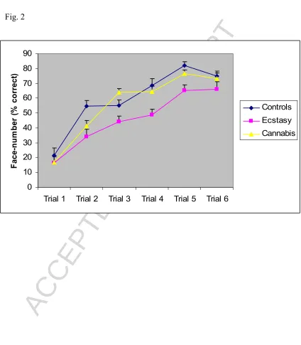
Face-number performance for the 3 groups is shown in Figure 2. A 3 (group) by 6
(trial) repeated measures analysis of variance (ANOVA) found a significant main
effect of trial (F=84.4, df= 5,133, p<0.001), reflecting an increase in memory performance over the first 6 trials. There was a significant group effect (F=5.8,
df=2,51, p<0.05) with post hoc tests showing ecstasy users had lower overall levels of
recall performance when compared to controls. There were no differences between
controls and cannabis users, and between ecstasy users and cannabis users. Analyses
showed no trial by group interaction (F=1.68, df=10,133 p= p>0.05), suggesting there
was no difference between groups in the gradient of the learning curves. In a separate
analysis on ecstasy users and controls there were no gender effects nor interactions
ACCEPTED MANUSCRIPT
2.2 Neuroimaging Results
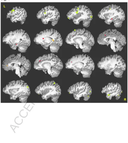
The t-test versus zero activation maps (p≤0.005) are shown in Figure 3. The encoding
and recall maps were quite similar with both activating bilateral PFC and bilateral
hippocampus. The activation patterns of ecstasy users were also similar to controls as
demonstrated, as noted earlier, by the lack of significant group differences on the
between-groups voxelwise t-tests.
Table 2 summaries the results of the conjunction analysis that was conducted for
ecstasy users, cannabis users, and controls. Three patterns were evident from the 18
clusters that were revealed (Figure 4). Ecstasy-specific increased brain activation
(ecstasy>cannabis=controls) was found in the left superior frontal gyrus, bilateral
middle frontal gyrus, left middle temporal gyrus, right tuber/declive, right inferior
parietal lobule, left inferior occipital gyrus, right inferior temporal gyrus, right middle
occipital gyrus, and right cuneus. Of these regions BOLD activation was greater
during encoding compared to recall in the left superior frontal gyrus, right
tuber/declive, right inferior parietal lobule, right inferior temporal gyrus, and right
cuneus. The left inferior frontal gyrus was the only region to show an interaction that
was driven by increased BOLD activation for ecstasy users compared to controls
during encoding face-number associations. For the second pattern, ecstasy-specific
decreased brain activation (ecstasy<cannabis=controls) was evident in a cluster that
included the left parahippocampal gyrus and the cingulate and in a second cluster that
fell in the right dorsal anterior cingulate. For these regions there were neither task
phase effects nor group X task phase interactions. For the third pattern both ecstasy
ACCEPTED MANUSCRIPT
medial frontal gyrus, left parahippocampal gyrus, left dorsal cingulate gyrus, and left
caudate. BOLD brain activation was greater during encoding compared to the recall
task in the left dorsal cingulate gyrus. In the separate analysis restricted to ecstasy
users and controls there were no gender effects nor interactions between gender and
group.
In ecstasy users the total number of lifetime ecstasy tablets consumed was positively
correlated with activation in the right middle temporal gyrus (51, 1, -19)
during encoding (r= 0.48, p≤0.05), with the right tuber/declive (51, -61,
-22) during recall (r= 0.49, p≤0.05), and with the left inferior occipital
ACCEPTED MANUSCRIPT
3. Discussion
In this hippocampal-dependant face-number paradigm ecstasy users had altered
memory performance compared to cannabis users and drug-naïve controls. The fMRI
results provide evidence that this compromised performance may be related to neural
alterations in a range of cortical and sub-cortical structures. This result may reflect the
widespread toxic effects of ecstasy on brain 5-HT neurons (Ricaurte et al., 1992a;
Fischer et al., 1995; Hatzidimitriou et al., 1999 Giorgi et al., 2005; Tamburini et al.,
2006; Frenzilli et al., 2007; Buscetti et al., 2008) and complements previous reports
of functional brain abnormalities during performance of memory tasks in ecstasy users
(Daumann et al., 2003a; Daumann et al., 2003b; Daumann et al., 2004a; Jacobsen et
al., 2004; Moeller et al., 2004; Daumann et al., 2005). Furthermore, the inclusion of a
cannabis group enabled us to separate ecstasy-specific effects (or effects that arise
from using both ecstasy and cannabis) from cannabis-specific effects. The results
identified a number of hypoactive brain regions, some of which were shared by the
cannabis and ecstasy groups and some that were unique to the ecstasy group.
Consistent with previous studies using a similar task, prominent bilateral prefrontal
and hippocampal activations were evident during the encoding and recalling of
task-number asociations (Zeineh et al., 2003; Morgan et al., 1999; Gouzoulis-Mayfrank et
al., 2000). Ecstasy-specific dysregulations were found in the left superior frontal
gyrus, bilateral middle frontal gyrus, left middle temporal gyrus, right tuber/declive,
right inferior parietal lobule, left inferior occipital gyrus, right inferior temporal gyrus,
left parahippocampal gyrus, left ACC, right middle occipital gyrus, and right cuneus.
In both ecstasy and cannabis groups brain activation was altered in the right medial
ACCEPTED MANUSCRIPT
Despite reports suggesting that females are particularly vulnerable to the chronic
effects of ecstasy (Bolla et al 1998; Topp et al 1999; Lynch et al 2002; Von Gersau et
al 2004) none of the ecstasy-related effects observed here were modulated by sex, a
null result that should be tempered by noting that there were just ten male and ten
female ecstasy users in the current sample where the drug use variables did not differ
between the sexes.
Both ecstasy and cannabis users demonstrated hypoactivity in the left
parahippocampal gyrus, bilateral dorsal ACC, and left caudate during the learning and
recall phases, when compared to control participants. Historically, the basal ganglia
have been implicated in higher-order motor control (Graybiel et al., 1995) but there
have been a number of demonstrations of a role for the caudate in learning and
memory (Graybiel et al., 1995; Packard et al., 1990, 1994). The parahippocampus has
previously been demonstrated to be involved in the formation of face-name
associations (Zeineh et al. 2003; Kirwin and Stark 2004) while the dorsal ACC has a
broad role in on-line monitoring of behaviour in cognitive tasks (Carter et al., 1998;
Mango et al., 2006) and, indeed, the dorsal ACC has been shown to activate with the
hippocampus in learning from one’s errors (Hester et al. 2008). Consequently,
hypoactivity in this network of areas may result in compromised memory abilities:
moreover, there is growing evidence that cannabis (Yucel et al. 2008) and ecstasy
(Cowan et al., 2003; Daumann et al., 2004b) induce structural hippocampal
abnormalities. The lack of memory performance impairments in the cannabis group is
surprising given previous results (Nestor et al. 2008) but may reflect sampling
characteristics, and may also indicate that dysregulated functional brain activity might
ACCEPTED MANUSCRIPT
et al. (2007). Hippocampal hypoactivity in ecstasy users is consistent with two
previous fMRI studies (Jacobsen et al., 2004; Daumann et al., 1995) where reduced
hippocampal activity during working memory tasks was reported, although this deficit
was apparent in the absence of performance differences in the latter.
Ecstasy-specific effects were associated with left superior frontal, bilateral middle
frontal, right inferior parietal lobule, bilateral temporal, and bilateral occipital
hyperactivity. Ecstasy related hyperactivity is suggestive that ecstasy users are placing
greater demands in these brain regions by working harder to perform the task,
suggesting pathological effects within these areas. Consistent with the frontal
hyperactivity reported here, an fMRI study found that ecstasy users showed greater
activation in PFC compared with non-drug-using controls during performance of an
immediate and delayed working memory task (Moeller et al., 2004). Furthermore,
there is a substantial body of work associating ecstasy with serotonergic frontal
deficits (O’Hearn et al,. 1988; Wilson et al., 1989; Fischer et al., 1995; Hatzidimitriou
et al., 1999). The prefrontal cortex has been implicated in retrieval effort or success
(Buckner et al., 1998; Zeineth et al., 2003), and although we did not find any
associations between memory performance and the PFC, frontal dysregulation in
ecstasy users may underlie their worse performance. Giorgi et al. (2005) demonstrated
the occurrence of metabolic hippocampal hyperexcitability measured by
2-deoxyglucose uptake using experimental models of MDMA administration. Although
hippocampal hyperactivation was not evident, the pattern of ecstasy-related
ACCEPTED MANUSCRIPT
Ecstasy-specific hyperactivity in the right inferior parietal lobule and bilateral
temporal regions is also consistent with reports of these regions being affected by
chronic ecstasy use during working memory in two previous studies (Daumann et al.,
2003a; Daumann et al., 2003b). Activity in the left middle temporal gyrus (BA 21)
was correlated with total lifetime number of ecstasy tablets consumed and this same
left region was found to have reduced cortical grey matter concentrations in ecstasy
consumers compared to drug-naïve controls (Cowan et al., 2003). The ecstasy-specific
occipital cortex hyperactivity fell within secondary visual cortex known to be
involved in forming visual associations and processing visual imagery and memory
(Sperling et al., 2001; Stock et al., 2008). Activity in the bilateral occipital gyrus was
correlated with total lifetime number of ecstasy tablets consumed which coincides
with this same bilateral BA18 region being previously reported to have reduced
cortical grey matter concentrations in ecstasy users compared to controls (Cowan et
al., 2003). Although we did not find an inverse relationship between the regions that
showed hyperactive frontal activations and the regions that revealed hypoactivity, we
cannot rule out the possibility of a neural compensatory mechanism, whereby
widespread hyperactivity is compensating for the ecstasy-related lack of hippocampal
involvement in memory formation. There are reports of widespread neurotoxicity of
ecstasy in both isocortical and allocortical brain regions, with a major emphasis placed
on serotonergic neurotoxicity (Fischer et al., 1885; Ricaurte et al., 1985; Schmidt
1987a; O Hearn 1988; Ricaurte et al., 1992;Mc Cann et al., 1994 ;Hatzidimitriou et
al., 1999; Giorgi et al., 2005; Tamburini et al., 2006; Frenzilli et al., 2007; Buscetti et
al., 2008). Given this widespread neurotoxicity and evidence that the serotonergic
ACCEPTED MANUSCRIPT
the hippocampal and frontal cortex (Buhot et al. 2000) we speculate that serotonergic
toxicity may be at least partly attributable to ecstasy-related deficits.
It should be noted that rather than scanning abstinent ecstasy users in whom persistent
neurotoxic effects of ecstasy might be evaluated, the present scans are of recreational
drug users who primarily use ecstasy and who have differing periods of abstinence
from ecstasy and other illicit drugs. A notable characteristic of this sample is that a
large proportion of ecstasy participants tested positive for cannabis and some had also
used ecstasy within two-three days prior to fMRI scanning. Negative subacute effects
of MDMA on cognition and mood have been previously characterized (Parrott and
Lasky, 1998; Parrott, 2001; Parrott, 2004)., Amphetamines have been reported to
cause toxicity (Berman et al., 2009) and some of the participants also used
amphetamines up to a total of 15 times in their lifetime. A large proportion of ecstasy
users were nicotine smokers, and a moderate proportion of the controls reported
nicotine use, whereas none of the cannabis users were nicotine users. We did not
record if participants smoked nicotine on the day of testing but as participants were
not requested to abstain from nicotine prior to testing the potential influence of
nicotine as a cofounding factor cannot be eliminated. Consequenty, the current results
might best be interpreted as showing the neurocognitive functioning of current
polydrug users, albeit users who vary in their primary drug of use, rather than
necessarily demonstrating persistent neurotoxic effects of those drugs.
Despite methological limitations, the findings of this study demonstrate alterations in
a network of brain regions that are related to ecstasy use or to the combined use of
ACCEPTED MANUSCRIPT
which effects might be attributed to which drug and suggest that there are a number of
regional deficits unique to the ecstasy group. From a systems-level neuroscience
perspective, the results indicate that ecstasy users showed unique cingulate and left
parahippocampal activity decreases and, we speculate, a compensatory hyperactivity
in mainly fronto-parietal cortical areas. Their performance was significantly impaired
suggesting that this is a sub-optimal functional neuroanatomy. Whether these results
preceded or arose from their ecstasy use will require longitudinal investigations.
4. Experimental Procedure
4.1 Participants
The ecstasy group included 20 chronic users of ecstasy and the drug-naïve group was
comprised of 20 participants with no history of illicit drug use. Participants were
recruited by poster recruitment and by the snowballing method. Participants in the
drug-naïve group were required to have never used any illicit substance. Participants
in the ecstasy group were required to be current users of ecstasy and to have
consumed at least 40 ecstasy tablets over a period of a year, but not necessarily over
the immediately preceding year. With the exception of cannabis, participants in the
ecstasy group were excluded if they used any other illicit drugs on more than ten
occasions in their lifetime (or more than fifteen times if the substance had not been
used in the five years that preceded the study) and were required to be abstinent of
ACCEPTED MANUSCRIPT
groups were also excluded if they had reported either past or present neurological or
psychiatric problems. Given the fact that daily smoking of cannabis is part of the
lifestyle of most club drug users (Daumann et al 2003), cannabis users who were also
ecstasy users were not excluded from the study or required to abstain from smoking
cannabis prior to participation. All ecstasy participants who reported cannabis use (last
use 0.5-12 days since last use) with the exception of one participant who had reported
use 3 years prior to study participation tested positive for cannabis. Ecstasy users were
requested to abstain from ecstasy for at least 48 hours prior to study participation.
Given this abstinence period, all participants provided a negative urine sample for
ecstasy. Additional urine analysis screening for methadone, benzodiazepines, cocaine,
opiates, barbiturates and tricyclic antidepressants (Cozart Rapiscan, UK) revealed
negative results in both groups. All participants gave informed consent and the study
was approved by the School of Psychology in Trinity College Dublin. In addition to
an ecstasy and control group the cannabis group from Nestor et al. (2008) were
incorporated into our analysis.
4.2 Demographics and drug use
Table 1 shows the group demographics and drug use histories for the ecstasy users,
cannabis users and controls. ANOVAs’ revealed that the groups did not differ in terms
of verbal IQ as assessed by the National Adult Reading Test (NART), age, years of
education, alcohol or other illicit drugs with the expected exception of ecstasy and
cannabis as specified in the selection criteria. As specified in the selection criteria the
control group did not use cannabis and the ecstasy users were polydrug users, but the
ACCEPTED MANUSCRIPT
compared to cannabis users. The heavier cannabis use of the cannabis group is not an
ideal match for the cannabis use of the ecstasy users but does provide a strong test of
any ecstasy-user group effects being ecstasy-specific (or arising from an interaction of
combined ecstasy and cannabis use). The ecstasy and cannabis groups self-reported
higher BDI scores compared to controls (p’s =0.05) and the two drug-using groups did
not differ from one another.
4.3 Stimuli and Behavioural Protocol
The face-number task used for the imaging procedure was adapted from that used by
Zeineh et al. (2003); associations were made between faces and two-digit numbers
which could be entered on a keypad during recall thereby avoiding possible head
motion associated with verbal responses. The task was programmed using E-Prime
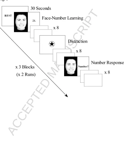
version 1.1 (Psychology Software Tools, Pittsburgh, USA). The task consisted of twoi runs of three blocks, with each block contining rest, learning, distraction and recall
phases (see Figure 1). The beginning of each block commenced with a 30 second rest
period. During the learning phase, participants were required to learn eight serially
presented face-number pairs that were presented for three seconds each. The numbers
paired with each face were randomly selected from the set: 11, 12, 13, 14, 21, 22, 23
and 24. During recall, these numbers were selected using two handheld response
keypads (left keypad contained the one and two keys, right keypad contained the three
and four keys). Following the presentation of each face-number pair, a fixation
crosshair was presented for between one to seven seconds. The face-number
association remained consistent throughout each learning phase, with each learning
phase being 60 seconds in duration. Eight distracter trials appeared between each
ACCEPTED MANUSCRIPT
participants to press the three key on the right hand-held keypad each time a black star
appeared in the center of a centrally displayed empty circle (see Figure 1). The eight
distracter trials were separated by variable delay intervals between three to five
seconds. During each recall trial, in random order participants were presented with
one of the eight ‘learning phase’ faces for three seconds, and required to respond
using the key-pad with the correct number for each face. A fixation crosshair of
variable duration between one to seven seconds followed the presentation of each
face. Each recall phase lasted 60 seconds, with a different presentation order of faces
administered during each of the six recall blocks. Each run of the face-memory task
was 570 seconds in duration. Dependent measures for the behavioural task were the
learning curve (percentage correct recall on each of the six recall trials).
4.5 Imaging parameters
All scanning was conducted on a Philips Intera Achieva 3.0 Tesla MR system (Best,
The Netherlands) equipped with a mirror that reflected a 640 x 480 pixel display,
projected on a panel placed behind the subject’s head outside the magnet. The mirror
was mounted on the head coil in the participants' line of vision. Imaging started with
31.5 seconds of standard scout images to adjust head positioning, followed by a
reference scan to resolve sensitivity variations. Imaging used a parallel SENSitivity
Encoding (SENSE) approach (Pruessmaan et al. 1999) with reduction factor 2. 180
high-resolution T1-weighted anatomic MPRAGE axial images (FOV 230 mm,
thickness 0.9 mm, voxel size 0.9 x 0.9 x 0.9) were then acquired (total duration 6
minutes), to allow subsequent activation localization and spatial normalization.
ACCEPTED MANUSCRIPT
were collected using a T2* weighted echo-planar imaging sequence (TE = 35 ms, TR
= 2000 ms, FOV 224 mm, 64 x 64 mm matrix size in Fourier space).
4.6 Time-series analyses
The fMRI data were analysed using the AFNI software package (Cox, 1996).
Time-series data were motion-corrected using 3D volume registration (least-squares
alignment of three translational and three rotational parameters). Activation outside
the brain was removed using edge detection algorithms. A block analysis was
conducted to estimate the activation for the learning and recall periods separately.
These ON-OFF block regressors were convolved with a standard haemodynamic
response to accommodate the lag time of the blood oxygen level-dependent (BOLD)
response. Multiple regression analyses were then used to derive the average level of
block activation as a percentage change relative to the distracter periods over the two
runs of the paradigm.
Activation maps were warped into a standard stereotaxic space (Talairach et al., 1998)
and spatially blurred with a 4.2-mm full-width at half-maximum isotropic Gaussian
filter after performing a second edge detection on the skull stripped brain. Encode and
recall activation maps for both ecstasy users and drug-naïve controls were determined
with one-sample t tests against the null hypothesis of zero activation changes (i.e., no
change relative to distracter-task activity). Significant voxels passed a voxelwise
statisticalthreshold (t = 8.94, P ≤ 0.005) and were required to bepart of a larger 286 µl
ACCEPTED MANUSCRIPT
Carlo simulationsand resulted in a 5% probability of a cluster surviving dueto chance
(Figure 3). Voxelwise independent-samples t-tests comparing ecstasy users and
drug-naïve controls were performed separately for both encode and recall activations using
similar thresholding to the previous t-tests.
No clusters survived the voxelwise between-group comparison. In addition, very
similar brain activation patterns were evident for both encoding and recall phases of
the task (see Figure 3). Consequently, in order to increase the statistical power of the
between-group comparisons, we performed a conjunction analysis that combined the
between-group comparisons of the encoding and recall periods. For this conjunction
analysis significant voxels from the between-groups t-tests passed a voxelwise
statisticalthreshold (t = 2.25, P ≤ 0.003 (derived from the square root of 0.001)) for
both the learning and recall periods and were required to bepart of a larger 141 µl
cluster of contiguous significantvoxels. Thresholding was determined through Monte
Carlo simulations and resulted in a 1% probability of a cluster surviving due to
chance. The mean activation for each separate cluster of the conjunction map was
calculated for all three groups (note that cannabis group is included here but this group
did not contribute to the construction of the conjunction map) for both recall and
encode task phases, and cluster level statistics were performed in SPSS (SPSS Inc).
4.7 Statistical analysis
Behavioural data are expressed as mean ± SEM and analyzed with the computerized
package SPSS (version 12). On the first six trials of the face-number task trial effects,
ACCEPTED MANUSCRIPT
repeated measures analysis of variance (ANOVA) followed by
Student-Newman-Keuls post hoc tests. As the cannabis group consisted of only 2 females it was excluded from a separate 2 (group) X 6 (trial 1-6) X 2 (gender) repeated measures
ANOVA which was carried out to investigate gender effects and gender interactions.
For the conjunction analysis a 3 (Group: ecstasy, cannabis, and controls) X 2 (encode,
recall) repeated measures analysis of variance (ANOVA) was performed for each
cluster of activation. Within the ecstasy group Pearsons correlations’ were carried out
to investigate relationships between behavioural performance, drug use and brain
activation. A separate 2 (group) X 2 (encode/recall) X 2 (gender) repeated measures
ANOVA was carried out to investigate gender effects and gender interactions for each
cluster.
Acknowledgements
NIDA. Grant number DA14100.
ACCEPTED MANUSCRIPT
References
Berman, S.M., Kuczenski, R., McCracken, J.T., London, E.D., 2009. Potential adverse effects of amphetamine treatment on brain and behavior: a review. Mol
Psychiatry. 14, 123-42.
Bhattachary, S., Powell, J.H., 2001. Recreational use of
3,4-methylenedioxymethamphetamine (MDMA) or 'ecstasy': evidence for
cognitive impairment. Psychol Med. 31, 647-58.
Bolla, K.I., McCann, U.D., Ricaurte, G.A., 1998. Memory impairment in abstinent
MDMA ("Ecstasy") users. Neurology. 51, 1532-7.
Buckner, R.L., Koutstaal, W., Schacter, D.L., Wagner, A.D., Rosen, B.R., 1998. Functional-anatomic study of episodic retrieval using fMRI. I. Retrieval effort
versus retrieval success. Neuroimage. 7, 151-62.
Busceti, C.L., Biagioni, F., Riozzi, B., Battaglia, G., Storto, M., Cinque, C., Molinaro, G., Gradini, R., Caricasole, A., Canudas, A.M., Bruno, V., Nicoletti, F., Fornai, F., 2008. Enhanced tau phosphorylation in the hippocampus of mice
treated with 3,4-methylenedioxymethamphetamine ("Ecstasy"). J Neurosci. 28,
3234-45.
Carta, G., Nava, F., Gessa, G.L., 1998. Inhibition of hippocampal acetylcholine release after acute and repeated Delta9-tetrahydrocannabinol in rats. Brain Res. 809, 1-4.
Carter, C.S., Braver, T.S., Barch, D.M., Botvinick, M.M., Noll, D., Cohen, J.D., 1998. Anterior cingulate cortex, error detection, and the online monitoring of
performance. Science. 280, 747-9.
Collins, D.R., Pertwee, R.G., Davies, S.N., 1995. Prevention by the cannabinoid antagonist, SR141716A, of cannabinoid-mediated blockade of long-term
potentiation in the rat hippocampal slice. Br J Pharmacol. 115, 869-70.
Cowan, R.L., Lyoo, I.K., Sung, S.M., Ahn, K.H., Kim, M.J., Hwang, J., Haga, E., Vimal, R.L., Lukas, S.E., Renshaw, P.F., 2003. Reduced cortical gray matter density in human MDMA (Ecstasy) users: a voxel-based morphometry study.
Drug Alcohol Depend. 72, 225-35.
Cox, R.W., 1996. AFNI: software for analysis and visualization of functional
magnetic resonance neuroimages. Comput Biomed Res. 29, 162-73.
Crane, J., Milner, B., 2002. Do I know you? Face perception and memory in patients
with selective amygdalo-hippocampectomy. Neuropsychologia. 40, 530-8.
Daumann, J., Fimm, B., Willmes, K., Thron, A., Gouzoulis-Mayfrank, E., 2003a. Cerebral activation in abstinent ecstasy (MDMA) users during a working memory task: a functional magnetic resonance imaging (fMRI) study. Brain
Res Cogn Brain Res. 16, 479-87.
Daumann, J., Schnitker, R., Weidemann, J., Schnell, K., Thron, A., Gouzoulis-Mayfrank, E., 2003b. Neural correlates of working memory in pure and
polyvalent ecstasy (MDMA) users. Neuroreport. 14, 1983-7.
Daumann, J., Fischermann, T., Pilatus, U., Thron, A., Moeller-Hartmann, W., Gouzoulis-Mayfrank, E., 2004a. Proton magnetic resonance spectroscopy in
ecstasy (MDMA) users. Neurosci Lett. 362, 113-6.
Daumann, J., Fischermann, T., Heekeren, K., Henke, K., Thron, A., Gouzoulis-Mayfrank, E., 2005. Memory-related hippocampal dysfunction in poly-drug ecstasy (3,4-methylenedioxymethamphetamine) users. Psychopharmacology
ACCEPTED MANUSCRIPT
Daumann, J., Jr., Fischermann, T., Heekeren, K., Thron, A., Gouzoulis-Mayfrank, E., 2004b. Neural mechanisms of working memory in ecstasy (MDMA) users who continue or discontinue ecstasy and amphetamine use: evidence from an 18-month longitudinal functional magnetic resonance imaging study. Biol
Psychiatry. 56, 349-55.
Fischer, C., Hatzidimitriou, G., Wlos, J., Katz, J., Ricaurte, G., 1995. Reorganization of ascending 5-HT axon projections in animals previously exposed to the recreational drug (+/-)3,4-methylenedioxymethamphetamine (MDMA, "ecstasy"). J Neurosci. 15, 5476-85.
Frenzilli, G., Ferrucci, M., Giorgi, F.S., Blandini, F., Nigro, M., Ruggieri, S., Murri, L., Paparelli, A., Fornai, F., 2007. DNA fragmentation and oxidative stress in the hippocampal formation: a bridge between
3,4-methylenedioxymethamphetamine (ecstasy) intake and long-lasting behavioral
alterations. Behav Pharmacol. 18, 471-81.
Gessa, G.L., Mascia, M.S., Casu, M.A., Carta, G., 1997. Inhibition of hippocampal acetylcholine release by cannabinoids: reversal by SR 141716A. Eur J
Pharmacol. 327, R1-2.
Giorgi, F.S., Pizzanelli, C., Ferrucci, M., Lazzeri, G., Faetti, M., Giusiani, M., Pontarelli, F., Busceti, C.L., Murri, L., Fornai, F., 2005. Previous exposure to (+/-) 3,4-methylenedioxymethamphetamine produces long-lasting alteration in limbic brain excitability measured by electroencephalogram spectrum analysis,
brain metabolism and seizure susceptibility. Neuroscience. 136, 43-53.
Gouzoulis-Mayfrank, E., Daumann, J., Tuchtenhagen, F., Pelz, S., Becker, S., Kunert, H.J., Fimm, B., Sass, H., 2000. Impaired cognitive performance in drug free
users of recreational ecstasy (MDMA). J Neurol Neurosurg Psychiatry. 68,
719-25.
Gouzoulis-Mayfrank, E., Thimm, B., Rezk, M., Hensen, G., Daumann, J., 2003. Memory impairment suggests hippocampal dysfunction in abstinent ecstasy
users. Prog Neuropsychopharmacol Biol Psychiatry. 27, 819-27.
Graybiel, A.M., 1995. The basal ganglia. Trends Neurosci. 18, 60-2.
Hales, J.B., Israel, S.L., Swann, N.C., Brewer, J.B., 2008. Dissociation of Frontal and Medial Temporal Lobe Activity in Maintenance and Binding of Sequentially Presented Paired Associates. J Cogn Neurosci.
Hatzidimitriou, G., McCann, U.D., Ricaurte, G.A., 1999. Altered serotonin innervation patterns in the forebrain of monkeys treated with
(+/-)3,4-methylenedioxymethamphetamine seven years previously: factors influencing
abnormal recovery. J Neurosci. 19, 5096-107.
Hester, R., Barre, N., Murphy, K., Silk, T.J., Mattingley, J.B., 2008. Human medial
frontal cortex activity predicts learning from errors. Cereb Cortex. 18,
1933-40.
Jacobsen, L.K., Mencl, W.E., Pugh, K.R., Skudlarski, P., Krystal, J.H., 2004. Preliminary evidence of hippocampal dysfunction in adolescent MDMA ("ecstasy") users: possible relationship to neurotoxic effects.
Psychopharmacology (Berl). 173, 383-90.
Jager, G., Van Hell, H.H., De Win, M.M., Kahn, R.S., Van Den Brink, W., Van Ree, J.M., Ramsey, N.F., 2007. Effects of frequent cannabis use on hippocampal
activity during an associative memory task. Eur Neuropsychopharmacol. 17,
289-97.
9-ACCEPTED MANUSCRIPT
tetrahydrocannabinol: possible mediation by glucocorticoid systems. Brain
Res. 443, 47-62.
Laws, K.R., Kokkalis, J., 2007. Ecstasy (MDMA) and memory function: a
meta-analytic update. Hum Psychopharmacol. 22, 381-8.
Lynch, W.J., Roth, M.E., Carroll, M.E., 2002. Biological basis of sex differences in
drug abuse: preclinical and clinical studies. Psychopharmacology (Berl). 164,
121-37.
Magno, E., Foxe, J.J., Molholm, S., Robertson, I.H., Garavan, H., 2006. The anterior
cingulate and error avoidance. J Neurosci. 26, 4769-73.
Matochik, J., Eldreth, D., Cadet, J., KI., B., 2005. Altered brain tissue composition in
heavy marijuana users. Drug Alcohol Depend. 77, 23-30.
McCann, U.D., Eligulashvili, V., Ricaurte, G.A., 2000.
(+/-)3,4-Methylenedioxymethamphetamine ('Ecstasy')-induced serotonin neurotoxicity:
clinical studies. Neuropsychobiology. 42, 11-6.
McCardle, K., Luebbers, S., Carter, J.D., Croft, R.J., Stough, C., 2004. Chronic
MDMA (ecstasy) use, cognition and mood. Psychopharmacology (Berl). 173,
434-9.
Moeller, F.G., Steinberg, J.L., Dougherty, D.M., Narayana, P.A., Kramer, L.A., Renshaw, P.F., 2004. Functional MRI study of working memory in MDMA
users. Psychopharmacology (Berl). 177, 185-94.
Morgan, M.J., 1999. Memory deficits associated with recreational use of "ecstasy"
(MDMA). Psychopharmacology (Berl). 141, 30-6.
Morgan, M.J., 2000. Ecstasy (MDMA): a review of its possible persistent
psychological effects. Psychopharmacology (Berl). 152, 230-48.
Nava, F., Carta, G., Colombo, G., Gessa, G.L., 2001. Effects of chronic Delta(9)-tetrahydrocannabinol treatment on hippocampal extracellular acetylcholine concentration and alternation performance in the T-maze.
Neuropharmacology. 41, 392-9.
Nestor, L., Roberts, G., Garavan, H., Hester, R., 2008. Deficits in learning and memory: Parahippocampal hyperactivity and frontocortical hypoactivity in
cannabis users. Neuroimage. 40, 1328-39.
O'Hearn, E., Battaglia, G., De Souza, E.B., Kuhar, M.J., Molliver, M.E., 1988. Methylenedioxyamphetamine (MDA) and methylenedioxymethamphetamine (MDMA) cause selective ablation of serotonergic axon terminals in forebrain:
immunocytochemical evidence for neurotoxicity. J Neurosci. 8, 2788-803.
Packard, M.G., White, N.M., 1990. Lesions of the caudate nucleus selectively impair
"reference memory" acquisition in the radial maze. Behav Neural Biol. 53,
39-50.
Packard, M.G., Cahill, L., McGaugh, J.L., 1994. Amygdala modulation of
hippocampal-dependent and caudate nucleus-dependent memory processes.
Proc Natl Acad Sci U S A. 91, 8477-81.
Parrott, A.C., Lasky, J., 1998. Ecstasy (MDMA) effects upon mood and cognition: before, during and after a Saturday night dance. Psychopharmacology (Berl). 139, 261-8.
Parrott, A.C., 2001. Human psychopharmacology of Ecstasy (MDMA): a review of 15
years of empirical research. Hum Psychopharmacol. 16, 557-577.
Parrott, A.C., 2004. MDMA (3,4-Methylenedioxymethamphetamine) or ecstasy: the neuropsychobiological implications of taking it at dances and raves.
ACCEPTED MANUSCRIPT
Parrott, A.C., Milani, R.M., Gouzoulis-Mayfrank, E., Daumann, J., 2007. Cannabis and Ecstasy/MDMA (3,4-methylenedioxymethamphetamine): an analysis of their neuropsychobiological interactions in recreational users. J Neural
Transm. 114, 959-68.
Pruessmann, K.P., Weiger, M., Scheidegger, M.B., Boesiger, P., 1999. SENSE:
sensitivity encoding for fast MRI. Magn Reson Med. 42, 952-62.
Reneman, L., Lavalaye, J., Schmand, B., de Wolff, F.A., van den Brink, W., den Heeten, G.J., Booij, J., 2001. Cortical serotonin transporter density and verbal memory in individuals who stopped using
3,4-methylenedioxymethamphetamine (MDMA or "ecstasy"): preliminary
findings. Arch Gen Psychiatry. 58, 901-6.
Ricaurte, G.A., Martello, A.L., Katz, J.L., Martello, M.B., 1992. Lasting effects of (+-)-3,4-methylenedioxymethamphetamine (MDMA) on central serotonergic neurons in nonhuman primates: neurochemical observations. J Pharmacol Exp
Ther. 261, 616-22.
Rodgers, J., 2000. Cognitive performance amongst recreational users of "ecstasy".
Psychopharmacology (Berl). 151, 19-24.
Scallet, A.C., 1991. Neurotoxicology of cannabis and THC: a review of chronic
exposure studies in animals. Pharmacol Biochem Behav. 40, 671-6.
Schmidt, C.J., 1987. Neurotoxicity of the psychedelic amphetamine,
methylenedioxymethamphetamine. J Pharmacol Exp Ther. 240, 1-7.
Smart, R.G., Ogborne, A.C., 2000. Drug use and drinking among students in 36
countries. Addict Behav. 25, 455-60.
Sperling, R.A., Bates, J.F., Cocchiarella, A.J., Schacter, D.L., Rosen, B.R., Albert, M.S., 2001. Encoding novel face-name associations: a functional MRI study.
Hum Brain Mapp. 14, 129-39.
Stock, O., Roder, B., Burke, M., Bien, S., Rosler, F., 2008. Cortical Activation Patterns during Long-term Memory Retrieval of Visually or Haptically Encoded Objects and Locations. J Cogn Neurosci.
Talairach and, T., 1998. J talairach and P. Tournoux, C0-planar Stereotaxic Atlas of the human Brain./ thieme, New York (1998).
Tamburini, I., Blandini, F., Gesi, M., Frenzilli, G., Nigro, M., Giusiani, M., Paparelli, A., Fornai, F., 2006. MDMA induces caspase-3 activation in the limbic system
but not in striatum. Ann N Y Acad Sci. 1074, 377-81.
Topp, L., Hando, J., Dillon, P., Roche, A., Solowij, N., 1999. Ecstasy use in Australia:
patterns of use and associated harm. Drug Alcohol Depend. 55, 105-15.
von Geusau, N.A., Stalenhoef, P., Huizinga, M., Snel, J., Ridderinkhof, K.R., 2004. Impaired executive function in male MDMA ("ecstasy") users.
Psychopharmacology (Berl). 175, 331-41.
Wilson, M.A., Ricaurte, G.A., Molliver, M.E., 1989. Distinct morphologic classes of serotonergic axons in primates exhibit differential vulnerability to the
psychotropic drug 3,4-methylenedioxymethamphetamine. Neuroscience. 28,
121-37.
Yucel, M., Solowij, N., Respondek, C., Whittle, S., Fornito, A., Pantelis, C., Lubman, D.I., 2008. Regional brain abnormalities associated with long-term heavy
cannabis use. Arch Gen Psychiatry. 65, 694-701.
ACCEPTED MANUSCRIPT
Zeineh, M.M., Engel, S.A., Thompson, P.M., Bookheimer, S.Y., 2003. Dynamics of the hippocampus during encoding and retrieval of face-name pairs. Science.
ACCEPTED MANUSCRIPT
Table 1. Mean and Range for ecstasy, cannabis and control groups on demographics and drug use history. *= p < 0.05 Vs control group , += p < 0.05
versus ecstasy group.# = on the day of testing one participant from the control group reported using 1 line of cocaine on 4 occasions (last use was 2 years prior to testing). Within the ecstasy group ecstasy and cannabis use measures did not differ between males and females( p > 0.05). The n given in parentheses refers to the number of subjects reporting a non-zero value while the mean and ranges are for the entire group.
Table 2 Cerebral Foci for conjunction analysis of face-number task. Phase X group interaction driven by increased activation for ecstasy users during encoding (p < 0.05) , ** = p < 0.01, ** = p < 0.001. E+C↓=ecstasy +cannabis decreased brain activation. ↑= increased activation for Encode condition, ↓= decreased activation for recall condition. Analysis was conducted on ecstasy, cannabis, and control groups.+ = Increased activation for males (as only two females were tested in the cannabis group, this group was eliminated from the gender analysis).
Fig. 1 Face-number protocol. Face memory task administered during fMRI data collection. Participants were required to learn and recall face-number pairs over a series of 6 blocks.
Fig. 2 Face-number performance. Mean performance on trials 1-6 in controls, ecstasy users and cannabis users (means and SEM). Ecstasy users had poorer performance compared to controls and cannabis users.
Fig. 3 Sagittal sections showing regions involved in the face-number task.
Examination of t-tests (p=0.005) for encoding (left column) and recalling (right column) between control participants (upper row) and ecstasy using participants (bottom row) demonstrate considerable overlap between the two conditions and qualittatively similar activations for both groups.
Fig. 4Cerebral Foci from conjunction analysis of face-number task.
ACCEPTED MANUSCRIPT
Table. 1
Ecstasy Cannabis Controls
(n=20) (n=14) (n=20)
Age 22.4 (18. 1 28.4) 22.4 (18.3 32.0) 22.5 (18.0 29.0)
Years of education 15.8 (11.0 19.0) 16.0 (12.0 19.0) 16.9 (11.0 21.0) Verbal intelligence score (NART) 122.1 (114.8 129.7) 122.9 (119.2 128.3) 123.3 (114.8 129.4) Beck Depression Inventory II score 5.8 (0 20)* 6.1 (0 14)* 3.2 (0 10)
Females/males 10/10 2/12 10/10
Ecstasy use in the last month (no. times) 2.3 (1.0 8.0) 0 0
Pills in last month (number) 10.7 (3.0 50.0) 0 0
Last ecstasy use (days) 16.1 (2.4 32.3) 960 (na 1835) (n=8) 0 Lifetime pills (number) 406.5 (50.0 1500.0) 18.8 (0 40) (n=8) 0
Pills in last year (number) 109 (15 600) 0 0
Years of cannabis use 6.8 (0 12.3) (n=15) 7.2 (2.5 16.0) 0
Days of use in last month (number) 16.1 (0 30.0) (n=15) 19.1 (7.0 30.0) 0 Joints in last month (number) 43.2 (0 180.0) (n=15) 82.8 (10.5 210.0) 0 Last cannabis use (days) 102.5 (na 1500.3) (n=15) 3.6 (0.1 4.5) 0 Lifetime joints (number) 2479 (0 10000.0) (n=15) 7925.9 (1000 33653) 0
Years of alcohol use 7.6 (3.6 13.3) 9.0 (4.5 18.0) 6.32 (2.0 10.5)
Alcohol use in the last month (no. days) 9 (0 20) 2.9±0.3 (0 20.0) 6.1 (2.0 15.0) Average units of alcohol per week 14.4 (1 35.5) 12.5± (5.0 20.0) 10.6 (5.0 21.0)
Years of nicotine use 5 (0 10) (n=14) 0 5.3± (0 9.0) (n= 6)
Years of amphetamine use 3.1 (0 8.0) (n=11) 1.5 (0 2.5) (n=7) 0 Last amphetamine use (days) 362.1± (na 1500.0) (n=11) 415 (na 2520) (n=7) 0 Amphetamine use (no. times) 6.3±1.7 (0 15.0) (n=11) 10.4 (0 10) (n=7) 0
Years of cocaine use 3.0 (0 6.1) (n=17) 0.5 (0 3) (n=9) #
ACCEPTED MANUSCRIPT
Effects
Structure Broadmann area Hemisphere Volume (µl) X Y Z Encode > recall Phase X Group Pattern Frontal lobe
Superior frontal gyrus 6, 9 L 2083 -46 4 28 *** ** E?
Middle frontal gyrus + 6 L 428 -36 -5 47 ns ns E?
Middle frontal gyrus 11 R 281 42 45 -11 ns ns E?
Middle frontal gyrus 10, 11 L 207 -41 55 -10 ns ns E?
Superior frontal gyrus 6 L 192 -12 16 69 ns ns E?
Parietal lobe
Tuber/declive 7 R 458 51 -61 -22 ** ns E?
Inferior parietal lobule 40 R 442 40 -53 52 *** ns E?
Temporal lobe
Middle temporal gyrus 21 L 576 51 1 -19 ns ns E?
Inferior temporal gyrus 20 R 309 61 -44 -18 ** ns E?
Occipital lobe
Inferior occipital gyrus 18 L 409 -38 -84 -1 ns ns E?
Middle occipital gyrus 18 R 285 44 -78 2 ns ns E?
Subcortical
Cuneus 19 R 244 30 -83 29 ** ns E?
Frontal lobe
Dorsal anterior cingulate 32 R 240 6 40 4 ns ns E?
Subcortical
Parahippocampal gyrus/cingulate 30 L 303 -17 -44 8 ns ns E?
Frontal lobe
Medial frontal gyrus 8, 32 R 574 13 32 34 ns ns E+C?
Dorsal cingulate gyrus 32 L 305 -7 24 38 ** ns E+C?
Subcortical
Parahippocampal gyrus L 368 -28 -16 -16 ns ns E+C?
Caudate L 284 -20 15 19 ns ns E+C?
Centre of mass
ACCEPTED MANUSCRIPT
Fig. 1
Face-Number Learning
x 8
Distraction
x 8
x 8
Number Response
30 Seconds
[image:30.595.110.518.109.599.2]ACCEPTED MANUSCRIPT
Fig. 2
0 10 20 30 40 50 60 70 80 90
Trial 1 Trial 2 Trial 3 Trial 4 Trial 5 Trial 6
F
ac
e-n
u
m
b
er
(
%
c
o
rr
ec
t)
[image:31.595.101.525.111.596.2]ACCEPTED MANUSCRIPT
Encode
Recall
Control
Ecstasy
R L
ACCEPTED MANUSCRIPT
Fig. 4
L
[image:33.595.104.532.110.589.2]