Original Article
Hyperbaric oxygen treatment promotes antioxidative
effects on rats with acute carbon monoxide poisoning
via NF-E2-related factor 2 activation
Rong Li1*, Yu Zhang2*, Ya Li3, Li Xue3, Wenlan Wang3
1Department of Internal Medicine, School of Clinical Medicine, Xi’an Medical University, Xi’an, China; 2Department
of Anesthesiology, The Second Affiliated Hospital of Shaanxi University of Chinese Medicine, Xianyang, China;
3Department of Aerospace Hygiene, School of Aerospace Medicine, The Fourth Military Medical University, Xi’an,
China. *Equal contributors.
Received November 16, 2017; Accepted May 3, 2018; Epub December 15, 2018; Published December 30, 2018
Abstract: Oxidative stress plays an important role in brain injuries after carbon monoxide (CO) poisoning. The aim of this study was to investigate the antioxidative effects of hyperbaric oxygen (HBO) treatment on rats with acute CO poisoning and elucidate underlying mechanisms associated with this event. Sprague-Dawley male rats were sub-jected to CO poisoning and HBO treatment. Their cognitive function was tested to show the effects of CO poisoning and HBO treatment. Antioxidative effects were investigated by determination of antioxidant enzymes. Expression of NF-E2-related factor 2 (Nrf2) and its cytoplasm binding protein kelch-like ECH-associated protein 1 (Keap1) was evaluated by immunohistochemical staining and Western blot. Additionally, Nrf2 transfer into nuclear and combi-nation with ARE were assessed by Western blot and Electrophoretic Mobility Shift Assay (EMSA). Results showed that HBO treatment increased levels of T-AOC, T-SOD, CuZnSOD, GPx, GR, and CAT, decreased by CO poisoning, and decreased MDA that had been increased by CO poisoning. Nrf2 disassociated from Keap1 in cytoplasm and transferred into nuclear to combine with ARE in the brains of HBO-treated CO-poisoned rats. These results suggest that HBO treatment increased antioxidant enzymes via the Nrf2-Keap1-ARE pathway. Therefore, Nrf2-mediated antioxidative effects may be one of the cellular mechanisms responsible for HBO treatment of brain injuries after acute CO poisoning.
Keywords: Carbon monoxide poisoning, hyperbaric oxygen, Nrf2-Keap1-ARE pathway
Introduction
Acute carbon monoxide (CO) poisoning is a common disease with high incidence and mor-tality. It is estimated that 3%-30% of patients will develop delayed neuropathological sequel-ae (DNS), despite adequate treatment [1]. It has been reported that the major causes of long-term mortality, after CO poisoning, are mental and psychiatric disorders [2]. The mech-anism of brain injuries and psychiatric disor-ders, after acute CO poisoning, has been asso-ciated with free radicals [3], oxidative stress [4], apoptosis [5], and immune reaction [6]. Hyperbaric oxygen (HBO) treatment is an effec-tive traditional therapy for acute CO poisoning and its delayed neuropathology [7]. However, it has been reported that HBO has the potential
for significant free radical generation, inducing
oxidative stress [7-9].
levels of Nrf2. In contrast, Nrf2 and Keap1 sep-arate and combine with ARE in the nucleus, thus, regulating cytoprotective genes [16]. The aim of this study was to further explain the mechanisms of HBO treatment association with antioxidative effects by establishing a rat model of acute CO poisoning and observing learning and memory abilities, antioxidant ca- pacity, and expression of associated protein in Keap1-Nrf2-ARE pathways.
Materials and methods
Experimental groups
After a week of adaptive feeding, 99 SD male rats (2-month-old) were randomly divided into three groups: normal control (NC) group (9 rats), acute carbon monoxide poisoning (CO) group (45 rats), and hyperbaric oxygen treat-ment (HBO) group (45 rats). The CO group and HBO group had 5 time points (1 day, 3 days, 7 days, 14 days, and 21 days after CO poisoning). NC group had no poison and no HBO treatment. CO group had carbon monoxide poisoning. HBO group had CO poisoning followed by daily bouts of HBO treatment. Each group had 9 rats. Six rats in each group were used for cognitive func-tion testing, antioxidant enzyme determinafunc-tion, and Western blot, while the other 3 rats were used for histopathological experiments after
perfusion-fixation. All rats were provided by the
Experimental Animal Center of the Fourth Mi- litary Medical University. This study was ap- proved by the Animal Ethics Committee of the Fourth Military Medical University. All experi-mental procedures were conducted in confor-mity with guidelines for the care and use of laboratory animals formulated by the Ministry of Science and Technology of the People’s Republic of China.
Animal manipulations
CO exposure was performed, according to pub-lished protocol, in a small temperature-con-trolled (22~24°C) animal hyperbaric chamber [15]. CO detectors in the chamber were used to determine the concentration of CO gas. Rats breathed 1,000 ppm CO gas for 40 minutes, then 3,000 ppm for 20 minutes. Rats were then removed to room air. Rats of the HBO group were given HBO treatment immediately
after CO exposure. For HBO treatment, rats were put into the same hyperbaric chamber used for CO exposure. HBO sessions began with a progressive increase in pressure for 30 minutes, followed by 60 minutes of continuous exposure to 100% oxygen at 2.5 ATA. After 60 minutes of exposure, pressure in the chamber was slowly reduced over a 30 minute period. After the procedure, animals were placed in single cages in the animal care room. Control rats and CO-poisoned rats were placed outside the hyperbaric chamber in the same room. After cognitive function tests, rats were anes-thetized with 2% pentobarbital sodium and
sac-rificed by left ventricle perfusion of 0.9% physi -ological saline. Brains were removed and washed with physiological saline and deionized water, repeatedly. All surgery was performed under pentobarbital sodium anesthesia and all efforts were made to minimize suffering.
Tissue preparation
Tissue preparation for Western blotting and
antioxidant capacities: The rats were anesthe-
tized and sacrificed by left ventricle perfusion
of 0.9% physiological saline. To determine anti-oxidant capacities in brain tissues, brains were removed from the skulls and washed with phys-iological saline and deionized water, repeated-ly. Isolated brains were homogenized in 1 mL ice-cold saline. After centrifugation (2000 rev/ minute for 5 minutes), the pellet was discarded and clear supernatant liquid was obtained. Total antioxidant capacities (T-AOC) activities, glutathione peroxidase (GSH-PX) activities, glu-tathione reductase (GR) activities, catalase (CAT) activities, superoxide dismutase (SOD) activities, and malondialdehyde (MDA) concen-tration in the brains were evaluated by spectro-photometric method. For Western blot, total protein of the cerebral cortex and hippocampus were obtained using Tissue Protein Extraction
reducing agent (Invitrogen), and boiled at 70°C for 10 minutes, prior to use. These proteins were separated by gel electrophoresis using NuPAGE Novex 4-12% Bis-Tris precast gels and
transferred to polyvinylidene fluoride mem -branes blotted with primary antibodies (dilu-tion: Nrf2 polyclonal antibody 1:500, anti-Keap1 polyclonal antibody 1:500 and anti- beta-actin monoclonal antibody 1:1000). Hor- seradish peroxidase-conjugated goat anti-mouse/rabbit IgG (1:5000) was used as sec-ondary antibody, followed by detection with enhanced chemiluminescence substrate for 5
minutes. After exposure for 1 minute, the film
was developed and total density of sample
pro-teins was quantified with Image-Pro Plus soft -ware. Results were calculated as: density of sample band/density of control band.
Tissue preparation for histopathological
experi-ment:Rats were anesthetized and sacrificed by
left ventricle perfusion of 0.9% saline followed by 4% paraformaldehyde in 100 mM phosphate buffer. Brains were then removed from the
skulls and postfixed in 4% paraformaldehyde at
4°C for 48 hours. Afterward, the brains were cut into 2 mm blocks. The 2 mm sections were
then paraffin-embedded and cut into 5 mm
sections, using a microtome, and mounted on poly-L-lysine-coated slides. Prior to staining, sections were de-waxed in xylene and dehy-drated through graded alcohol. For immunohis-tochemical staining, sections were incubated in 0.5% hydrogen peroxide (H2O2) in methanol to block endogenous peroxidase activity and then incubated in 0.2% Triton X-100 for 30 minutes. They were rinsed and incubated in phosphate-buffered saline, containing 1% bovine serum albumin and 10% normal calf serum, for 1 hour at room temperature. Incubation with primary antibodies (anti-Nrf2 polyclonal antibody 1:500 and anti-Keap1 polyclonal antibody 1:500) was performed for 24 hours at 4°C. The sections were then incubated with biotinylated goat anti-mouse/rabbit IgG (1:100) followed by ABC com-plex with 3, 3’-diaminobenzidine. Negative con-trols were included in each experiment where the primary antibody was omitted. No immuno-reactivity was detected in brain sections from the CO group in the absence of primary anti-bodies. The sections were then dehydrated, cleared, and mounted for microscopic analy- sis.
Cognitive function tests
Morris water maze: The Morris water maze
experiment is based on the fact that rodents placed in water have a strong motivation to escape the water environment using the fast-est and most direct route. Rats escaping from
the water environment reflect an ability to learn.
Rats swimming to a safe place (platform) in the
surrounding environment reflect an ability to
use spatial memory. Morris water maze experi-ment was performed in a tank, 122 cm in diam-eter, with water temperature maintained at 21°C. The water was tinted with black ink to obscure the platform. A 10 cm × 10 cm plat- form was hidden 1 cm below the surface of the water in quadrant 1. Entry points were
random-ly assigned to quadrant 2, 3, or 4. Briefrandom-ly, the
familiarization procedure was performed 1 day and 2 days before CO poisoning. Acute carbon monoxide poisoning occurred after the familiar-ization session and testing procedure was per-formed 1 day, 3 days, 7 days, 14 days, and 21 days after CO poisoning. Each trial lasted until
the rats found the fixed platform or for a maxi -mum of 60 seconds. Rats were allowed to rest on the platform for 10 seconds and each rat was given four trials per day. Place navigation ability was determined by recording the latency period before searching for the platform. After place navigation testing (21 days after CO poi-soning), the platform was removed to create a spatial probe test. Swimming times and swim-ming distances in searching for the platform in quadrant 1 were recorded, representing spatial probe ability.
Open field test
Open Field test is based on the exploratory behavior and spontaneous activity of rodents entering an open environment. Movement
dis-tances and speed of the rats reflect their fear. Rats activities in the central zone reflect their
anxiety. Open Field testing was performed on the 10th day after CO poisoning. Briefly, the apparatus consisted of a gray square of 70 cm × 70 cm × 40 cm. It was divided into 35 × 35 equal squares, which had been drawn in the
floor of the arena. The testing room was dimly
illuminated. A single rat was placed in the
the central zone, and stay period in the central zone were counted, automatically, for 15 min-utes by an image collecting and processing sys-tem. After each test, the arena was cleaned with 90% alcohol solution.
High plus maze
Elevated plus maze (EPM) consists of a device with two opposite open arms (50 × 10 cm) and two closed arms (50 × 10 cm), elevated 45 cm
from floor level and a central platform (10 × 10
cm). The high plus maze was performed on the 6th day after CO poisoning. Rats were placed, individually, in the center of maze with the head turned to one of the closed arms. Their behav-ior was evaluated for 5 minutes. Each trial was video recorded by a digital camera linked to a computer in an adjacent room. The recordings
were analyzed, offline, by a highly-trained
researcher using X-PLO-RAT (version 3.3). Behavioral parameters evaluated in this test were the number of entries and length of stay of the animals in the open arms, closed arms, and center area of the equipment. Results are expressed as mean ± SD for each area and analyses were performed by one-way ANOVA. Between groups, variance was determined using LSD-T test. P value < 0.05 was
consid-ered statistically significant.
Electrophoretic mobility shift assays (EMSA)
ARE binding activity of Nrf2 was measured with electrophoreticmobility shift assays. Nuclear fraction was isolated, as above. A double-stranded 27-mer oligonucleotide with the Nrf2 consensus sequence 5’-TGGGGAACCTGTGCT- GAGTCACTGGAG-3’ (Santa Cruz) was used for Nrf2-ARE binding activity. These probes were
end-labeled using [γ-32P] ATP (3000 Ci/mmol,
Amersham) and T4 polynucleotide kinase, and
purified with a G-25 Sephadex column.
Statistical analysis
The number of staining cells in each field was counted at high magnification (× 400). Five high-power fields of the cerebral cortex and hip -pocampus were photographed in each section.
These high-power fields were separated by at
least 60 mm to avoid double counting of cells. Positive cells, in each sample, were taken as
the average count from five brain sections and
number of values are presented as mean ± SD.
Statistical analysis was performed with SPSS Statistics 17.0, using one-way analysis of vari-ance to establish whether differences were
sta-tistically significant. Between groups, variance was determined by least significant difference
t-test. P value < 0.05 was considered statisti-
cally significant.
Results
HBO treatment increases cognitive function of rats with acute CO poisoning
HBO treatment increases learning and memory ability of rats with acute CO poisoning: In the place navigation experiment with the Morris water maze, escape latency in the CO group was longer than the NC group on each record-ing day. Compared with the CO group, HBO treatment shortened escape latency. Times of the HBO group were not different, however, from that of the NC group (Figure 1A). Escape latency and swimming locus of the three groups
(Figure 1B) showed that HBO treatment
improved place navigation ability that had been lessened by acute CO poisoning.
In spatial probe testing with the Morris water maze, swimming times and distances of the CO group in quadrant IV were shorter than that of the NC group. In the HBO group, swimming times and distances were longer than that in the CO group but differences between the HBO
group and NC group were not significant (Figure 1C). Swimming times and swimming locus of the three groups (Figure 1D) showed that HBO treatment enhanced their spatial probe abili- ty which had been lowered by acute CO poisoning.
HBO treatment increases adaptive capacity of rats with acute CO poisoning in a novel environment: In open field testing, the total
distance and distance in the central zone in the CO group were both longer than those in the NC group. Average speeds were faster in the CO group than the NC group. Stay periods in the central zone in the CO group were longer than the NC group. In the HBO group, total distance was shorter, average speed was slower, and stay period in the central zone was shorter than those in the CO group. Differences in distance in the central zone between the HBO group and
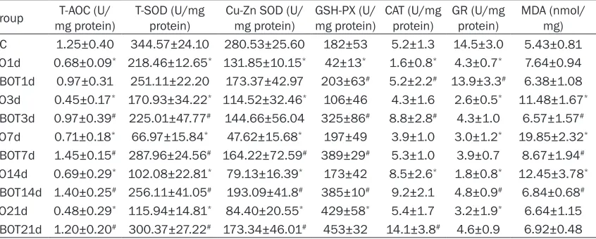
Table 2. Activities of antioxidant enzymes and content of MDA in brain (n=6, ±s)
Group mg protein)T-AOC (U/ T-SOD (U/mg protein) Cu-Zn SOD (U/mg protein) GSH-PX (U/mg protein) CAT (U/mg protein) GR (U/mg protein) MDA (nmol/mg) NC 1.25±0.40 344.57±24.10 280.53±25.60 182±53 5.2±1.3 14.5±3.0 5.43±0.81 CO1d 0.68±0.09* 218.46±12.65* 131.85±10.15* 42±13* 1.6±0.8* 4.3±0.7* 7.64±0.94 HBOT1d 0.97±0.31 251.11±22.20 173.37±42.97 203±63# 5.2±2.2# 13.9±3.3# 6.38±1.08 CO3d 0.45±0.17* 170.93±34.22* 114.52±32.46* 106±46 4.3±1.6 2.6±0.5* 11.48±1.67* HBOT3d 0.97±0.39# 225.01±47.77# 144.66±56.04 325±86# 8.8±2.8# 4.3±1.0 6.57±1.57# CO7d 0.71±0.18* 66.97±15.84* 47.62±15.68* 197±49 3.9±1.0 3.0±1.2* 19.85±2.32* HBOT7d 1.45±0.15# 287.96±24.56# 164.22±72.59# 389±29# 5.3±1.0 3.9±0.7 8.67±1.94# CO14d 0.69±0.29* 102.08±22.81* 79.13±16.39* 173±42 8.5±2.6* 1.8±0.8* 12.45±3.78* HBOT14d 1.40±0.25# 256.11±41.05# 193.09±41.8# 385±10# 9.2±2.1 4.8±0.9# 6.84±0.68# CO21d 0.48±0.29* 115.94±14.81* 84.40±20.55* 429±58* 5.4±1.7 3.2±1.9* 6.64±1.15 HBOT21d 1.20±0.20# 300.37±27.22# 173.34±46.01# 453±32 14.1±3.8# 4.6±0.9 6.92±0.48
*P < 0.05, compared with NC group; #P < 0.05, compared with CO group.
Figure 1. Results of cognitive function in each group. In the place navigation test, escape latency in CO group was longer than that in NC group and HBO treatment made it shorter than CO group (A, B). In the spatial probe test, the swimming time in quadrant 1 in the CO group was shorter than that in the NC group and the HBO group was longer than that in the CO group (C, D). In open field test, total distance and distance in the central zone in the CO group were both longer than that in the NC group. In the HBO group, total distance was shorter, average speed was slower, and stay period in the central zone was shorter than in the CO group (E). In the elevated plus maze, CO poisoned rats significantly approached less in open arms compared with control rats. After HBO treatment, open arm retention time and distance were significantly greater than that of CO group, the number of entry open arm was significantly more than that of CO group (F, G). Compared with NC group, *p < 0.05; compared with the CO group, #p < 0.05.
Figure 1E). HBO treatment enhanced their adaptive capacity in a novel environment.
HBO treatment decreases anxiety status of
rats with acute CO poisoning: In the elevated
plus maze test, CO-poisoned rats spent less time and contributed less distance in the open arms, compared with controls. As for the
num-ber of entries, CO-poisoned rats significantly
approached less in open arms, compared with control rats. After HBO treatment, open arm
retention times and distances were significant -ly greater than that of CO poisoning group (P < 0.05) and number of entry of open arms was
significantly more than the CO poisoning group
(P < 0.05) (Figure 1F and 1G). HBO treatment decreased rat anxiety status.
HBO treatment enhances antioxidant activities of rats with acute CO poisoning
Antioxidant enzyme activities and MDA concen-tration in brain tissues might determine antioxi-dant capacities after acute CO poisoning. This idea raised the question of how HBO treatment affects antioxidant capacities. Compared with the control group, the CO group had lower T-AOC activities and higher MDA content of brain tis-sue (P < 0.05). GSH-PX, GR, CAT, T-SOD, and CuZnSOD activities in the CO group decreased to different degrees (P < 0.05). Compared with the CO group, the HBO group increased T-AOC activity and decreased MDA content of brain tissue. GSH-PX, GR, CAT, T-SOD, and CuZnSOD activities in the HBO group increased to
differ-Table 1. Results in the open field test (n=6)
Group Total distance (cm) Average speed (cm/s) Distance in central zone (cm) Stay period in central zone (s)
NC 2271.35±551.25 2.52±0.61 2.84±31.33 7.01±6.63
CO 3540.42±592.44* 4.26±0.52* 133.75±86.71* 26.02±12.80*
H2 2865.98±794.86 3.19±0.88# 21.95±20.82# 3.69±2.71#
*P < 0.05, compared with NC group; #P < 0.05, compared with CO group.
[image:6.612.99.522.301.471.2]Figure 3. Immunohistochemical staining and Western blot of Nrf2. Nrf2-positive cells in the CO group were signifi-cantly less than that of the NC group, at all time points, and HBO treatment increased expression of Nrf2 protein (A-E). However, the results of Western blot for Nrf2 showed that Nrf2 protein in the CO group was not significantly different in cerebral cortex and hippocampus compared to the NC group, at all time points, expect for the 7th day. At 7th day, the Nrf2 from brain of hippocampus of CO poisoned rats was significantly decreased compared to that of the NC group. HBO treatment increased Nrf2 in cortex in 7th day and hippocampus in 1st and 3rd days (F, G). Compared with the NC group, *p < 0.05; compared with the CO group, #p < 0.05.
ent degrees (P < 0.05, Table 2). These data
confirm that HBO treatment enhances the anti -oxidant capacity of rats with acute CO poisoning.
HBO treatment changed Keap1 and Nrf2 pro-tein expression in brains of CO poisoned rats
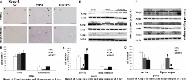
Protein expression levels of Keap1 and Nrf2 were examined by immunohistochemical and Western blot analyses. In immunohistochemi-cal analysis, cells that stained positive for Keap1 and Nrf2 were found in the brain isolat-ed from NC, CO-poisonisolat-ed, and HBO rats. Keap1-positive cells, in the cerebral cortex tis-sue and hippocampus from rats of CO group,
were not significantly different than that of NC
group on the 1st day (Figure 2B) and 7th day (Figure 2D). However, samples from the CO group showed less staining of Keap-1 on the 3rd day compared with NC group (P < 0.05) (Figure 2C). After HBO treatment, Keap1-positive cells in the cerebral cortex and hippocampus from
brain sections of rats were significantly more
than that in the CO group on the 1st day (Figure 2B) and 3rd day (Figure 2C) (P < 0.05). In con-trast, samples from HBO group showed less than that from the CO group (P < 0.05) on the 7th day (Figure 2A and 2D). Total protein of the cerebral cortex and hippocampus was used for Western blot analysis. In samples from the cerebral cortex and hippocampus of rats after
CO exposure, Keap1 protein was not signifi -cantly different relative to samples from either NC group or HBO, at any time point (Figure 2E
and 2F). The results indicate that there was no
significant tendency of Keap-1 among each
group.
Nrf2-positive cells in the cerebral cortex and
hippocampus of CO group were significantly
less than the NC group, at all time points, and HBO treatment increased expression of Nrf2 (P < 0.05, Figure 3A-E). However, results of Western blotting for Nrf2 showed that Nrf2
pro-teins in CO group were not significantly differ
-ent in the cerebral cortex and hippocampus compared to NC group, at all time points, expect for the 7th day (Figure 3F and 3G). On the 7th day, Nrf2 from brains of hippocampus of
CO-poisoned rats significantly decreased com -pared to that of NC group (P < 0.05, Figure 3G). HBO treatment increased Nrf2 in the cortex at the 7th day and the hippocampus in 1st and 3rd days (Figure 3F and 3G).
Taken together, these findings indicate that pro
-tein expression of Keap1 had no significant ten
-dency among each group. Nrf2 was significant -ly decreased in the brains of CO rats and HBO treatment can increase expression.
HBO treatment increased nuclear transloca-tion of Nrf2 in brain of CO rats
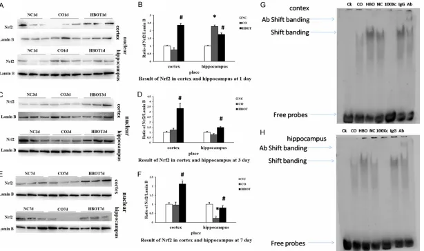
Nuclear translocation of Nrf2 was assessed by evaluating protein expression levels of Nrf2 in nuclear proteins. Nrf2 was assessed, quantita-tively, by comparing Nrf2 band density with the band density of Lamin B, which was used as loading control. As shown in Figure 4, Nrf2
pro-teins in nuclear of CO group were no significant -ly different in the cerebral cortex and hippo-campus compared to the NC group, at all time points, expect for the 1st day (Figure 4C-F). On the 1st day, Nrf2 in nuclear protein in the hip-pocampus of rats, after CO exposure, was
sig-nificantly increased (P < 0.05, Figure 4A and
4B). HBO treatment increased Nrf2 in nuclear in the cortex and hippocampus, at all time points, expect for hippocampus at the 1st day (Figure 4A-F). Taken together, these findings
indicate that CO poisoning induces Nrf2 disso-ciation with Keap1, subsequently promoting nuclear translocation of Nrf2 in the brain tissue of CO rats. HBO treatment decreases translo- cation.
HBO treatment increased activity of Nrf2 bind-ing to ARE in the brains of CO rats
HBO-treated rats. As shown in Figure 4G and
4H, CO rats displayed a decrease in activity of Nrf2 binding to ARE over levels observed in
control rats. However, HBO treatment signifi -cantly increased Nrf2 binding activity in
CO-poisoned rats. Specific binding of Nrf2 in CO rats was confirmed by the addition of a
100-fold molar excess of unlabeled Nrf2 competitor DNA into the EMSA reaction. Unlabeled Nrf2 DNA competed for binding in brain tissue extracts prepared from CO rats. Furthermore, an antibody super shift assay was used to dem-onstrate the presence of Nrf2 in protein
com-plexes, as Nrf2 antibody binding significantly
shifted major Nrf2 binding complexes.
Discussion
The purpose of this study was to assess the effects of HBO treatment on cognitional func-tion, antioxidant ability, and Nrf2 pathways in rat models with acute carbon monoxide poisoning.
Data from human studies has revealed that up to 44% of patients with carbon monoxide poi-soning have cognitive problems with memory, attention, and concentration [6]. These prob-lems could be improved by HBO treatment [16]. This present study used the Morris Water Maze, Open Field Test, andHigh Plus Maze to reflect neurophysiological and cogni tive deficits.
Re-sults of this study revealed that the CO group
demonstrated significant impairment in learn -ing and memory ability, adaptive capacity, and anxiety status. The results also suggest that HBO treatment can improve these cogni tive
deficits.
Under normal conditions, CO is an endogenous generated gas, in humans, and a cytoprotective molecule[17]. It has been reported that low concentrations of CO play an important role in
treatment of ischemic strokes and its beneficial
effects could be partially mediated by activa-tion of Nrf2pathways [18]. However, since CO can promote the formation of ROS, overdose of CO may result in overproduction of ROS, possi-bly playing a critical role in the reduction of anti-oxidant capacity of brain tissues from rats with CO poisoning. In this study, comparing antioxi-dant abilities between the NC group and CO group, it was revealed that rats with CO poison-ing had lower T-AOC, T-SOD, GSH-PX, GR, and
CAT activities and higher MDA concentration in brain tissues. This present study also demon-strated that overdose of CO decreased Nrf2 protein accumulation in the nucleus after CO poisoning and that low Nrf2 protein levels in the nucleus is associated with lower antioxi-dant capacity.
Additionally, high concentrations of oxygen may exacerbate ROS damage after HBO treatment [8]. In this study, CO promoted the generation of ROS. Rats were then given HBO treatment. It was found that T-AOC and activities of
antioxi-dant enzymes were significantly improved and
MDA concentrations reduced after HBO ment. These results indicate that HBO treat-ment increases the antioxidant capacity and reduces oxidative stress of brain tissues in rats. Furthermore, nuclear accumulation of Nrf2 was observed in the HBO group. In this study, it was also found that changes of Nrf2 proteins only occur at the 85~115 kDa range
with no alteration in levels of other nonspecific
bands or any migratory species of 55~65 kDa. This phenomenon had been mentioned, previ-ously, in other studies [19, 20].
As mentioned above, Keap1-Nrf2-ARE is an important pathway which can regulate oxida-tive stress. This present study demonstrated that HBO-induced ROS activated Nrf2 and made Nrf2, released from Keap1 and trans-ferred to nucleus to combine with ARE, to acti-vate expression of antioxidant enzymes. In con-clusion, HBO treatment improves the antioxi-dant ability of rats with CO poisoning by activat-ing Keap1-Nrf2-ARE pathways.
Acknowledgements
This work was supported by the Scientific and
Technological Research and Development Projects of Shaanxi Province (Grant No: 2014KJXX-59).
Disclosure of conflict of interest
None.
References
[1] Hampson NB, Weaver LK. Carbon monoxide poisoning: a new incidence for an old disease. Undersea Hyperb Med 2007; 34: 163-168. [2] Hampson NB, Rudd RA, Hauff NM. Increased
long-term mortality among survivors of acute carbon monoxide poisoning. Crit Care Med 2009; 37: 1941-1947.
[3] Lopez IA, Acuna D, Beltran-Parrazal L, Espi- nosa-Jeffrey A, Edmond J. Oxidative stress and the deleterious consequences to the rat co-chlea after prenatal chronic mild exposure to carbon monoxide in air. Neuroscience 2008; 151: 854-867.
[4] Wang P, Zeng T, Zhang CL, Gao XC, Liu Z, Xie KQ, Chi ZF. Lipid peroxidation was involved in the memory impairment of Carbon Monoxide-induced delayed neuron damage. Neurochem Res 2009; 34: 1293-1298.
[5] Piantadosi CA, Zhang J, Levin ED, Folz RJ, Schmechel DE. Apoptosis and delayed neuro-nal damage after carbon monoxide poisoning in the rat. Exp Neurol 1997; 147: 103-114. [6] Wang W, Li J, Chang Y, Xie X, Ren J, Wang X, Li
Y. Effects of immune reaction in rats after acute carbon monoxide poisoning. Undersea Hyperb Med 2011; 38: 239-246.
[7] Weaver LK, Hopkins RO, Chan KJ, Churchill S, Elliott CG, Clemmer TP, Orme JF Jr, Thomas FO, Morris AH. Hyperbaric oxygen for acute carbon monoxide poisoning. N Engl J Med 2002; 347: 1057-1067.
[8] Roderique JD, Josef CS, Feldman MJ, Spiess BD. A modern literature review of carbon mon-oxide poisoning theories, therapies, and po-tential targets for therapy advancement. Toxicology 2015; 334: 45-58.
[9] McMonnies CW. Hyperbaric oxygen therapy and the possibility of ocular complications or contraindications. Clin Exp Optom 2015; 98: 122-125.
[10] Shih AY, Johnson DA, Wong G, Kraft AD, Jiang L, Erb H, Johnson JA, Murphy TH. Coordinate regulation of glutathione biosynthesis and re-lease by Nrf2-expressing glia potently protects neurons from oxidative stress. J Neurosci 2003; 23: 3394-3406.
[11] Itoh K, Chiba T, Takahashi S, Ishii T, Igarashi K, Katoh Y, Oyake T, Hayashi N, Satoh K, Ha- tayama I, Yamamoto M, Nabeshima Y. An Nrf2/ small Maf heterodimer mediates the induction of phase II detoxifying enzyme genes through antioxidant response elements. Biochem Biophys Res Commun 1997; 236: 313-322. [12] Ishii T, Itoh K, Takahashi S, Sato H, Yanagawa
T, Katoh Y, Bannai S, Yamamoto M. Tran- scription factor Nrf2 coordinately regulates a group of oxidative stress-inducible genes in macrophages. J Biol Chem 2000; 275: 16023-16029.
[13] Muthusamy VR, Kannan S, Sadhaasivam K, Gounder SS, Davidson CJ, Boeheme C, Hoidal JR, Wang L, Rajasekaran NS. Acute exercise stress activates Nrf2/ARE signaling and pro-motes antioxidant mechanisms in the myocar-dium. Free Radic Biol Med 2012; 52: 366-376. [14] Hybertson BM, Gao B, Bose SK, McCord JM.
Oxidative stress in health and disease: the therapeutic potential of Nrf2 activation. Mol Aspects Med 2011; 32: 234-246.
[15] Reuland DJ, Khademi S, Castle CJ, Irwin DC, McCord JM, Miller BF, Hamilton KL. Upre- gulation of phase II enzymes through phyto-chemical activation of Nrf2 protects cardiomy-ocytes against oxidant stress. Free Radic Biol Med 2013; 56: 102-111.
[16] Yeh ZT, Tsai CF, Yip PK, Lo CY, Peng SM, Chen SY, Kung LY. Neuropsychological performance in patients with carbon monoxide poisoning. Appl Neuropsychol Adult 2014; 21: 278-287. [17] Motterlini R, Foresti R. Biological signaling by
carbon monoxide and carbon monoxide-re-leasing molecules. Am J Physiol Cell Physiol 2017; 312: C302-C313.
[18] Wang B, Cao W, Biswal S, Doré S. Carbon mon-oxide-activated Nrf2 pathway leads to protec-tion against permanent focal cerebral isch-emia. Stroke 2011; 42: 2605-2610.
[19] Lau A, Tian W, Whitman SA, Zhang DD. The predicted molecular weight of Nrf2: it is what it is not. Antioxid Redox Signal 2013; 18: 91-93. [20] Done AJ, Traustadóttir T. Nrf2 mediates redox