Original Article
The expressions and a correlation analysis of miR-155
and miR-27b in mycobacterium brain abscess
Tianyu Fan, Yunfeng Huang, Jiangliu Yin, Yong Gong, Yiping Deng, Shuai Zhang, Peng Xiang
Department of Neurosurgery, Changsha Central Hospital, Changsha 410004, Hunan, P. R. China
Received July 18, 2019; Accepted September 10, 2019; Epub November 15, 2019; Published November 30, 2019
Abstract: miR-27b is associated with inflammation, and miR-155 is involved in the regulation of mycobacterium tuberculosis. However, the expressions and correlation of miR-155 and miR-27b in mycobacterial brain abscess remain unclear. New Zealand white rabbits were randomly divided into a control group and a brain abscess group. 1×10⁷ CFU/mL Mycobacterium tuberculosis suspension was injected intracranially through the skull followed by an
analysis of the cerebrospinal fluid and the expressions of miR-155 and miR-27b by real time PCR. The correlations between miR-155 and miR-27b and cerebrospinal fluid pressure, cerebrospinal fluid cell number, protein, sugar, and chloride content were analyzed. In the brain abscess group, the cerebrospinal fluid pressure, the cerebrospinal fluid cell number, and the protein content significantly increased, and the sugar and chloride content decreased with
an increased miR-155 level and decreased miR-27b level compared with the control group (P < 0.05). miR-155 was
positively correlated with cerebrospinal fluid pressure, the cerebrospinal fluid cell number, the protein content and
negatively correlated with the sugar and chloride content (P < 0.05). On the other hand, miR-27b had a negative
correlation with the cerebrospinal fluid pressure, the cerebrospinal fluid cell number, and the protein content, and
a positive correlation with sugar and chloride content (P < 0.05). In addition, miR-155 and miR-27b were negatively correlated. The miR-155 expression was increased and miR-27b was decreased in mycobacterial brain abscess.
miR-155 and miR-27b were negatively correlated. The combined detection of miR-155 and miR-27b was beneficial
to the diagnosis of mycobacterial brain abscess.
Keywords: Mir-155, Mir-27b, mycobacteria, brain abscess, correlation
Introduction
Mycobacteria are a type of actinomycete and contain mycolic acid. They are classified into three groups: Mycobacterium tuberculosis com- plex, nontuberculous mycobacteria, and Myco- bacterium leprae with bacilli being more com-mon [1]. Tuberculosis caused by Mycobacterium tuberculosis is one a worldwide health prob-lem, and it is a serious threat to human life [2]. In recent years, the incidence of tubercular infectious diseases has risen, and with the wide application of antibiotic drugs, the number of patients with multi-drug resistant tuberculo-sis and extensive drug-retuberculo-sistant tuberculotuberculo-sis has increased [3, 4]. The emergence of drug-resistant strains and HIV co-infection leads to an increase in the incidence of tuberculosis and the difficulty in diagnosis and treatment [5]. According to statistics, about 9 million ac- tive tuberculosis patients are diagnosed each year, and more than one million patients die
MicroRNAs (miRNAs, also known as microR-NAs) are widely found in animals, plants and even eukaryotes such as viruses, at 22-23 nucleotides in length [13]. MicroRNA plays a role in the endogenous negative regulation of gene expression mainly at the post-transcrip-tional level, which binds mRNA by targeting complementary pairing, thereby promoting mR- NA degradation or translational inhibition [14]. MiRNAs can participate in the regulation of var-ious diseases, and they also participate in the regulation of the occurrence and development of tuberculosis [15]. miR-155 has been shown to be involved in the regulation of tuberculosis, and miR-27b is involved in the inflammatory response [16, 17]. However, the expressions and correlation of miR-155 and miR-27b in mycobacterial brain abscesses have not been elucidated.
Materials and methods
Experimental animals
Healthy, male New Zealand white rabbits, aged 6-8 weeks, weighing 2.5 kg ± 0.5 kg, of SPF grade, were purchased from the experimental animal center of this unit and fed in in the SPF animal experiment center at a temperature of 21 ± 1°C and a relative humidity of 50-70% under constant temperature and constant hu- midity conditions, ensuring a 12-hour day/night cycle. Animal experiments were performed in strict accordance with the experimental design and performed by experienced technicians to minimize animal suffering. This study was app- roved by the Ethics Committee of our hospital.
Main reagents and instruments
Pentobarbital was purchased from Shanghai Medical Reagent Co., Ltd. The Mycobacterium tuberculosis suspension H37RV was supplied by the laboratory and stored in liquid nitrogen. A Medlab-U automatic biochemical analyzer was purchased from Nanjing Meiyi Technology Co., Ltd. Other commonly used reagents were purchased from Shanghai Shenggong Biological
mental animals
Twenty male New Zealand white rabbits were randomly divided into two groups (n=10 in each group), a control group and a brain abscess group, in which the rabbits were anesthetized with pentobarbital and fixed on the operating table followed by an intracranial injection of 1×10⁷ CFU/mL through the skull and subse-quent infection with H37Rv standard strain
Mycobacterium tuberculosis. The model was established successfully if the brain abscess cavity occurred.
Specimen collection
After 4 weeks of treatment, 5 ml blood was col-lected from the tail vein, centrifuged at 3000 rpm for 15 minutes, and then the serum was placed in a -80°C refrigerator. The white rabbits were sacrificed, and the cerebrospinal fluid was separated and stored in a -80°C refrigerator. The brain tissue was collected and quickly fro-zen in liquid nitrogen for 2 hours, and then stored in a -80°C refrigerator for use.
Cerebrospinal fluid routine and biochemical testing
The routine and biochemical tests of cerebro-spinal fluid in each group of white rabbits were analyzed by automatic biochemical analyzer. The analysis included the cerebrospinal fluid pressure, the cerebrospinal fluid cell number, and the protein, sugar, and chloride content.
Real time PCR analysis of the expressions of miR-155 and miR-27b
Total RNA was extracted using Trizol reagent, and DNA reverse transcription synthesis was performed according to the kit’s instructions. The primers were designed by Primerpremier 6.0 according to each gene sequence and syn-thesized by Shanghai Yingjun Biotechnology Co., Ltd. (Table 1). Real-time PCR was per-formed for detection of the gene of interest using the conditions as follows: 92°C 30 S, 58°C 45 S, 72°C 35 S, for a total of 35 cycles. GAPDH was selected as a reference. According Table 1. Primer sequences
Gene Forward 5’-3’ Reverse 5’-3’
GAPDH ACCAGGTATCTTGGTTG TAACCATGTCAGCGTGGT Mir-155 AGCTTGCAGTCGTCTCTTG CAGCAGTTAACACCATGTGC Mir-27b TCGTCACCATGTCTGTTAA GTCCTTGGTAACACGTCT
chased from ABI, USA. Surgical micros-copy equipment was purchased from the Suzhou Medical Instrument Factory. The Labsystem Version 1.3.1 microplate rea- der was purchased from the Bio-Rad Corporation of the United States.
experi-cycle number (CT) of all the samples and stan-dards was calculated. Based on the standard CT value, a standard curve was drawn and then a semi-quantitative analysis was carried out using the 2-ΔCt method.
Statistical analysis
The data were analyzed using SPSS 19.0 soft-ware. The measurement data were expressed as the mean ± standard deviation, and the comparisons among multiple groups was per-formed using a one-way analysis of variance. The correlation analysis was performed using the Pearson correlation analysis. P < 0.05 indi-cates a significant statistical difference.
Results
Conventional and biochemical analysis of the cerebrospinal fluid
In the rabbit model of mycobacterial brain abscess, the cerebrospinal fluid pressure, the
cerebrospinal fluid cell number, and the protein content increased, but the sugar and chloride content decreased. Compared with the control group, the differences were statistically signifi-cant (P < 0.05) (Table 2).
The expressions of miR-155 and miR-27b in brain tissue
A real-time PCR analysis of 155 and miR-27b expressions in the brain tissue of rabbit models of mycobacterial brain abscess con-firmed that the expression of miR-155 in the brain tissue of the Mycobacterium tuberculosis
rabbit model was significantly increased, and the expression of miR-27b was significantly decreased, compared with the rabbits in the control group (P < 0.05) (Figure 1).
miR-155 and miR-27b expressions in the cere-brospinal fluid
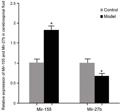
A real-time PCR analysis of 155 and miR-27b expressions in the cerebrospinal fluid of the rabbit model of mycobacterial brain abscess showed that the expression of miR-155 in the cerebrospinal fluid of the rabbit model of mycobacterial brain abscess was sig-nificantly increased, and the expression of miR-27b was decreased compared with the rabbits in the control group (P < 0.05) (Figure 2).
miR-155 and miR-27b expressions in the blood
[image:3.612.90.522.97.163.2]A real-time PCR analysis of 155 and miR-27b expressions in the blood of the rabbit model of mycobacterial brain abscess revealed that the expression of miR-155 was significant-ly increased in the rabbit model of mycobacte-rial brain abscess, and the expression of miR-27b was decreased. Compared with the control group, the differences were statistically signifi-cant (P < 0.05) (Figure 3).
Table 2. Conventional and biochemical analysis of cerebrospinal fluid in a rabbit model of mycobacte-rial brain abscess
Group Cerebrospinal fluid pressure (mmH 2O)
Cell number
(×105/L) content (g/L)Protein (mmol/L)Sugar (mmol/L)Chloride
Control 121.25±17.05 87.61±9.05 0.41±0.06 3.32±0.98 145.31±2.98 Model 186.75±22.20* 132.55±0.09* 1.35±0.67* 2.12±0.17* 102.11±1.16*
P value < 0.0001 < 0.0001 0.0003 0.0013 < 0.0001 Student’s t test. Compared with control group, *P < 0.05.
[image:3.612.88.287.197.381.2]Correlation analysis of routine and biochemi-cal indicators with miR-155 and miR-27b
miR-155 was positively correlated with cerebro-spinal fluid pressure, the cerebrocerebro-spinal fluid cell number, and protein content, and negative-ly correlated with sugar and chloride content (P < 0.05). However, miR-27b was negatively cor-related with the cerebrospinal fluid pressure, the cerebrospinal fluid cell number, and protein content, and sugar and chloride content was positively correlated (P < 0.05) (Table 3).
Correlation analysis between miR-155 and miR-27b in cerebrospinal fluid, brain tissue, and blood of the rabbit model of mycobacterial brain abscess
miR-155 and miR-27b were negatively correlat-ed in the cerebrospinal fluid, brain tissue, and blood of the rabbit model of mycobacterial brain abscess (P < 0.05) (Table 4; Figure 4).
Discussion
Among the central nervous system infectious diseases, mycobacterial brain abscess is an important disease sub-type. Due to the blood-borne dissemination of the lungs, the cellular immune function of the patient decreases, leading to a mycobacterial infection of the brain tissue [11]. The relevant response to mycobac-teria depends mainly on the immune status of
[image:4.612.88.288.71.254.2]the individual to the mycobacteria, the location and number of infections of the infected bacte-ria, as well as the state of the patient’s treat-ment [18]. Infecting the body, the inoculation of a small amount of mycobacteria can lead to the formation of infectious diseases such as tuber-culosis. For example, the administration of a large number of bacilli may lead to an excessive exudation of a large amount of cheese, accom-panied by the infiltration of inflammatory cells, resulting in the formation of pus, which may be accompanied by tuberculous necrosis, tubercu-losis. Bacteria and their products need to be present in brain tissue to form brain abscesses [19, 20]. Mycobacterial brain abscess lacks typical giant cell and epidermoid granuloma-tous reactions and is often difficult to distin-guish from purulent brain abscess. Patients may have focal neurological deficits correspon- ding to the degree of peripheral edema, the onset of the process is slow, and the appear-ance of brain signs depends on the location of the abscess [21]. The diagnosis of mycobacte-rial brain abscess mainly depends on clinical symptoms and signs, bacterial tests, and imag-ing analysis such as MRI or CT. However, be- cause of other causes of brain abscess, the bacterial detection process takes a long time, leading to misdiagnosis, and the delayed treat-ment of patients [22]. Therefore, it is necessary to find a rapid and effective molecular target to assist in the diagnosis of mycobacterial brain Figure 2. miR-155 and miR-27b expressions in the
[image:4.612.321.520.71.260.2]cerebrospinal fluid of the rabbit model of mycobacte -rial brain abscess. Compared with the control group, *P < 0.05.
abscess, which is conducive to the diagnosis and treatment of the disease.
MicroRNAs are a class of highly conserved sin-gle-stranded 18-25 nt small RNA molecules that regulate the expression of target mRNAs [23]. Recent studies have shown that serum miRNAs are stable to repeated freeze-thaw cycles and thermal, acidic and alkaline condi-tions, among other extremes. It may be a use-ful biomarker for disease diagnosis, treatment outcome, and prognosis [24, 25]. Previous studies have suggested that microRNAs are involved in several diseases, including cancer and heart, immune and infectious diseases
miR-155 is increased and miR-27b is decreased in mycobacterial brain abscess. miR-27b and miR-155 are negatively correlated. The detec-tion of mir-27b is beneficial to mycobacterial brain abscess diagnosis.
Acknowledgements
This study was supported by the Natural Science Foundation of Hunan Proince, China (Grant No. 2019dk2009).
Disclosure of conflict of interest
[image:5.612.89.373.98.174.2]None. Table 3. Correlation analysis of routine and biochemical indica-tors with miR-155 and miR-27b
Cerebrospinal
fluid pressure numberCell contentProtein Sugar Chloride miR-155 0.62* 0.78* 0.69* -0.58* -0.88*
P values 0.031 0.023 0.032 0.043 0.021 miR-27b -0.61* -0.79* -0.59* 0.77* 0.51*
[image:5.612.90.371.221.260.2]P values 0.026 0.041 0.036 0.015 0.032 Pearson correlation analysis. *P < 0.05.
Table 4. Correlation analysis between miR-155 and miR-27b
Cerebrospinal fluid Brain tissue Blood
miR-155 -0.73* -0.85* -0.64*
miR-27b
Pearson correlation analysis. *P < 0.05.
Figure 4. Correlation analysis of miR-155 with miR-27b.
[26]. It has been reported that microRNA levels change follow-ing mycobacterial infections [27]. A previous study con-firmed that miR-155 is increa- sed in tuberculosis, and miR-27b is involved in immune sta-tus and autoimmune diseases [16]. Therefore, this study ana-lyzed the expression of both in mycobacterial brain abscess and their correlation with dis-ease. The results showed that the expression of miR-155 in brain tissue, cerebrospinal flu- id, and the blood of mycobac-terial brain abscess was increa- sed, but the expression of miR-27b was decreased. miR-155 and miR-27b were positively or negatively correlated with the cerebrospinal fluid pressure, the cerebrospinal fluid cell nu- mber and protein content, re- spectively, but negatively or positively correlated with the sugar and chloride content. In addition, 27b and miR-155 were negatively correlat-ed. This result suggests that miR-27b and miR-155 are ab- normally expressed in myco-bacterial brain abscess and may be a promising molecular marker for mycobacterial brain abscess.
[image:5.612.92.376.284.525.2]Address correspondence to: Dr. Peng Xiang, Depart- ment of Neurosurgery, Changsha Central Hospital, No. 161, Shaoshan South Road, Changsha 4100- 04, Hunan, P. R. China. Tel: +86-0731-85667907; Fax: +86-0731-85667907; E-mail: lianrang08583- gy@126.com
References
[1] Ramshini E, Alaei H, Reisi P, Naghdi N, Afrozi H,
Alaei S, Alehashem M and Eftekharvaghefi S.
Effect of intracerebroventricular injection of GABA receptors antagonists on morphine-in-duced changes in GABA and GLU transmission within the mPFC: an in vivo microdialysis study. Iran J Basic Med Sci 2019; 22: 246-250. [2] Kilpelainen A, Saubi N, Guitart N, Moyo N, Wee
EG, Ravi K, Hanke T and Joseph J. Priming with recombinant BCG expressing novel HIV-1 con-served mosaic immunogens and boosting with recombinant ChAdOx1 is safe, stable, and
elic-its HIV-1-specific T-cell responses in BALB/c
mice. Front Immunol 2019; 10: 923.
[3] Chiappini E, Storelli F, Tersigni C, Venturini E, de Martino M and Galli L. QuantiFERON-TB Gold In-Tube test performance in a large pedi-atric population investigated for suspected tu-berculosis infection. Paediatr Respir Rev 2019; [Epub ahead of print].
[4] Fatima R, Yaqoob A, Qadeer E, Hinderaker SG, Heldal E, Zachariah R, Harries AD and Kumar AMV. Building sustainable operational re-search capacity in Pakistan: starting with tu-berculosis and expanding to other public health problems. Glob Health Action 2019; 12: 1555215.
[5] Lal A, Al Hammadi A and Rapose A. Latent Tu-berculosis Infection, Treatment initiation and completion rates in Persons seeking immigra-tion and healthcare workers. Am J Med 2019. [6] Jin C, Wu X, Dong C, Li F, Fan L, Xiong S and
Dong Y. EspR promotes mycobacteria survival in macrophages by inhibiting MyD88 mediated
inflammation and apoptosis. Tuberculosis (Ed -inb) 2019; 116.
[7] Indira Krishnan AK, Mini GK and Aravind LR. Evidence based interventions and implemen-tation gaps in control of tuberculosis: a sys-tematic review in low and middle-income coun-tries with special focus on India. Indian J Tuberc 2019; 66: 268-278.
[8] Hemavarneshwari S, Shaikh RB, Naik PR and Nagaraja SB. Strategy to sensitize private prac-titioners on RNTCP through medico-social
workers in urban field practice area of a Medi -cal College in Bengaluru, Karnataka. Indian J Tuberc 2019; 66: 253-258.
[9] Li Z, Wu W, Chen R, Chen X and Lin J. Periop-erative management of spinal tuberculosis in
patients with end-stage renal disease: a pre-liminary report on a patient series. World Neu-rosurg 2019; 129: e452-e457.
[10] Machida M, Toyoda K, Matsuda M, Sumida K, Yamamoto A, Sakurai K and Oba H. Extensive perivascular dissemination of cerebral miliary tuberculomas: a case report and review of the literature. Acta Radiol Open 2018; 7: 2058460118817918.
[11] Singh J and Dinkar A. Positive outcome of pul-monary tuberculosis associated with extraordi-nary extensive extrapulmoextraordi-nary tuberculosis in an immunocompetent adult. Int J Mycobacte-riol 2018; 7: 183-185.
[12] Lee HS, Kim JH, Kim YH and Lee S. Surgically treated community-acquired brain abscess: bacteriological analysis based on predisposing infections. Jpn J Infect Dis 2018; 71: 191-196. [13] Marjaneh RM, Khazaei M, Ferns GA, Avan A
and Aghaee-Bakhtiari SH. The role of microR-NAs in 5-FU resistance of colorectal cancer: possible mechanisms. J Cell Physiol 2019; 234: 2306-2316.
[14] Neudecker V, Brodsky KS, Kreth S, Ginde AA and Eltzschig HK. Emerging roles for MicroR-NAs in perioperative medicine. Anesthesiology 2016; 124: 489-506.
[15] Cerro-Herreros E, Sabater-Arcis M, Fernandez-Costa JM, Moreno N, Perez-Alonso M, Llamusi B and Artero R. miR-23b and miR-218 silenc-ing increase Muscleblind-like expression and alleviate myotonic dystrophy phenotypes in mammalian models. Nat Commun 2018; 9: 2482.
[16] Ding S, Qu Y, Yang S, Zhao Y and Xu G. A novel miR-1958 is a promoter towards Mtb survival in RAW264.7 cells by inhibiting autophagy via Atg5. J Microbiol Biotechnol 2019; 29: 989-998.
[17] Huang J, Jiao JH, Xu WH, Zhao HY, Zhang CX, Shi Y and Xiao ZJ. miR-155 is upregulated in patients with active tuberculosis and inhibits apoptosis of monocytes by targeting FOXO3. Mol Med Rep 2015; 12: 7102-8.
[18] Sathyapalan D, Balachandran S, Kumar A, Mangalath Rajamma B, Pillai A and Menon VP. Long term outcome of medical and surgical co-management of craniospinal aspergillosis in an immunocompromised patient. Med Mycol Case Rep 2016; 14: 33-37.
[19] Greninger AL, Langelier C, Cunningham G, Keh C, Melgar M, Chiu CY and Miller S. Two rapidly growing mycobacterial species isolated from a
brain abscess: first whole-genome sequences
of mycobacterium immunogenum and myco-bacterium llatzerense. J Clin Microbiol 2015; 53: 2374-2377.
C and Kolsch U. Late-onset disseminated my-cobacterium avium intracellulare complex in-fection (MAC), cerebral toxoplasmosis and sal-monella sepsis in a german caucasian patient with unusual anti-interferon-gamma IgG1 auto-antibodies. J Clin Immunol 2015; 35: 361-365.
[21] Chung YT, Pasquinelli V, Jurado JO, Wang X, Yi N, Barnes PF, Garcia VE and Samten B. Elevat-ed cyclic AMP Inhibits mycobacterium tubercu-losis-stimulated T-cell IFN-gamma secretion through type I protein kinase A. J Infect Dis 2018; 217: 1821-1831.
[22] Joshi V, Germano I, Meenakshi R and Doshi A. Paradoxical evolution of a cerebellar tubercu-losis abscess after surgical drainage and anti-biotic therapy. Surg Neurol Int 2014; 5: 143. [23] Golby P, Villarreal-Ramos B, Dean G, Jones GJ
and Vordermeier M. MicroRNA expression
pro-filing of PPD-B stimulated PBMC from M.
bovis-challenged unvaccinated and BCG vaccinated cattle. Vaccine 2014; 32: 5839-5844.
[24] Zhou MY, Yu GY, Yang XT, Zhu CM, Zhang ZZ and Zhan X. Circulating microRNAs as bio-markers for the early diagnosis of childhood tuberculosis infection. Mol Med Rep 2016; 13: 4620-6.
[25] Li M, Cui J, Niu W, Huang J, Feng T, Sun B and Yao H. Long non-coding PCED1B-AS1 regu-lates macrophage apoptosis and autophagy by sponging miR-155 in active tuberculosis. Bio-chem Biophys Res Commun 2019; 509: 803-809.
[26] Etna MP, Sinigaglia A, Grassi A, Giacomini E, Romagnoli A, Pardini M, Severa M, Cruciani M, Rizzo F, Anastasiadou E, Di Camillo B, Barzon L, Fimia GM, Manganelli R and Coccia EM. My-cobacterium tuberculosis-induced miR-155 subverts autophagy by targeting ATG3 in hu-man dendritic cells. PLoS Pathog 2018; 14: e1006790.