GENE EXPRESSION ANALYSIS OF CARDIOMYOCYTES DIFFERENTIATED FROM HUMAN BONE
MARROW DERIVED MESENCHYMAL STEM CELLS
Ramesh K. Garg, Sudarsanam,
Department of Advanced Zoology and Biotechnology, School of Genomics and Bioinformatics, Loyola College, Chennai
ARTICLE INFO ABSTRACT
Adult human bone marrow (BM) is the major source of mesenchymal stem cells (MSC) for cell based therapies. This study focuses to identify, isolate and characterize the mesenchymal stem cells that are believed to be present
collected. The key attributes like isolation efficiency, cell yield, colony (CFU-F) frequency,
determined for the isolated MSCs from bone marrow specimens. It was observed that the bone marrow possessed a good number of nucleated cells and had a high yield of adherent cells. Moreover, the characteristics of BM
morphology, colony forming unit and immunophenotype were observed.
were analyzed for their specific marker expression using flowcytometry, they were found to be positive for CD29, CD73,
respectively. Apart from this, it was observed that those MSCs, when induced to differentiate into cardiomyocytes, they successfully differentiated into cardiomyocyte like cells with flat, wi myotube like structure with sarcomeric organization, which were later confirmed to be cardiomyocytes by qRT
marrow and subsequently differentiated into cardiomyocytes with 100% success
study suggests that human bone marrow is a potential source of mesenchymal stem cells that could be differentiated in to ccardiomyocytes and used for cell based therapies for treating various cardiovascular diseases.
Copyright © Ramesh K Garg et al. This is an open access article distributed under the Creative Commons Attribution License, which permits unrestricted use, distribution, and reproduction in any medium, provided the original work is properly cited.
INTRODUCTION
Mesenchymal stem cells (MSC) are of great therapeutic potential due to their capacity of self-renewal and multilineage
differentiation (Mosca et al., 1999 and Ortiz
Reyes, 2002). Currently, bone marrow (BM) represents the
major source of MSC for cell therapy. Cardiovascular diseases are the major cause of morbidity and mortality in developed countries. Despite the advances in modern medical science, many diseases are not amenable to modern medical and surgical therapy. Coronary bypass after myocardial infarction causes fibrous bands around the infarcted area. This fibrous tissue restricts the cardiac contractility which causes the reduction of cardiac output of blood pumped through the aorta. Presently heart transplantation remains the only alternative for successful cardiac therapy, but due to donor shortage, the
therapy still remains in elusion. Given the nee
the limitations of these treatments, cell -based therapy has emerged in the past 2 decades as a novel regenerative
*Corresponding author: Praveena, P.
Department of Advanced Zoology and Biotechnology, School of Genomics and Bioinformatics, Loyola College, Chennai – 600 034, South India
ISSN: 0975-833X
International Journal of Current Research Vol. 6, Issue, 10, pp.9410
Article History:
Received 10th
July, 2014 Received in revised form 15th
August, 2014 Accepted 04th
September, 2014 Published online 25th
October,2014
Key words:
Mesenchymal Stem Cells, Human Bone-Marrow, Cardiomyocytes, Flowcytometry
and Potential Source of Stem Cells.
RESEARCH ARTICLE
GENE EXPRESSION ANALYSIS OF CARDIOMYOCYTES DIFFERENTIATED FROM HUMAN BONE
MARROW DERIVED MESENCHYMAL STEM CELLS
Garg, Sudarsanam, D., Jennifer M. Ambrose and
*Praveena
Department of Advanced Zoology and Biotechnology, School of Genomics and Bioinformatics, Loyola College, Chennai – 600 034, South India
ABSTRACT
Adult human bone marrow (BM) is the major source of mesenchymal stem cells (MSC) for cell based therapies. This study focuses to identify, isolate and characterize the mesenchymal stem cells that are believed to be present in BM. In the present study, six samples of human bone
collected. The key attributes like isolation efficiency, cell yield, colony
F) frequency, phenotypic characteristics, and multi-lineage differentiation capaci determined for the isolated MSCs from bone marrow specimens. It was observed that the bone marrow possessed a good number of nucleated cells and had a high yield of adherent cells. Moreover, the characteristics of BM-MSC, such as adherent capacity to surface of the T flask, fibroblastic morphology, colony forming unit and immunophenotype were observed.
were analyzed for their specific marker expression using flowcytometry, they were found to be positive for CD29, CD73, CD90, and CD105 and negative for CD34 and CD45 stem cell markers respectively. Apart from this, it was observed that those MSCs, when induced to differentiate into cardiomyocytes, they successfully differentiated into cardiomyocyte like cells with flat, wi myotube like structure with sarcomeric organization, which were later confirmed to be cardiomyocytes by qRT-PCR. Thus, this study shows that MSCs were isolated from human bone marrow and subsequently differentiated into cardiomyocytes with 100% success
study suggests that human bone marrow is a potential source of mesenchymal stem cells that could be differentiated in to ccardiomyocytes and used for cell based therapies for treating various cardiovascular diseases.
is an open access article distributed under the Creative Commons Attribution License, which permits unrestricted use, distribution, and reproduction in any medium, provided the original work is properly cited.
Mesenchymal stem cells (MSC) are of great therapeutic renewal and multilineage
Ortiz-Gonzalez and
2002). Currently, bone marrow (BM) represents the major source of MSC for cell therapy. Cardiovascular diseases are the major cause of morbidity and mortality in developed e the advances in modern medical science, many diseases are not amenable to modern medical and surgical therapy. Coronary bypass after myocardial infarction causes fibrous bands around the infarcted area. This fibrous lity which causes the reduction of cardiac output of blood pumped through the aorta. Presently heart transplantation remains the only alternative for successful cardiac therapy, but due to donor shortage, the
Given the need to overcome based therapy has 2 decades as a novel regenerative
Department of Advanced Zoology and Biotechnology, School of Genomics and 034, South India.
therapy for ischemic heart disease. When human MSCs from
adult bone marrow were transplanted directly into the adult murine myocardium, they differentiated into cardiomyocytes
(Toma et al., 2002, Rangappa et al., 2002). Another strategy is
to differentiate adult bone marrow MSCs into cardiomyocytes in vitro prior to implantation. Because there was little information about the differentiation of adult human MSCs in vitro, the current study was conducted to investigate MSC differentiation into cardiomyocytes after treatment with 5 azacytidine (Polychronis Antonitsisa,
advances in stem cell biology, the autologous cells derived from the human bone-marrow become the best source of therapy for cardiac diseases (Beyersdorf
study aimed to isolate mesenchymal ste
bone marrow specimen and determine their key attributes in terms of isolation efficiency, cell yield, colony
fibroblast (CFU-F) frequency, phenotypic characteristics and lineage differentiation capacity. In addition to this, differentiation potential of those MSCs into a specific lineage cell types were also determined in this study.
International Journal of Current Research
Vol. 6, Issue, 10, pp.9410-9414, October,2014
OF CURRENT RESEARCH
GENE EXPRESSION ANALYSIS OF CARDIOMYOCYTES DIFFERENTIATED FROM HUMAN
BONE-Praveena, P.
Department of Advanced Zoology and Biotechnology, School of Genomics and Bioinformatics,
Adult human bone marrow (BM) is the major source of mesenchymal stem cells (MSC) for cell-based therapies. This study focuses to identify, isolate and characterize the mesenchymal stem cells
in BM. In the present study, six samples of human bone-marrow were collected. The key attributes like isolation efficiency, cell yield, colony-forming unit-fibroblast
lineage differentiation capacity were determined for the isolated MSCs from bone marrow specimens. It was observed that the bone marrow possessed a good number of nucleated cells and had a high yield of adherent cells. Moreover, to surface of the T flask, fibroblastic-like morphology, colony forming unit and immunophenotype were observed. Further, when those MSCs were analyzed for their specific marker expression using flowcytometry, they were found to be CD90, and CD105 and negative for CD34 and CD45 stem cell markers respectively. Apart from this, it was observed that those MSCs, when induced to differentiate into cardiomyocytes, they successfully differentiated into cardiomyocyte like cells with flat, wide, myotube like structure with sarcomeric organization, which were later confirmed to be PCR. Thus, this study shows that MSCs were isolated from human bone-marrow and subsequently differentiated into cardiomyocytes with 100% success rate. And hence, this study suggests that human bone marrow is a potential source of mesenchymal stem cells that could be differentiated in to ccardiomyocytes and used for cell based therapies for treating various
is an open access article distributed under the Creative Commons Attribution License, which permits unrestricted use,
When human MSCs from adult bone marrow were transplanted directly into the adult murine myocardium, they differentiated into cardiomyocytes 2002). Another strategy is to differentiate adult bone marrow MSCs into cardiomyocytes in vitro prior to implantation. Because there was little information about the differentiation of adult human MSCs in vitro, the current study was conducted to investigate MSC differentiation into cardiomyocytes after treatment with 5-
2007). Due to the
advances in stem cell biology, the autologous cells derived marrow become the best source of
therapy for cardiac diseases (Beyersdorf et al., 2005). So, this
study aimed to isolate mesenchymal stem cells from human bone marrow specimen and determine their key attributes in terms of isolation efficiency, cell yield, colony-forming
unit-F) frequency, phenotypic characteristics and lineage differentiation capacity. In addition to this, the differentiation potential of those MSCs into a specific lineage cell types were also determined in this study.
MATERIALS AND METHODS
Initial Processing and Culture
Six male patients aged between 25 and 45 years, scheduled for coronary artery bypass graftingwere recruited for the study after informed consent. The study received institutional Ethical committee approval as well. Bone marrow was aspirated from the posterior iliac crest after induction of anesthesia. 20 ml of bone-marrow aspirate was collected in heparinized centrifuge tube and transferred to the laboratory immediately under aseptic condition. The sample was processed within one hour to isolate mononuclear cells (MNCs) using Ficoll-paque density gradient separation method and the other mature blood cell lineages were removed. Mononuclear cells were then washed in PBS and resuspended in DMEM F-12 supplemented with 10% fetal bovine serum (FBS) and 1% antibiotic (Gibco),
plated at a density of 1×10⁶ cells/cm2. The cells were incubated
at 37°C with 5% CO2 and 95% humidity. This was followed by
recovery and expansion of cells that adhered to plastic surface of the culture flask, in standard serum-containing medium (passage zero cells). The non-adherent cells were removed subsequently and the medium was changed every 3 days. The cells were expanded up to 5 passages (P5).
Flow cytometry
Cells at fifth passage (P5) were stained with phycoerythrin (PE)-conjugated antibodies against CD29, CD73, CD90 and
CD105. Fluorescein isothiocyanate (FITC)-conjugated
antibodies against CD34, CD45 and HLA- DR. Mouse isotypic antibodies served as the control. All the antibodies were purchased from Becton Dickinson (San Diego, CA, USA). The labeled cells were analyzed by flow cytometry with a FACS Aria using BD FACS Diva Software (Becton Dickinson).
Colony Forming Unit Fibroblast (Cfu-F) Assay
The frequency of CFU-F was measured using the method of Castro-Malaspina with slight modification (Castro-Malaspina
et al., 1980). In brief, 1×10⁶ nucleated cells, isolated from BM (n=6), were seeded in bone marrow growth medium (BM-GM) in T-25 flasks and incubated in a humidified atmosphere with 5% CO2at 37°C. The medium was completely renewed every
3days. The fibroblast colonies in culture were counted on 7th
day. Cell clusters containing > 50 cells were scored as CFU-F colonies.
Cardiomyocyte Differentiation
Cells at fifth passage (P5) were plated in six-well plates at a
density of 4X10³cells/cm2. 15 ml of the cardiomyocyte
differentiation medium (Millipore, USA) was added to the plates containing MSCs to induce differentiation. The medium
was changed every 3 days and the experiment was terminated four weeks after the treatment.
Total rna Isolation and Qrt-Pcr
RNA was extracted from cultured MSCs at fifth passage (P5) and the differentiated cardiomyocytes, using TRIzol method. 100ng of total RNA was subjected to cDNA synthesis (SuperScript™ First Strand Synthesis System, Invitrogen) following manufacturer’s recommendations. Contaminating genomic DNA was removed by treatment with DNAse I amplification grade (Invitrogen). The endogenous ‘house-keeping’ gene glyceraldehydes-3-phosphate dehydrogenase (GAPDH) was used to evaluate the efficiency of reverse transcription. PCR primers were specifically designed for cardiac specific genes NKX2.5, DESMIN (GSM2), MYH-7 AND CARDIAC TROPONIN-T(cTnT) (Table 1). Quantitative real time PCR was performed using the SYBR Green master mix (Invitrogen), eppendorf instrument and realplex software version 2.2, to measure the expression of 6 significant cardiac genes. RNA samples were run individually for all the 6 genes. The thermal profile for PCR was 95° C for 2 min, followed by 40 cycles of 20 s at 95° C, with 15 s annealing intervals (55° C for NKX 2.5 and cTnT, 53° C for MYH7 and GSM2), followed by 20 s extension at 72° C.
RESULTS
Isolation and Culture of Mesenchymal Stem Cells
Cells with fibroblastic morphology could be observed as early as 72 hours. A monolayer of homogenous bipolar spindle-like cells was formed within 2 weeks. The morphology of BM-derived cells was fibroblastic-like spindle shaped cells with one nucleus (Figure 1a-b).
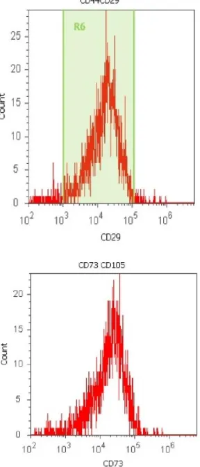
[image:2.612.136.472.339.392.2]Those cells began to proliferate at about day 10 and gradually grew to form small colonies. As growth of the cells continued, colonies gradually expanded in size with the adjacent ones inter-connected to each other. The colonies reached confluence at 15 days after initial plating, depending on the proliferating ability of each sample. Passaged MSCs behaved in a similar fashion as that of cells in primary culture. Two types of cells observed were small spindle-like and triangle-like cells. The spindle- and triangle-like MSCs were gradually transformed into broad flattened cells. The cells were finally isolated from all the six BM samples, which represented 100% harvesting efficiency. Flowcytometric analysis revealed that the MSCs obtained were positive for MSC markers namely CD29, CD44, CD73, CD90 and CD105 and negative for hematopoietic lineage markers such as CD34 and CD45 (Figures 2 and 3).
Table 1. Primer characteristics used for qRT-PCR
Gene Forward primer Reverse primer Amplicon size
NKX-2.5 GGTGGAGCTGGAGAAGACAG AGATCTTGACCTGCGTGGAC 197
GSM-2 ATTCCCTGATGAGGCAGATG CTCAGAACCCCTTTGCTCAG 290
MYH-7 TGTGTCACCGTCAACCCTTA TGGCTGCAATAACAGCAAAG 238
Figure 1a). Mesenchymal stem cells (MSCs) in culture on 10th day and b) Spindle-shaped mesenchymal stem cells with 80% confluency on15th day
[image:3.612.97.266.48.306.2]Figure 2. Flowcytometric profile of MSCs derived from bone marrow, being positive for CD29, CD44, CD73 and CD105
Figure 3. Flow cytometric characterization of mesenchymal stem cells with cell surface markers expressed positive for CD90 and negative for CD34
Cardiomyocyte Differentiation
After the induction of hMSCs to cardiomyocyte differentiation, the cells began to differentiate from third day onwards. After a week, it was observed that the cells extended their cytoplasmic processes with adjacent cells. Then, the cells aggregated and gradually increased in size to form a stick-like appearance (Figure 4), which then became enlarged and showed a number of branches in two weeks’ time. In three weeks they formed myotube-like structures by connecting with adjoining cells. Finally, they formed wide and flat sarcomeric striations in four weeks, while un-induced cells maintained their fibroblast-like morphology. qRT-PCR analysis for the expression of cardiomyocyte-specific genes showed that both induced and un-induced cells expressed NKX2.5, Desmin (GSM2), cardiac beta-myosin heavy chain (MYH-7) and cardiac troponin-T (cTnT) (Table 2).
Table 2. qRT-PCR results showing average ct value for genes up-regulated and down-regulated during cardiomyocyte differentiation
Bone- Marrow Sample
Actin
(Control) NKX2.5 GSM 2 MHY 7 cTnT
MSCs (Con) 20.05 29.28 26.06 35.59 28.54
CMG PRO 15.53 29.92 26.03 33.81 28.25
[image:3.612.109.253.351.686.2] [image:3.612.319.548.663.712.2](a)
(b)
Figure 4. a) Cells with stick-like appearance one week after induction and b) Cells forming flat sarcomeric structures four weeks after induction
DISCUSSION
In the present study, we successfully isolated fibroblastic-like mesenchymal cells from all the six human Bone-marrowspecimens that were collected and demonstrated their characteristics for MSCs in terms of their morphology, immune-phenotype and multi-lineage differentiation potentials
(Dennis and Charbord 2002; Jackson et al., 2001; Minguell
et al., 2001 and Wexler et al., 2003). Several research groups reported that MSCs were able to proliferate and potentially differentiate in vitro. The data presented above demonstrate that the ability of human MSCs to proliferate remains strong at
Passage five (P5) (Campagnoli et al., 2001 and Martin et al.,
2002). Shim et al., 2004 showed that hMSCs can be
independently differentiated towards a cardiomyogenic lineage with defined myocardial characteristics using Dulbecco’s Modified Eagle’s Medium (DMEM) supplemented with 10% FBS, insulin, transferring, sodium selenite, albumin, linoleic
acid, ascorbate phosphate and dexamethasone (Shim et al.,
2004). In the recent years, various studies report that human mesenchymal stem cells from adult bone marrow can be differentiated in to cardiac like muscle cellsby treating the cells with various agents like 5-azacytidine, laminin, oxytocin, DMSO etc., In our study, cells at passage five (P5)were treated with cardiomyocyte differentiation medium to induce cardiomyogenic differentiation. Here in this study, theoptimal quantity of cardiomyocyte specific differentiation medium (Millipore, USA) that was used to induce the differentiation process in hMSCs was 3ml and the medium was changed every three days. Three weeks after induction of differentiation process, myotube-like structures were found to be formed. The changes in morphology with the characteristic appearance of ‘stick-like’ cells might be associated with the expression of
proteins maintaining cell morphology (Makino et al., 1999;
Saito et al., 1995). The results of our study also confirmed this
characteristic feature of the differentiating and differentiated cardiomyocytes. Bone marrow cells are recently reported as able tomigrate into skeletal and cardiac muscle and then differentiate into the skeletal and cardiac muscle cells.
[image:4.612.141.467.481.681.2]Thissuggests that such a process may normally contribute
totissue maintenance or regeneration (Orlic et al., 2001; Ferrari
et al., 1998 and Barry, 2003). Thus, our results indicate that MSCs derived from adult human bone marrow could be directed towards a cardiomyogenic phenotype in vitro. In this study, ACTIN was used as a housekeeping gene (HKG) in qRT-PCR experiments with the relative quantification method. In every well, the qRT-PCR experiment measured the expression intensity of a certain gene from a sample under specific biological conditions. This measurement was expressed in Cycles to the Threshold (Ct) of PCR, a relative value that represented the cycle number at which the amount of amplified DNA reached the threshold level. The ct values recorded were for all the four significant genes expressed in cardiomyocytes. A graphical representation of the significant cardiac genes expressed in tall he MSCs, cardiomyocytes-progenitors and differentiated cardiomyocytes is given below (Graph1).
Conclusion
We have developed a model to demonstrate the differentiation potential of human bone marrow-derived mesenchymal stem cells to differentiate in to cardiomyocytes by treating the MSCs with the cardiomyocyte differentiation medium under suitable in vitro culture conditions. All those precursor MSCs,
cardiomyocytes-progenitors and the differentiated
cardiomyocytes were analysed for their expression of significant genes involved in cardiomyogenesis. And hence, our results suggest that human bone marrow-derived mesenchymal stem cells could be differentiated in to cardiomyocytes in vitro and used for the therapeutic purposes for treating various cardiovascular diseases. Further studies need to be carried out in order to understand the molecular mechanisms that regulate cardiomyocyte differentiation process.
REFERENCES
Barry FP. 2003. Mesenchymal stem cell therapy in joint
disease. Novartis Found Symp., 249:86–96.
Beyersdorf F., Doenst T., Velazquez EJ., et al. 2005. To
STICH or not to STICH: we know the answer, but do we
understand the question? J Thorac Cardiovasc Surg.,
129:246 –9.
Campagnoli C, Roberts IA, Kumar S, Bennett PR, Bellantuono I, Fisk NM. 2001. Identification of mesenchymal stem/progenitor cells in human first-trimester fetal blood,
liver, and bone marrow. Blood Oct., 15; 98 (8): 2396-402.
Castro-Malaspina H, et al. 1980. Characterization of human
bone marrow fibroblast-colony forming cells (CFU-F) 56:289-301.
Dennis JE, Charbord P. 2002. Origin and differentiation of
human and murinestroma. Stem Cells, 20:205–214.
Ferrari, G., Cusella-De Angelis, G., Coletta, M., Paolucci, E., Stornaiuolo, A., Cossu, G., Mavilio, F. 1998. Muscle
regeneration by bone marrow-derivedmyogenic
progenitors. Science, 279:1528–1530.
Jackson, KA., Majka, SM., Wang, H., Pocius, J., Hartley, CJ., Majesky, MW., Entman, ML., Michael, LH., Hirschi, KK., Goodell, MA. 2001. Regeneration of ischemic cardiac
muscle and vascular endothelium by adult stem cells. J
Clin Invest., 107:1395–1402.
Makino S., Fukuda K., Miyoshi S., Konishi F., Kodama H., Pan J., Sano M., Takahashi T., Hori S., Abe H., Hata J.,
Umezawa A., Ogawa S. 1999. Clin. Invest., 103:697–705.
Martin DR., Cox NR., Hathcock TL. et al. 2002. Isolation and
characterization of multipotential mesenchymal stem cells
from feline bone marrow. Exp. Hematol., 30: 879-886.
Minguell JJ., Erices A., Conget P. 2001. Mesenchymal stem
cells. Exp. Biol. Med., 226:507–520.
Mosca JD., Pittenger MF., Mackay AM., Beck SC., Jaiswal RK., Douglas R., Moorman MA., Simonetti DW., Craig S., Marshak DR. 1999. Multilineage potential of adult human
mesenchymal stem cells. Science, 284 (5411):143-7.
Orlic D., Kajstura J., Chimenti S., Jakoniuk I., Anderson SM., Li B., Pickel J., McKay R., Nadal-Ginard B., Bodine DM., Leri A., Anversa P. 2001. Bonemarrow cells regenerate
infracted myocardium. Nature, 410:701–705.
Ortiz-Gonzalez X.R., Reyes M. et al. 2002. Pluripotency of
mesenchymal stem cells derived from adult marrow.
Nature, 418, 41–9.
Polychronis Antonitsisa, Elisavet Ioannidou-Papagiannakib,
Aikaterini Kaidoglouc and Christos Papakonstantinoua. 2007. In vitro cardiomyogenic differentiation of adult human bone marrow mesenchymal stem cells and the role
of 5-azacytidine. Interactive CardioVascular and Thoracic
Surgery, 6, 593–597.
Rangappa S., Reddy VG., Bongso A., Lee EH., Sim EKW. 2002. Transformation of the adult human mesenchymal stem cells into cardiomyocyte- like cells in vivo.
Cardiovasc Eng., 2:7–14.
Saito T., Dennis JE., Lennon DP. 1995. Myogenic expression of mesenchymalstem cells within myotubes of mdx mice in
vitro and in vivo. Tissue Eng., 1:327–343.
Shim WS., Jiang S., Wong P., Tan J., Chua YL., Tan YS., Sin YK., Lim CH., Chua T., The M., Liu TC., Sim E. 2004. Ex vivo differentiation of human adult bone marrow stem cells
into cardiomyocyte-like cells. Bioch. Biophys. Res.
Commun., 324:481–488.
Toma C., Pittenger MF., Cahill KS., Byrne BJ., Kessler PD. 2002. Human mesenchymal stem cells differentiate to a cardiomyocyte phenotype in the adult murine heart.
Circulation, 105:93–98.
Wexler, S.A., Donaldson, C., Denning-Kendall, P., Rice, C., Bradley, B. and Hows, J.M. 2003. Adult bone marrow is a rich source of human mesenchymal ‘stem’ cells but
umbilical cord and mobilized adult blood are not. British
Journal of Haematology, 121,368–374.