Original Article
Significant hypomethylation of
TNFAIP8
and increased
expression in the placenta and peripheral blood cells
from early-onset preeclamptic patients
Liang Lin1,2, Yanhong Yu1, Zhenhai Zhang1, Yin Yang2
1Department of Obstetrics, Affiliated Southern Hospital of Southern Medical University, Guangzhou 510515, Guangdong, China; 2Provincial Clinical College of Fujian Medical University, Fuzhou 350001, Fujian, China Received November 23, 2015; Accepted February 10, 2016; Epub June 15, 2016; Published June 30, 2016
Abstract: Objective: To investigate the methylation and expression of tumor necrosis factor α-induced protein 8
(TNFAIP8) in the placenta and peripheral blood from preeclamptic patients. Study design: Placental and peripheral blood samples were obtained from women with early-onset and late-onset preeclampsia (EOPE, LOPE) and healthy pregnant women. TNFAIP8 methylation, mRNA and protein expression and its localization in the placenta were examined by pyrosequencing, quantitative RT-PCR, Western blotting, immunohistochemistry, respectively. Results:
We observed significant hypomethylation of TNFAIP8 gene in placentas of EOPE patients compared with those of
LOPE and control patients. TNFAIP8 was mainly localized in syncytiotrophoblasts and vascular endothelial cells of
placenta. Moreover, TNFAIP8 mRNA and protein expression in placenta and peripheral blood cells significantly de
-creased following the order of EOPE→LOPE→control patients. Conclusions: Significant hypomethylation and high
expression of TNFAIP8 in the placenta and peripheral blood cells were observed in EOPE, suggesting TNFAIP8 may be associated with the pathogenesis of preeclampsia.
Keywords: Preeclampsia, methylation, TNFAIP8, placenta, peripheral blood
Introduction
Preeclampsia (PE) is a critical pregnancy-relat-ed disease that is clinically characterizpregnancy-relat-ed by hypertension and proteinuria [1]. PE affects 3-5% of all pregnancies and is the leading cause for maternal mortality, morbidity, perina-tal death, preterm birth, and intrauterine growth restriction [2, 3]. Pathological changes in the placenta with PE mainly include vascular lesions in the decidua, infarction, placental abruption, villus dysplasia, and incremental syncytial knots [1, 4]. Increasing evidence has suggested that PE results from placental dys-function during the first trimester, which is pri-marily caused by the shallow invasion of extravillous trophoblast cells and impaired vas-cular development in the placental bed [5]. All PE-associated symptoms resolve after the delivery of the placenta. PE has been classified into two types: early-onset (<34 weeks) and late-onset (>34 weeks). Early-onset PE (EOPE) is a distinct and more severe clinical entity
compared with late-onset PE (LOPE). High pro-teinuria is the most frequent symptom in EOPE [6]. It has been reported that placental lesions associated with maternal underperfusion occur more frequently in EOPE than in LOPE [7]. Moreover, these two types of PEs have been proposed to have different pathophysiological mechanisms and significant differences in pla-cental findings, but the differences at the molecular level are still not well clarified.
DNA methylation is the most commonly studied epigenetic process, which is primarily observed in CpG dinucleotides that are underrepresent-ed in the genome at low density, but are enriched in the promoter regions of genes [8, 9]. Elevated DNA methylation in one particular gene is generally associated with gene inactivi-ty, whereas a lower level of DNA methylation indicates the potential for gene expression [9]. Aberrant gene expression in the placenta has been linked to PE [10]. Tumor necrosis factor
SCC-S2, GG2-1, and MDC-3.13) is a 21-kDa cytosolic protein that is induced by TNF-αand transcription factor nuclear factor-κB activation [11]. TNFAIP8 primarily functions as an onco-genic and antiapoptotic molecule by enhancing cell survival and inhibiting the activity of apop-totic proteins caspase 3 and caspase 8 [12, 13]. Previous studies have demonstrated that TNFAIP8 is overexpressed in various tumors and is correlated with chemotherapy resistance in tumors and its clinical outcomes [14, 15]. Moreover, Liu et al. reported that TNFAIP8 overexpression is positively correlated to high-er histological grade, deep myometrial inva-sion, lymphovascular space invainva-sion, lymph node metastasis, and recurrence in endome-trial cancer [16]. Because the occurrence of PE is closely associated with the shallow invasion of extravillous trophoblast cells into the endo-metrium, we assume that TNFAIP8 may play a specific role in the pathogenesis of PE. However, the potential roles of TNFAIP8 and its methyla-tion in the placenta and placenta-related dis-eases like PE have never been investigated. Therefore, in the present study, we aimed to investigate the methylation of TNFAIP8 in the placenta and examine TNFAIP8 expression in the placenta and peripheral blood of patients with EOPE and LOPE.
Materials and methods
Patient enrollment and sample collection
A total of 64 pregnant women who underwent labor delivery at Fujian Provincial Hospital between March 2013 and August 2014 were recruited for this study. They included 23 preg-nant women with EOPE (<34 weeks, mean age 29.39±5.69 years), 16 pregnant women with LOPE (≥34 weeks, mean age 28.00±6.55 years), and 25 healthy pregnant women who delivered at full term (control, mean age 28.32±4.55 years). The diagnosis of PE was made according to previously described criteria [17]. The exclusion criteria for pregnant women included: (1) stillbirth and fetal malformation; (2) kidney disease, diabetes, immune disease,
cancer, epilepsy, and other obstetrical compli-cations; (3) multiple pregnancies (≥3 fetuses); and (4) alcohol and drug addiction. This study was approved by the local ethics committee of Fujian Provincial Hospital, and written consent was obtained from all recruited participants. Peripheral blood samples (3 ml × 2 tubes) was collected into tubes with EDTA (e thylenediami-netetraacetic acid) from all PE patients and control participants before labor delivery and stored at -80°C. The blood cells were collected by centrifugation and used for RNA and protein extraction. Four placental tissue samples of 1.0 × 1.0 × 1.0 cm3 were collected from
differ-ent locations of every placdiffer-enta, washed with phosphate-buffered saline (PBS), and frozen at -80°C.
DNA and total RNA isolation
DNA isolation from placental issues was car-ried out using a DNA Extraction Kit (Qiagen, Cat No. 59124, Valencia, CA). The DNA concentra-tion was measured using a NanoDrop 1000 Spectrophotometer (Thermo Scientific, Tewks- bury, MA), and its quality was examined on an agarose gel. Total RNA from placental tissues and blood cells was isolated using the RNeasy Mini Kit (CWbio Co. Ltd., Cat No. CW0581, Beijing, China). The RNA quality was assessed by electrophoresis on a 1% denaturing agarose gel.
Evaluation of TNFAIP8 methylation by bisulfite pyrosequencing
DNA methylation of TNFAIP8 was confirmed by bisulfite pyrosequencing as previously descri- bed [18]. Methylation-unbiased pyrosequenc-ing primers (Table 1) were designed by Pyro- Mark Assay Design 2.0 to include the same CpG sites targeted by the Illumina probes. A total of 1-2 μg DNA was transformed using the EpiTect Plus DNA Bisulfite Kit (Qiagen) and used as a template for PCR. The assay was run on a Qiagen PyroMark Q24 system (Qiagen) follow-ing the manufacturer’s instructions. The quanti-tative levels of TNFAIP8 methylation for the CpG dinucleotide were evaluated using the Pyro Q-CpG software (Biotage, Uppsala, Swe- den).
Immunohistochemistry
[image:2.612.91.286.96.152.2]Placental samples were fixed in formalin and embedded in paraffin. Sections (4 m) were Table 1. Sequences of primers for
pyrose-quencing
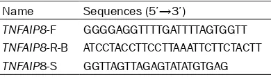
Name Sequences (5’→3’)
TNFAIP8-F GGGGAGGTTTTGATTTTAGTGGTT
TNFAIP8-R-B ATCCTACCTTCCTTAAATTCTTCTACTT
deparaffinized, rehydrated, and subjected to antigen retrieval at 95°C for 10 min in 0.05 mol/L Tris-EDTA solution (pH 6.0). Then sec-tions were treated with 4% hydrogen peroxide for 10 min in the dark at room temperature, blocked in normal goat serum, and then incu-bated with rabbit antihuman TNFAIP8 poly-clonal antibody (1:100 dilution, Bioss, China) at 37°C for 2 h. The sections were incubated with secondary antibody (goat anti-rabbit 1:1,000; Beijing Zhongshan Biotechnology) for 20 min at 37°C after three washes in PBS. The sections were counterstained with hematoxylin and mounted with a cover glass. The positive and negative images were captured using a BX51T-PHD-J11 microscope (Olympus Corp., Tokyo, Japan).
Quantitative reverse transcription polymerase chain reaction (RT-PCR)
The cDNA synthesis was conducted using HiFi-MMLV cDNA kit (CWbio. Co. Ltd., Cat. No. CW- 0744). The primer sequences for quantitative PCR (qPCR) are listed in Table 2. qPCR was car-ried out with an ABI prism 7500 Sequence Detection System (Applied Biosystems, Foster City, CA). After heating at 95°C for 5 min, the samples were run for 45-48 cycles at 94°C for 30 sec, 56°C for 30 sec, and 72°C for 30 sec, and then incubated at 72°C for 10 min. The relative quantification of gene expression was calculated using the 2-ΔΔCT method and using
β-actin gene expression as an endogenous control.
Western blotting
The placenta tissues were homogenized, and blood cells were lysed in lysis buffer containing 20 mM Tris (pH 7.5), 150 mM NaCL, 1% NP-40, 10 mM EDTA, 1 mM phenylmethane sulfonyl fluoride, 50 mM NaF, 40 mM β-glycerophosphate, 200 μM sodium orthovanadate, 40 mM benza-midine, and protease inhibitor cocktail (EMD, Gibbstown, NJ) on ice for 20 min. The lysates were collected and centrifuged at 12,000×g for 10 min at 4°C. The concentration of protein in
lysates was detected using the Pierce BCA Protein Assay Kit (Life Technologies, Grand Island, NY). Aliquots of 30 μg protein were re- solved by sodium dodecyl sulfate (SDS)-pol- yacrylamide gel electrophoresis (PAGE) and transferred onto nitrocellulose membranes (Bio-Rad, Hercules, CA). The membranes were blocked with Tris-buffered saline (TBS) contain-ing 0.05% Tween-20 (TBST) and 5% skim milk powder. The membranes were then incubated with antibodies to TNFAIP8 (1:100 dilution, Bioss) and rabbit polyclonal β-actin (loading control, 1:100 dilution, Beijing Zhongshan Bio- technology) with gentle shaking at room tem-perature for 1 h. After a few washes with TBST, the membranes were incubated with second-ary antibody for 1 h, washed, and then devel-oped using an ECL Western Blotting Detection System (Amersham Biosciences, Buckingha- mshire, England). The intensities of the bands were scanned using ImageJ software (National Institutes of Health, Bethesda, MD).
Statistical analysis
The data are presented as mean ± standard deviation (SD) and were analyzed using SPSS 19.0 software (SPSS, Inc., Chicago, IL). The data among three groups with normal distribu-tion were analyzed by single factor analysis of variance (ANOVA) and using the least signifi-cant difference (LSD) method. Linear regres-sion was used to evaluate the correlation between TNFAIP8 methylation in the placenta and mean arterial pressure (MAP) and fetal birth weight. A P value <0.05 was considered statistically significant.
Results
General characteristics of participants
[image:3.612.91.288.83.152.2]The general characteristics of the participants are presented in Supplementary Table 1. No significant differences were noted in maternal age or body mass index at early pregnancy among the three groups. The urine protein con-centration at 24 h did not differ significantly between EOPE and LOPE patients (P>0.05). Gestational age at delivery and fetal birth weight in the EOPE group were significantly lower than those in the LOPE and control groups (P<0.05). The MAP at delivery in PE patients was significantly higher than that in control par-ticipants (P<0.05). Twenty-one cases (91%) in the EOPE group and four cases (25%) in LOPE group delivered preterm.
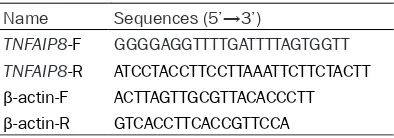
Table 2. Primer sequences for qPCR Name Sequences (5’→3’)
TNFAIP8-F GGGGAGGTTTTGATTTTAGTGGTT TNFAIP8-R ATCCTACCTTCCTTAAATTCTTCTACTT
β-actin-F ACTTAGTTGCGTTACACCCTT
TNFAIP8 methylation in the placentas of pa-tients with PE
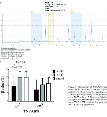
TNFAIP8 methylation was examined by bisulfite pyrosequencing, because Δβ was approximate-ly 14% when the EOPE group was compared with the LOPE and control groups. Two Illumina CpG sites in the 5’ untranslated region (UTR) and 1st exon of TNFAIP8 were selected to
ensure a comprehensive assessment of the gene region. The sequence analyzed was YGGTA- ATYGTTTTTGTAGTTGGTTAT. Moreover, two sig-nificant methylation sites in TNFAIP8 were observed in the placenta (Figure 1A). The methylation of site 1 was not detected (value
[image:4.612.93.520.67.532.2]=0) in four specimens in the EOPE group (17.4%), but was detected in all specimens from the LOPE and control groups. Likewise, no methylation of site 2 was found in five patients in EOPE group (21.7%), but only in one patient in each of the LOPE and control groups. We fur-ther compared the methylation percentage of the two sites in TNFAIP8 among the three groups. We found the methylation percentage of site 1 in the EOPE group was significantly lower than that in the LOPE and control groups (P<0.05), but there was no significant differ-ence in the methylation of site 1 between the LOPE and control groups (P>0.05, Figure 1B). Surprisingly, we did not observe a significant
difference in the methylation percentage of site 2 among the three groups (P>0.05, Figure 1B). These data suggest that the degree of
TNFAIP8 methylation in the placentas of EOPE patients is significantly less than that in placen-tas of LOPE and control patients.
TNFAIP8 mRNA expression in placenta and peripheral blood cells of patients with PE
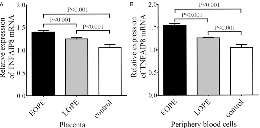
To assess the effect of TNFAIP methylation on its mRNA expression, the TNFAIP8 mRNA ex- pression in the placenta and peripheral blood samples from the three groups was examined by qRT-PCR. We observed that TNFAIP8 mRNA expression in the placenta of EOPE patients was significantly higher than that in the LOPE and control groups (P<0.01, Figure 2A). Also, TNFAIP8 mRNA expression in the placenta of LOPE patients was significantly higher than that in control participants (P<0.01, Figure 2A). Interestingly, we also examined the TNFAIP8 mRNA expression in the peripheral blood sam-ples in the three groups and found that its expression pattern in the peripheral blood cells was completely consistent with that in the pla-centa for the three groups. That is, TNFAIP8 mRNA expression in the peripheral blood cells from EOPE patients was significantly higher than that in LOPE and control patients (P<0.01, Figure 2B).
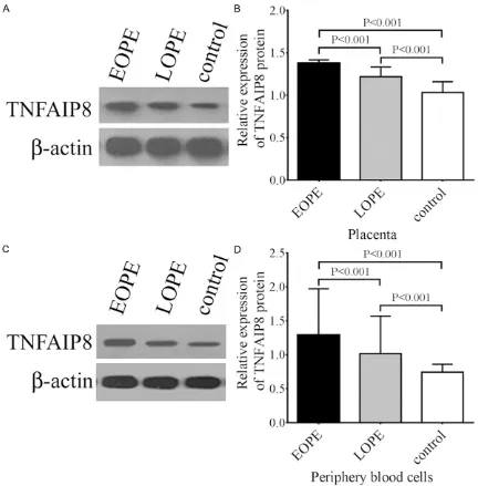
TNFAIP8 protein expression in placenta and peripheral blood cells of patients with PE
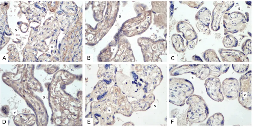
To further confirm the results of TNFAIP8 mRNA expression, we examined the protein expres-sion of TNFAIP8 in the placenta and peripheral blood cells by Western blotting. Similar to the trend in mRNA expression, TNFAIP8 protein expression in the placenta of EOPE patients was significantly higher than that in the LOPE and control groups (P<0.01, Figure 3A). Mor- eover, TNFAIP8 protein expression in the pla-centa of LOPE patients was significantly higher than that in control participants (P<0.01, Figure 3A). In the meantime, we also observed that TNFAIP8 protein expression in the peripheral blood cells decreased significantly according to the following order EOPE→LOPE→control pati- ents (P<0.01, Figure 3B).Immunohistochemical staining showed that positive TNFAIP8 expres-sion was mainly localized in the syncytiotropho-blasts and vascular endothelial cells of the pla-centa in all three groups, and the staining patterns also indicated that TNFAIP8 expres-sion gradually decreased following the trend of EOPE→LOPE→control patients (Figure 4A-F).
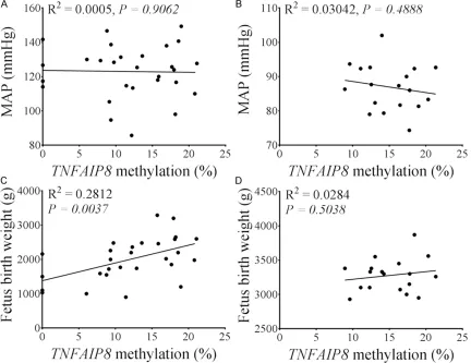
Associations between TNFAIP8 methylation in the placenta and clinical parameters
[image:5.612.93.522.71.283.2]Because we observed significant differences in MAP and fetal birth weight and TNFAIP8 meth-ylation (site 1) was significant lower in EOPE
Figure 2. TNFAIP8 mRNA expression in the placentas (A) and peripheral blood cells (B) from EOPE, LOPE, and control patients. The data are presented as mean ± SD (n=23 for EOPE, n=16 for LOPE, and n=25 for controls). The relative expression of TNFAIP8 mRNA was determined according to the ratio of TNFAIP8 mRNA expression to endogenous
patients, we sought to examine whether there is an association between TNFAIP8 methyla-tion in the placenta and fetal birth weight and MAP. Although the MAP in PE patients (includ-ing EOPE and LOPE) was significantly higher than that in control patients, we did not find any correlation between TNFAIP8 methylation in the placenta and MAP in both PE patients and control patients (Figure 5A and 5B). However, we observed a significant positive correlation
between TNFAIP8 methylation in the placenta and fetal birth weight in PE patients (including EOPE and LOPE; P<0.01, Figure 5C), and this correlation was not significant in control patients (P>0.05, Figure 5D) and the individual EOPE and LOPE groups (data not shown). Discussion
[image:6.612.90.522.70.509.2]Placental function is highly associated with fetal development and health, and placental
Figure 3. TNFAIP8 protein expression in the placentas and peripheral blood cells from EOPE, LOPE, and control patients. A. Representative image for TNFAIP8 protein expression in the placentas from EOPE, LOPE, and control patients. B. Statistical analysis of TNFAIP8 protein expression in the placentas. C. Representative image for TNFAIP8 protein expression in the peripheral blood cells from EOPE, LOPE, and control patients. D. Statistical analysis of TN-FAIP8 protein expression in the peripheral blood cells. The data are presented as mean ± SD (n=23 for EOPE, n=16 for LOPE, and n=25 for controls). The relative expression of TNFAIP8 protein was determined according to the ratio
dysfunction, particularly aberrant placental gene expression, has been linked to many preg-nancy-associated diseases including PE, intra-uterine growth restriction, gestational diabetes mellitus, and gestational trophoblastic disease [10, 19]. Placenta-specific methylation pat-terns have been demonstrated in the placenta and maternal plasma of PE patients. Previous studies have demonstrated that gene expres-sion is altered in pregnancies with PE and PE is associated with altered DNA methylation [18, 20]. Given our limited understanding of the epi-genetic alterations in PE, it is necessary to ver-ify the reported associations between DNA methylation and PE in further studies. There- fore, in this study, we examined TNFAIP8 meth-ylation in the placenta and its expression in the placenta and peripheral blood from EOPE, LOPE, and control patients. We found a lower level of TNFAIP8 methylation in the placenta from LOPE patients but higher expression of TNFAIP8 mRNA and protein in the placenta and peripheral blood from EOPE patients than in those from LOPE and control patients.
Because DNA methylation profiling in pre-eclamptic placentas has been reported, some researchers initiated studies of the methylation of specific genes or a group of genes in pre-eclamptic placentas and their functions in the pathogenesis of PE, especially EOPE. Xiang et
al. reported that promoter hypomethylation of
TIMP3 is associated with PE [21]. Moreover, hypomethylation of multiple genes such as GATA zinc finger domain containing 1 (GATAD1), [22] LEP, [23] and matrix metalloproteinase 9
[24] have been identified in preeclamptic pla-centas. In addition, hypermethylation of some genes such as SH3PXD2A [23] and human endogenous retrovirus (HERV) [25] is also observed in preeclamptic placentas. Here we report significantly greater hypomethylation of
TNFAIP8 in the placentas of EOPE patients compared with that in LOPE and control patients, although the significant difference was only observed in one of two methylated sites in TNFAIP8. These data implicate that hypomethylation of TNFAIP8 in the placentas of EOPE patients may influence its expression and regulate placental function during the patho-genesis of EOPE.
The hypomethylation of genes is associated with the activation of gene expression, and thus, it is reasonable to speculate that hypo-methylation of TNFAIP8 in the placentas of EOPE patients may lead to its high expression. Indeed, we observed that both mRNA and pro-tein expression of TNFAIP8 was significantly up-regulated in the placentas of EOPE patients compared with LOPE and control patients. Interestingly, we also found that the expression
of TNFAIP8 mRNA and protein in the placentas of LOPE patients was significantly higher than that in control participants, although no signifi-cant difference in TNFAIP8 methylation was observed between the two groups. These data suggest that TNFAIP8 may be associated with the severity of PE. It has been found that TNFAIP8 is highly related to the growth, inva-sion, metastasis, and clinical prognosis of some cancers, [12-16] but to the best of our knowledge, this is the first report to investigate the function of TNFAIP8 in the placenta and a placenta-associated disease like PE. Recent work has demonstrated that the production of TNF-α and other inflammatory cytokines is ele-vated in women with pE, [26] and TNF-α block-ade can partially attenuate the associated hypertension in the reduced uterine perfusion pressure (RUPP) animal model of PE [27].
These findings can help to explain the high expression of TNFAIP8 in the placentas of EOPE patients and its potential role in the pathogen-esis of PE. Moreover, PE is closely associated with defective trophoblast cell invasion, endo-thelial cell dysfunction, and dysregulated utero-placental vascularization [28]. It has been sug-gested that aberrant placental expression of matrix metalloproteinases (MMPs) may cause shallow cytotrophoblastic invasion and incom-plete remodeling of the spiral arteries, which is thought to link placental ischemia to the cardio-vascular alterations of PE [29]. Antisense inhi-bition of endogenous TNFAIP8 expression results in decreased expression of vascular endothelial growth factor (VEGF) receptor-2, MMP-1, and MMP-9 in tumor cells [30]. Here we observed that TNFAIP8 was predominantly present in the syncytiotrophoblasts and
[image:8.612.92.524.75.408.2]lar endothelial cells of the placenta. This evi-dence suggests that TNFAIP8 may cause aber-rant invasion of placenta villi via mediating the expression of MMPs. Additionally, consistent with the TNFAIP8 expression pattern in the pla-centa, we also observed higher mRNA and pro-tein expression of TNFAIP8 in the peripheral blood of EOPE patients compared with LOPE and control patients, which is consistent with the significantly elevated serum TNF-α concen-tration in women with severe PE [31]. High expression of TNFAIP8 in the peripheral blood cells of EOPE patients indicates that TNFAIP8 may be used as a potential biomarker for the diagnosis of EOPE. More importantly, we observed that the level of TNFAIP8 methylation in the placentas of PE patients was significantly correlated with fetal birth weight, a correlation that was not obvious in control patients. This evidence indicates that TNFAIP8 may be an important factor that affects placental function and fetal growth during the progression of PE, although this finding requires further investi- gation.
In conclusion, we observed significant hypo-methylation of TNFAIP8 in the placenta and increased expression of TNFAIP8 in the placen-ta and peripheral blood cells in EOPE patients. The level of TNFAIP8 methylation in the placen-ta was significantly correlated with feplacen-tal birth weight in PE patients. These findings demon-strate that TNFAIP8 may be a critical factor in the pathogenesis of PE.
Acknowledgements
This study was supported by the National Natural Science Foundation of China (No. 81270716). We are grateful to the medical staff of the Obstetrics Department of Fujian Provincial Hospital for sampling and to all par-ticipants who donated their placentas and blood samples.
Disclosure of conflict of interest
None.
Address correspondence to: Dr. Yanhong Yu, De-
partment of Obstetrics, Affiliated Southern Hospital
of Southern Medical University, 1838 North Guang- zhou Avenue, Guangzhou 510515, Guangdong, China. Tel: +86 61648687; Fax: +86 020-62787562; E-mail: yuyh1010@hotmail.com
References
[1] Sibai B, Dekker G and Kupferminc M. Pre-ec-lampsia. Lancet 2005; 365: 785-799. [2] Huppertz B. Placental origins of preeclampsia:
challenging the current hypothesis. Hyperten-sion 2008; 51: 970-975.
[3] Roberts JM and Escudero C. The placenta in preeclampsia. Pregnancy Hypertens 2012; 2: 72-83.
[4] Roberts DJ and Post MD. The placenta in pre-eclampsia and intrauterine growth restriction. J Clin Pathol 2008; 61: 1254-1260.
[5] Boulanger H and Flamant M. [New insights in the pathophysiology of preeclampsia and po-tential therapeutic implications]. Nephrol Ther 2007; 3: 437-448.
[6] Kucukgoz Gulec U, Ozgunen FT, Buyukkurt S, Guzel AB, Urunsak IF, Demir SC and Evruke IC.
Comparison of clinical and laboratory findings
in early- and late-onset preeclampsia. J Matern Fetal Neonatal Med 2013; 26: 1228-1233. [7] Ogge G, Chaiworapongsa T, Romero R, Hussein
Y, Kusanovic JP, Yeo L, Kim CJ and Hassan SS. Placental lesions associated with maternal un-derperfusion are more frequent in early-onset than in late-onset preeclampsia. J Perinat Med 2011; 39: 641-652.
[8] Bird A. DNA methylation patterns and epigen-etic memory. Genes Dev 2002; 16: 6-21. [9] Bird AP. CpG-rich islands and the function of
DNA methylation. Nature 1986; 321: 209-213. [10] Hoegh AM, Borup R, Nielsen FC, Sorensen S
and Hviid TV. Gene expression profiling of pla -centas affected by pre-eclampsia. J Biomed Biotechnol 2010; 2010: 787545.
[11] Liu L, Zhang X, Rong C, Rui C, Ji H, Qian YJ, Jia R and Sun L. Distinct DNA methylomes of hu-man placentas between pre-eclampsia and gestational diabetes mellitus. Cell Physiol Bio-chem 2014; 34: 1877-1889.
[12] Kumar D, Gokhale P, Broustas C, Chakravarty D, Ahmad I and Kasid U. Expression of SCC-S2, an antiapoptotic molecule, correlates with en-hanced proliferation and tumorigenicity of MDA-MB 435 cells. Oncogene 2004; 23: 612-616.
[13] You Z, Ouyang H, Lopatin D, Polver PJ and Wang CY. Nuclear factor-kappa B-inducible death effector domain-containing protein sup-presses tumor necrosis factor-mediated apop-tosis by inhibiting caspase-8 activity. J Biol Chem 2001; 276: 26398-26404.
[14] Miao Z, Zhao T, Wang Z, Xu Y, Song Y, Wu J and Xu H. SCC-S2 is overexpressed in colon can-cers and regulates cell proliferation. Tumour Biol 2012; 33: 2099-2106.
and Kuwano H. TNFAIP8 overexpression: clini-cal relevance to esophageal squamous cell carcinoma. Ann Surg Oncol 2012; 19 Suppl 3: S589-596.
[16] Liu T, Gao H, Yang M, Zhao T, Liu Y and Lou G. Correlation of TNFAIP8 overexpression with the proliferation, metastasis, and disease-free sur-vival in endometrial cancer. Tumour Biol 2014; 35: 5805-5814.
[17] Faas MM, Spaans F and De Vos P. Monocytes and macrophages in pregnancy and pre-ec-lampsia. Front Immunol 2014; 5: 298.
[18] Yuen RK, Penaherrera MS, von Dadelszen P, McFadden DE and Robinson WP. DNA
methyla-tion profiling of human placentas reveals pro -moter hypomethylation of multiple genes in early-onset preeclampsia. Eur J Hum Genet 2010; 18: 1006-1012.
[19] Dieber-Rotheneder M, Beganovic S, Desoye G, Lang U and Cervar-Zivkovic M. Complex ex-pression changes of the placental endothelin system in early and late onset preeclampsia, fetal growth restriction and gestational diabe-tes. Life Sci 2012; 91: 710-715.
[20] Blair JD, Yuen RK, Lim BK, McFadden DE, von Dadelszen P and Robinson WP. Widespread DNA hypomethylation at gene enhancer re-gions in placentas associated with early-onset pre-eclampsia. Mol Hum Reprod 2013; 19: 697-708.
[21] Xiang Y, Zhang X, Li Q, Xu J, Zhou X, Wang T, Xing Q, Liu Y, Wang L, He L and Zhao X. Pro-moter hypomethylation of TIMP3 is associated with pre-eclampsia in a Chinese population. Mol Hum Reprod 2013; 19: 153-159.
[22] Geng J, Hu T, Wang B, Lu W and Ma S. Thyroid stimulating hormone levels and risk of coro-nary heart disease in patients with type 2 dia-betes mellitus. Int J Cardiol 2014; 174: 851-853.
[23] Xiang Y, Cheng Y, Li X, Li Q, Xu J, Zhang J, Liu Y, Xing Q, Wang L, He L and Zhao X. Up-regulated expression and aberrant DNA methylation of LEP and SH3PXD2A in pre-eclampsia. PLoS One 2013; 8: e59753.
[24] Wang Z, Lu S, Liu C, Zhao B, Pei K, Tian L and Ma X. Expressional and epigenetic alterations of placental matrix metalloproteinase 9 in pre-eclampsia. Gynecol Endocrinol 2010; 26: 96-102.
[25] Zhuang XW, Li J, Brost BC, Xia XY, Chen HB, Wang CX and Jiang SW. Decreased expression and altered methylation of syncytin-1 gene in human placentas associated with preeclamp-sia. Curr Pharm Des 2014; 20: 1796-1802. [26] Germain SJ, Sacks GP, Sooranna SR, Sargent
IL and Redman CW. Systemic inflammatory
priming in normal pregnancy and preeclamp-sia: the role of circulating syncytiotrophoblast microparticles. J Immunol 2007; 178: 5949-5956.
[27] LaMarca BD, Gilbert J and Granger JP. Recent progress toward the understanding of the pathophysiology of hypertension during pre-eclampsia. Hypertension 2008; 51: 982-988. [28] Escudero C, Celis C, Saez T, San Martin S,
Va-lenzuela FJ, Aguayo C, Bertoglia P, Roberts JM and Acurio J. Increased placental angiogenesis in late and early onset pre-eclampsia is associ-ated with differential activation of vascular en-dothelial growth factor receptor 2. Placenta 2014; 35: 207-215.
[29] Palei AC, Granger JP and Tanus-Santos JE. Ma-trix metalloproteinases as drug targets in pre-eclampsia. Curr Drug Targets 2013; 14: 325-334.
[30] Zhang C, Chakravarty D, Sakabe I, Mewani RR, Boudreau HE, Kumar D, Ahmad I and Kasid UN. Role of SCC-S2 in experimental metastasis and modulation of VEGFR-2, MMP-1, and MMP-9 expression. Mol Ther 2006; 13: 947-955.
Supplementary Table 1. Clinical characteristics of study participants
EOPE group (n=23) LOPE group (n=16) Control group (n=25)
Maternal age (years) 29.826±5.523 28.000±6.552 28.320±4.553
Gravidity, median (range) 2.522 (1-4) 1.938 (1-4) 1.840 (1-4)
Primiparity, n (%) 12 (52.174) 12 (75) 15 (60)
Early pregnancy BMI (kg/m2) 21.550±3.585 21.472±2.483 21.604±3.450
Gestational age at delivery (weeks) 33.161±2.727A,B 38.063±1.534 39.360±0.732
Mean arterial pressure (mmHg) 124.014±12.938A,B 119.125±14.246C 87.733±9.272
24 h urine protein (g/L) 5.310±4.430 2.913±2.259
-Birth weight (g) 1606.520±611.694A,B 2740.000±565.108C 3324.000±287.141 Data are presented as mean ± standard error of the mean (SEM). AEOPE group vs. LOPE group, BEOPE group vs. control group