Original Article
Regulatory effect of mangiferin on kidney function
and in serum uric acid through hyperuricemia
model mice experiment
Zhen-Kun Li1,2, Yan-Fen Niu1, Li-Hui Gao1, Xiang-Wei Xu1, Hua Lin1, Xu Liu1, Ling Li1
1Biomedical Engineering Research Center, Kunming Medical University, Kunming 650500, China; 2The Second
Affiliated Hospital of Kunming Medical University, Kunming, Yunnan, China
Received September 4, 2016; Accepted February 15, 2017; Epub April 15, 2017; Published April 30, 2017
Abstract: Objective: It analyze the effect of magiferin on uric acid metabolism and interference on the urate re-absorption and delivery in vivo. It further study the role of three effector molecules, glucose transporter 9 (GLUT9), organic anion transporter 10 (OAT10) and urate anion exchanger 1 (URAT1) in these progress; Methods: An animal model was established by once a day intraperitoneal injection of uric acid with dose of 300 mg·kg-1·d-1 for 14 days, which suffer from hyperuricemia in long time. Meanwhile, it used benzbromarone as positive control, and normal saline as negative control. The expression of GLUT9, OAT10, and URAT1 was detected by RT-PCR and Western blot,
respectively in gene and protein expression level; Results: Magiferin could significantly reduce the level of serum uric acid on model animals, which treated with mangiferin with dose of ≥ 3.0 mg·kg-1·d-1. But the benzbromarone almost needed more than dose of 25 mg·kg-1·d-1 to get the same effect in the parallel experiment. Furthermore, mangiferin gastric administration could inhibit the expression of GLUT9, but benzbromarone couldn’t; on the other hand, mangiferin did not interfere the expression of URAT1 and OAT10; Conclusion: Mangiferin could reduce GLUT9 expression both at gene and protein level on the hyperuricemic model mice, which result the reduction of re-absorp-tion and delivery of urate.
Keywords: Mangiferin, hyperuricemia, GLUT9, animal model
Introduction
Uric acid is a product of the metabolic break-down of purine nucleotides. High blood concen-trations of uric acid can lead to gout, which associated with other disease including diabe-tes and the formation of ammonium acid urate kidney stones [1].
Hyperuricemia is an abnormally high level of uric acid in the blood. In the low pH conditions, uric acid exists largely as urate, as ion form [1, 2]. The amount of urate in the body depends on the changes of the amount of purines eaten, the amount of urate synthesized, and the amount of urate excretion [2]. In humans, the upper limit tolerance of the normal range is 360 µmol/L (6 mg/dL) for women and 400 µmol/L (6.8 mg/dL) for men [3].
Many factors contribute to hyperuricemia, in- cluding: genetics, insulin resistance,
hyperten-sity, diet, usage of diuretics, and alcoholic drinking [4, 5]. Causes of hyperuricemia can be
classified into three types: [6] increased pro -duction of uric acid, decreased excretion of uric acid, and mixed type. Causes of increased pro-duction include high levels of purine in the diet and increased purine metabolism. Causes of decreased excretion include kidney disease, certain drugs, and competition for excretion between uric acid and other molecules. There are two contributing factors as uric acid excre-tion disorder and re-absorpexcre-tion of urate in the renal tubule for the mixed type [6]. It is a bal-ance between them to maintain the uric acid at normal levels. Once that balance was broken, it caused hyperuricemia or other disease.
cose homeostasis. SLC2A9 has also recently been found in transporting uric acid process, and genetic variants of the transporter, which link to increasing risk of development of hyper-uricemia, gout and Alzheimer’s disease.
Urate anion exchanger 1 (URAT1), encoded by the SLC22A12 gene, is a urate transporter and urate-anion exchanger which regulates the le- vel of urate in the blood. This protein is an inte-gral membrane protein primarily found in kid-ney tissue. Two transcript variants encoding dif-ferent forms have been found for this gene, which altered re-absorption of uric acid [3, 4]. It expression change contributed to hyperurice- mia.
An organic anion-transporting polypeptide (OA- TP) are present in the lipid bilayer of the cell membrane, acting as the cell’s gatekeepers. The organic anion transporter 10 (OAT10) has relationship with the uric acid metabolism. According to Le Chatelier’s principle, lower con-centration of uric acid would interfere crystals of uric acid existing in the blood, if the dissolved uric acid can be excreted opportune. In con-trast, the high concentration of uric acid crys-tals would accumulate in the joints and other tissues.
Medications most often used to treat hyperuri-cemia are of two kinds: xanthine oxidase tors and uricosurics. Xanthine oxidase inhibi-tors decrease the production of uric acid, by interfering xanthine oxidase. Uricosurics increa- se the excretion of uric acid, by reducing the re-absorption of uric acid once the kidneys have
filtered it out of the blood. Some of these medi -cations are used as indicated. Several other kinds of medications have potential usage in treating hyperuricemia. Mangiferin is a xan-thonoid molecule which is a glucoside of
nora-thyriol, finding in mangoes. Laboratory study has identified a variety of potential pharmacol -ogy associated with mangiferin, including anti-microbial and antioxidant activities, blocking
the type II 5α-reductase in the body.
It has variety of potential effect, including scav-enging oxides [9], reducing blood sugar levels [10], improving renal function [11], hepatopro-tective action [12] and inhibits tumor [13]. However, whether mangiferin could decrease the level of uric acid in the blood by promoting
uric acid excretion has not been established
[14]. In this study, firstly, it constructed a hyper -uricemia animal model by injection of large doses of uric acid. Then studied the effect of mangiferin on model animals. Furthermore, three genes (GLUT9, URAT1 and OAT10) as described above, related uric acid metabolism, were detected its expression level, and ana-lyzed its relationship with intervention effect of
mangiferin. All these findings might be useful
for understanding the molecular mechanism of mangiferin effect clearly.
Materials and methods
Reagents
Uric acid was purchased from Sigma (St. Louis, MO, USA); mangiferin was extracted from the Biomedical Engineering Research Center, Kun- ming Medical University (Kunming, Yunnan, China); Benzbromarone was purchased from Herman Big Pharma (Germany).
Animal model experiment
70 Kunming strain male mice, with weights ranging from 18-22 g, were purchased from Sichuan Chengdu Biological Technology Co.,
Ltd. (Certificate No. SCXK2013-24, Chengdu,
China). Animal model construction programs were carried out by intraperitoneal injection of mice with uric acid (pH 8.0) twice everyday at the dose of 150 mg·kg-1 for 14 days.
The intervention programs were as following: all the male mice (n = 70) were randomly divided into three groups, as negative group treated with phosphate buffer saline (PBS, pH 8.0), positive group treated with benzbromarone (dose of 25 mg·kg-1·d-1) and experiment group with four ladder concentration gradients. Those different concentrations were with 1.5 mg·kg -1·d-1, 3.0 mg·kg-1·d-1, 6.0 mg·kg-1·d-1 and 100.0 mg·kg-1·d-1. In addition, it took 0.5% sodium car-boxymethyl cellulose (CMC-Na) once a day for 14 days, in both experimental group and posi-tive group. All animals were under intragastric administration.
Tested uric acid concentration in serum and liver
then their blood and liver were collected to test the uric acid concentration by the phospho-tungstic acid method [15]. A reagent kit using reduction phosphotungstic acid was employed to determine the serum uric acid levels accord-ing to the Carroll’s method [15]. As for testaccord-ing for liver, it used 100 mg of homogenized frozen liver tissue samples. Additional, the 9-fold dilu-tion was necessary, by double distilled water. Used the reduction phosphotungstic acid kit for this experiment, according to instruction manual.
Pathological examination of renal tissue
After mice dissection, it separated the renal
tis-sue and fixed them in neutral formaldehyde
solution (40 g/L) for 24 h. Then those samples were taken hematoxylin and eosin (HE) staining in Department of Pathology of Basic Medical College, Kunming Medical University. This De- partment taken photograph and made a patho-logical diagnosis for these samples.
Detected gene expression by RT-PCR
Separated the renal tissue and stored in liquid nitrogen. Grind these tissues and extracted the total RNA. The RNA was extracted following the instruction of the RNA Mini kit (Qiagene, Ger- many). The concentration and purity of RNA were measured as absorbance value at 280 nm and 260 nm and the ratio of A260/A280 ratios with value of between 1.8 to 2.1 was used for subsequent experiments.
Based on the instruction of reverse transcrip-tion kit (TAKARA), reverse transcriptranscrip-tion reac-tion mixture was prepared (2 × miRNA RT
buf-fer 10 μL; 0.1% BSA 2 μL; miRNA reverse tran
-scriptase mixture 2 μL; RNA quantification to
0.5 μg, plus Rnase-free water to the total vol -ume of reaction solution 20 µL) with reaction at 37°C for 60 min, at 85°C for 5 s. The synthe-sized cDNA were stored at -20°C. All the used primers were list in Table 1. Internal control gene was used the GAPDH.
The PCR reaction conditions were as following: denatured at 94°C for 30 sec, annealed at 57°C for 35 sec, and extended at 70°C for 2 min. There were 30 cycles for mGLUT9 and mURAT1, but 29 cycles for mOAT10. The PCR product were electrophoresed on 1.5% agarose gels, visualized with Bio-Rad Gel Doc XR+, and
then quantified using Bio-Rad Quantity One 1-D
analysis software.
Tested the protein expression by western blot
Freeze the samples of renal tissue -80°C and thawed them at room temperature, for tour times. The mixture was centrifuged at 10000 g for 30 min at 4°C (Thermo, USA). The superna-tant was collected and analyzed using 12% sodium dodecyl sulfate polyacrylamide gel electrophoresis (SDS-PAGE). Then transferred
the proteins onto a polyvinylidene difluoride
(PVDF) membrane (Millipore; California). The membrane was blocked with 5% BSA Tris-HCl buffered saline and 0.05% Tween-20 for 2 h at 25°C. It incubated with primary antibodies of mouse anti target protein monoclonal antibody and secondary antibodies of rabbit anti mouse monoclonal antibodies. The primary antibody for GLUT9, URAT1 and OAT10 were got from Sigma (St. Louis, MO, USA). The secondary anti-bodies were got from Abmart, Inc. (Shanghai, China).
It observed the gray bands and calculated the protein expression, according to value of Wes- tern Blot bands gray, using an enhanced chemi-luminescence system (ECL, Bestbio, China),
fol-lowing the manufacturer’s protocol. The β-actin
was used as internal quality control. Statistical analysis of data
[image:3.612.91.288.94.217.2]All data were represented as means ± SD (± s) of three or more independent experiments. If the Data are homogenous, analysis of variance, Student-Newman-Keulsa and Pearson’s corre-lation will be used. If the data are not homoge-nous, Kruskal-Wallis, Games-Howell test, as well as Spearman Rank correlation analysis will Table 1. The primer sequence for RT-PCR
testing
Gene Primer
URAT1 F 5’-GCTACCAGAATCGGCACGCT-3; R 5’-CACCGGGAAGTCCACAATCC-3’ GLUT9 F 5’-GAGATGCTCATTGTGGGACG-3’;
R 5’-GTGCTACTTCGTCCTCGG-3’ OAT10 F 5’-GAAAGAACCTGAAGAAGACCTC-3’;
R 5’-TCCTGCTAACACCATCTGC-3’ GAPDH F 5’-CAAGGTCATCCATGACAACTTTG-3’;
be used. All the analyses were carried out using the SPSS19.0 software (SPSS Inc, Chicago, IL, USA). Values less than 0.05 were considered to
be statistically significant.
This study was carried out in strict accordance with the recommendations in the Guide for the Care and Use of Laboratory Animals of the National Institutes of Health. The protocol was approved by the Committee on the Ethics of Animal Experiments of the Kunming Medical University (Permit Number: KMUA201319- 337851).
Results
Successful modeling
Under intraperitoneal injection of uric acid twi- ce daily with dose of 150 mg·kg-1 for 14 days, the uric acid levels of model mice serum in- creased enough higher (P < 0.001) than before injection. It indicated that intraperitoneal injec-tion of uric acid could increase its concentra-tion in serum, and the program of construcconcentra-tion hyperuricemic animal model was success. Effective Intervention by mangiferin
The intervention of different dose of mangiferin was carried out on the hyperuricemic mice for
erin was 6.0 mg/kg. Even using the dose of 100 mg/kg in the experimental group, it can’t get the more effects. Furthermore, after com-pared with the positive control group with benz-romarone (dose of 25 mg/kg), treatment effect with mangiferin was more obvious.
The change of uric acid level in liver tissues
It separated the liver of mice after intervention, and detected the uric acid in liver tissues.
However, after calculation, there was no signifi -cant difference between control group and experimental group with four different doses of mangiferin in the change of serum uric acid lev-els. The benzbromarone couldn’t cause any change of that level too. The result was showed in Figure 2.
Histopathological test for kidney
[image:4.612.90.388.74.159.2]To investigate the improvement effect of man-giferin in kidney, renal tissues were separated and stained to be made pathological observa-tion. After detecting, the 5-10% thickening and volume hypertrophy of glomerular mesangial cells could be observed. It found that 1-5% glo-merular capillary expansion, renal tubular epi-thelial cell vacuoles degeneration, and renal Figure 1. Effects of mangiferin on serum uric acid level in the hyperuricemic
mice. (means ± SD) (means ± S.E.M). ***P > 0.001 vs. normal group, #P < 0.01, ###P < 0.001 vs. model group, &&&P < 0.001 vs. Benzbromarone (t-test).
Figure 2. Effects of mangiferin on hepatic homogenate uric acid levels in hyper-uricemic mice (P > 0.05) (means ± S.E.M).
14 days. It detected their uric acid level in serum and calculated the chan- ges trend. Figure 1 illustrat-ed that mangiferin could reduce the serum uric acid in some ways.
The benzromarone used for positive group, PBS for neg-ative group. It found that mangiferin intragastric ad- ministration can decrease the serum uric acid level in experimental group with dose of 3.0 and 6.0 mg/kg once a day in hyperurice-mic hyperurice-mice. There was signi-
ficant differences between
experimental group with
dose ≥ 3.0 mg/kg and neg -ative control group in de- creasing the uric acid level (**P < 0.01).
In addition, the efficiency
[image:4.612.91.388.223.335.2]mangif-tubular necrosis. Around 1-5% visible focal
fibrosis in renal papilla and focal lymphocy-tic infiltrates were observed in the mice mod -els, which indicated severe histopathological
changes. But renal functions were
significant-ly improved via intragastric administration of mangiferin and positive group. No renal tubular necrosis was observed when mangiferin was given at a dose of 6.0 mg·kg-1·d-1 (Figure 3). Mangiferin interfered target gene expression
After intervention by mangiferin, it detected the
URAT1, GLUT9 and OAT10. The expressions of their RNA were analyzed by RT-PCR. A dose-dependent trend was found in the experimental group with different mangiferin dose for the GLUT9 gene, but not for URAT1 and OAT10
gene. And there was a significant differences
between experimental group and other groups in GLUT9 gene expression (P < 0.05) (Figure 4). Western blot for target protein
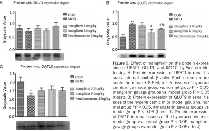
[image:5.612.86.528.72.222.2]To investigate the effects of mangiferin on the levels of target protein expression with URAT1, Figure 3. Renal function was improved via gavage of mangiferin and benzbromarone to hyperuricemic mice. A: Normal renal function presented in normal group mice. B: Damaged renal tissue presented in the model group:
[image:5.612.93.296.300.555.2]The black arrow represents renal tubular necrosis and the white arrow was indicates interstitial inflammatory cell infiltration. B+: Enlargement of it. C-F: Renal function improved via gavage mangiferin at a dose of 1.5, 3.0, 6.0, and 100 mg/kg. G: Renal function improved via gavage benzbromarone at a dose of 25 mg/kg.
ern blot assay for animal model after mangif-erin intervention. The result was showed in Figure 5. URAT1 and OAT10 protein expression did not differ from normal mouse renal tissues between after and before intervention (P > 0.05).
However, OAT10 was lower expression in the negative intervention group, compared with the normal health mice (P < 0.05). Just by the oppo-site, GLUT9 protein was higher expression (P < 0.01). As for GLUT9 protein, mangiferin could reduce its expression in animal model, com-pared with normal level (P < 0.05).
Discussion and conclusions
There are many treatment research about hy- peruricemia models animal using mangiferin. Hu has investigated the effects of mangiferin on the decrease in uric acid levels in hyperuri-cemic mice induced by intragastric administra-tion once daily with potassium oxonate for seven days, and their found that allopurinol decreased the levels of uric acid, suggested that mangiferin might enhance uric acid excre-tion [16]. However, the effect of mangiferin on uric acid production can’t be excluded.
In the present study, consecutive intraperito-neal injection of uric acid at a dose of 150 mg·kg-1 twice daily was used to construct hy-
peruricemic model mice, which had more ad- vantage.
On the other hand, the increase of blood uric acid could result in the increase of uric acid excretion under the condition of the same pro-duction rate of uric acid. These results showed that intragastric treatment with mangiferin
sig-nificantly decreased the serum urate levels of
hyperuricemic mice with different dosage. The effects of mangiferin with a dose of 3.0 and 6.0 mg·kg-1·d-1 were as good as the effects of benzbromarone.
The mangiferin could improve excretion and decrease serum uric acid levels through pre-venting renal tubular re-absorption uric acid. To our knowledge, this might be a novel report that
mangiferin can significantly decrease serum
uric acid levels in long term hyperuricemic exposure mice.
In addition, our findings offer insights into the beneficial effects of mangiferin on pathological
changes in hyperuricemic mice kidneys, and serious renal tubular necrotic lesions could be repaired by using mangiferin at a dose of 6.0 and 100 mg·kg-1·d-1.
[image:6.612.93.521.72.340.2]The kidney plays an important role in uric acid metabolism [6]. Abnormal uric acid excretion in the kidney results in clinical hyperuricemia. The Figure 5. Effect of mangiferin on the protein expres-sion of URAT1, GLUT9, and OAT10, by Western blot testing. A: Protein expression of URAT1 in renal
excretion of uric acid was mediated by renal
urate transporters such as URAT1. The first
report on the relation of urate transporters to renal excretion showed that it was located in the branches of proximal tubules and is mainly responsible for uric acid re-absorption [17]. However, the function of URAT1 was controver-sial. Urate and creatinine concentrations in
URAT1 knockout mice were significantly higher
than the normal mice, which suggest that URAT1 plays an important role in uric acid re-absorption. On the other hand, the URAT1 was not the only transporter for urate re-absorption [18]. Unlike mice, the presence of uricase in human result in relatively weak urate re-absorp-tion via URAT1 [19]. There was increasing evi-dence that URAT1 might not be as the predomi-nant urate transporter occurring in nature [17]. This study showed that intraperitoneal injection of uric acid 300 mg·kg-1·d-1 and intragastric administration of mangiferin once daily for 14 days did not induce changes of URAT1 gene and protein expression. These results suggest that other urate transporters might be more
efficient in urate re-absorption, which was simi -lar to the results of previous investigation [17]. Several common important urate re-absorption transporters other than URAT1 have been reported, including GLUT9 [7, 20-22]. Serum uric acid level was closely related to the GLUT9 gene expression [20]. This conclusion was sup-ported by other research studies, which
sug-gest that GLUT9 was a highly efficient urate transporter [21]. The efficiency of GLUT9 trans -porter was higher than URAT1 with eight-fold [7], which played a key role in uric acid re-absorption at the proximal renal tubule. GLUT9 transporter could maintain the balance bet- ween serum uric acid levels and uric acid trans-fers [20-24].
In this present study, GLUT9 gene and protein expression were up-regulated (P < 0.05, P < 0.01) in mice after intraperitoneal injection of uric acid with dose of 300 mg·kg-1·d-1 for 14 days. GLUT9 gene expression was effectively down-regulated by each dose of mangiferin and its effect of regulation was better than benzbro-marone (25 mg·kg·d-1). These results suggested that GLUT9 could be used as a target of mangif-erin to improve uric acid excretion and to decrease uric acid levels in the body.
OAT10 is a new urate transporter as confirmed
by RT-PCR and immunohistochemical analyses, which is located at the top cell membrane of renal proximal tubule cells [25]. Our results showed that OAT10 gene expression showed no obvious changes among all of the experimental groups. OAT10 protein expression was down-regulated in the hyperuricemia mouse model group (P < 0.05), while it was up-regulated in mangiferin intragastric administration groups (P > 0.05). However, the relationship of URAT1, GLUT9, and OAT10 transporters remains elu-sive and thus required further study.
Conclusions
The effects of mangiferin about regulation for serum uric acid and improvement renal patho-logical changes were better than using benz-bromarone. Mangiferin can decrease uric acid levels by regulating the gene and protein expression of the urate re-absorption trans-porter GLUT9, but no effects for URAT1 and OAT10.
Acknowledgements
The National Natural Science Foundation of China (81360505), and the Yunnan Provincial Science Foundation (2013FA017, 2013FZ076) supported this study.
Disclosure of conflict of interest
None.
Address correspondence to: Ling Li, Biomedical Engineering Research Center, Kunming Medical University, 1168 Western Chunrong Road, Yuhua Street, Chenggong New City, Kunming 650500, China. Tel: 5922743; Fax: +86-871-5922952; E-mail: [email protected]
References
[1] Jin M, Yang F, Yang I, Yin Y, Luo JJ, Wang H, Yang XF. Uric acid, hyperuricemia and vascular diseases. Front Biosci 2012; 17: 656-669. [2] Abbott RD, Brand FN, Kannel WB, Castelli WP.
Gout and coronary heart disease: the Framing-ham study. J Clin Epidemiol 1988; 41: 237-242.
[3] Choi HK, Mount DB, Reginato AM. Pathogene-sis of gout. Ann Intern Med 2005; 143: 499-516.
the Third National Health and Nutrition Exami-nation Survey. Arthritis Rheum 2007; 57: 109-115.
[5] Krishnan E, Baker JF, Furst DE, Schumacher HR. Gout and the risk of acute myocardial in-farction. Arthritis Rheum 2006; 54: 2688-2696.
[6] Sica DA, Schoolwerth AC. Renal handling of or-ganic anions and cations: excretion of uric acid. In: Brenner BM, editor. The Kidney. 6th edition. Philadelphia: WB Saunders; 2000. pp. 680-700.
[7] Sakurai H. Urate transporters in the genomic era. Curr Opin Nephrol Hypertens 2013; 22: 545-550.
[8] Telang M, Dhulap S, Mandhare A, Hirwani R. Therapeutic and cosmetic applications of mangiferin: a patent review. Expert Opin Ther Pat 2013; 23: 1561-80.
[9] Sánchez GM, Re L, Giuliani A, Núñez-Sellés AJ, Davison GP, León-Fernández OS. Protective ef-fects of Mangiferaindica L. extract, mangiferin and selected antioxidants against TPA-induced biomolecules oxidation and peritoneal macro-phage activation in mice. Pharmacol Res 2000; 42: 565-573.
[10] Garciaa D, Delgadob R, Ubeira FM. Modulation of rat macrophage function by the Mangifera-indica L. extracts vimang and mangiferin. Int Immunopharmacol 2002; 2: 797-806. [11] Muruganandan S, Gupta S, Kataria M, Lal J,
Gupta PK. Mangiferin protects the streptozoto-cin-induced oxidative damage to cardiac and renal tissues in rats. Toxicology 2002; 176: 165-173.
[12] Yoshikawa M, Ninomiya K, Shimoda H, Nishida N, Matsuda H. Hepatoprotective and antioxida-tive properties of Salaciareticulata: prevenantioxida-tive effects of phenolic constituents on CCl4-in-duced liver injury in mice. Biol Pharm Bull 2002; 25: 72-76.
[13] Yoshimi N, Matsunaga K, Katayama M, Yama-da Y, Kuno T, Qiao Z, Hara A, Yamahara J, Mori H. The inhibitory effects of mangiferin, a natu-rally occurring glucosylxanthone, in bowel car-cinogenesis of male F344 rats. Cancer Lett 2001; 163: 163-170.
[14] Niu Y, Lu W, Gao L, Lin H, Liu X, Li L. Reducing effect of mangiferin on serum uric acid levels in mice. Pharm Biol 2012; 50: 1177-1182. [15] Carroll JJ, Coburn H, Douglass R, Babson AL. A
simplified alkaline phosphotungstate assay for
uric acid in serum. Clin Chem 1971; 17: 158-160.
[16] Hu QH, Zhang X, Wang Y, Kong LD. Mangiferin promotes uric acid excretion and kidney func-tion improvement and modulates related renal transporters in hyperuricemic mice. Yao Xue Xue Bao 2010; 45: 1239-46.
[17] Enomoto A, Kimura H, Chairoungdua A, Shige-ta Y, JuShige-tabha P, Cha SH, Hosoyamada M, Take-da M, Sekine T, Igarashi T, Matsuo H, Kikuchi Y, Oda T, Ichida K, Hosoya T, Shimokata K, Niwa
T, Kanai Y, Endou H. Molecular identification of
a renal urate anion exchanger that regulates blood urate levels. Nature 2002; 417: 447-452.
[18] Hosoyamada M, Takiue Y, Morisaki H, Cheng J, Ikawa M, Okabe M, Morisaki T, Ichida K, Ho-soya T, Shibasaki T. Establishment and analy-sis of SLC22A12 (URAT1) knockout mouse. Nucleosides Nucleotides Nucleic Acids 2010; 29: 314-20.
[19] Eraly SA, Vallon V, Rieg T, Gangoiti JA, Wikoff WR, Siuzdak G, Barshop BA, Nigam SK. Multi-ple organic anion transporters contribute to net renal excretion of uric acid. Physiol Genom-ics 2008; 33: 180-192.
[20] Li S, Sanna S, Maschio A, Busonero F, Usala G, Mulas A, Lai S, Dei M, Orrù M, Albai G, Bandi-nelli S, Schlessinger D, Lakatta E, Scuteri A, Najjar SS, Guralnik J, Naitza S, Crisponi L, Cao A, Abecasis G, Ferrucci L, Uda M, Chen WM, Nagaraja R. The GLUT9 gene is associated with serum uric acid levels in Sardinia and Chi-anti cohorts. PLoS Genet 2007; 3: e194. [21] Caulfield MJ, Munroe PB, O’Neill D, Witkowska
K, Charchar FJ, Doblado M, Evans S, Eyhera-mendy S, Onipinla A, Howard P, Shaw-Hawkins S, Dobson RJ, Wallace C, Newhouse SJ, Brown M, Connell JM, Dominiczak A, Farrall M, Lath-rop GM, Samani NJ, Kumari M, Marmot M, Brunner E, Chambers J, Elliott P, Kooner J, Laan M, Org E, Veldre G, Viigimaa M, Cappuc-cio FP, Ji C, Iacone R, Strazzullo P, Moley KH, Cheeseman C. SLC2A9 is a high-capacity urate transporter in humans. PLoS Med 2008; 5: e197.
[22] Wang MX, Liu YL, Yang Y, Zhang DM, Kong LD. Nuciferine restores potassium
oxonate-in-duced hyperuricemia and kidney inflammation
in mice. Eur J Pharmacol 2015; 747C: 59-70. [23] Stark K, Reinhard W, Neureuther K, Wiedmann
S, Sedlacek K, Baessler A, Fischer M, Weber S, Kaess B, Erdmann J, Schunkert H, Hengsten-berg C. Association of common polymorphisms in GLUT9 gene with gout but not with coronary artery disease in a large case-control study. PLoS One 2008; 3: e1948.
[24] Wu XH, Ruan JL, Zhang J, Pallidifloside D. A sa -ponin glycoside constituent from Smilax ripar-ia, resist to hyperuricemia based on URAT1 and GLUT9 in hyperuricemic mice. J Ethno-pharmacol 2014; 157: 201-5.
[25] Bahn A, Hagos Y, Reuter S, Balen D, Brzica H, Krick W, Burckhardt BC, Sabolic I, Burckhardt