Original Article
MiR-15a promotes phenotypic and
functional maturation of LPS-induced dendritic
cells through down-regulating DDX39 expression
Demin Xu, Hua Li, Yun Zhao, Chunsheng Wang
Department of Cardiac Surgery, Zhongshan Hospital, Fudan University, Shanghai 200032, China
Received July 13, 2016; Accepted September 2, 2016; Epub February 15, 2017; Published February 28, 2017
Abstract: Chronic rejection after heart transplantation was mainly caused by adaptive T cell response activated by
mature dendritic cells (DCs), is the main reason influencing the long-term survival rate which is still not satisfactory.
While there is little known about the mechanism of dendritic cells maturation. In this study, we evaluated the miR-15a expression in lipopolysaccharides (LPS) induced DCs maturation, and the results showed that miR-miR-15a was highly expressed in LPS-induced DCs. MiR-15a inhibitor inhibited the DCs activation via suppressing the surface markers expression of activated DCs (CD80, CD86, MHC-II) and cytokines levels secreted by activated DCs (12,
IL-6, IL-1β and TNF-α). Besides, MiR-15a inhibitor increased the endocytic activity and weakened allostimulatory activ -ity of LPS-induced DCs. DDX39, predicted as a target gene of miR-15a, had the contrary effect on LPS-induced DCs, and overexpression of DDX39 inhibited the phenotype and functional maturation of LPS-induced DCs. Furthermore,
DDX39 is the downstream effector of miR-15a in regulating DCs maturation. These findings help understanding the
mechanism of DCs maturation.
Keywords: Dendritic cells, maturation, LPS, miR-15a, DDX39
Introduction
Chronic rejection after heart transplantation is the major factor causing graft inactivation and
influencing the patients’ long-term survival [1,
2]. The main pathological characteristics of chronic rejection after heart transplantation are cardiac allograft vasculopathy (CAV), a
sig-nificant rise of infiltrates cells in myocardium
mesenchyme, myocyte hypertrophy and
inter-stitial fibrosis [3, 4]. It plays an important role
on the development of CAV that activated T cells and macrophages gather around the blood vessels in the transplanted heart tissue
[5]. Besides, the number of T cells and antigen presenting cells (APC) specifically increased in
the transplanted tissues can stimulate and
intensify inflammation development and results in graft function damage [6].
The activation of T cells not only needs the
interaction between antigen specific T cell
receptor (TCR) and the antigen peptide-MHC complex on the APCs, but also needs the
co-stimulating signals [7]. Dendritic cells (DCs) as
an important APC were the sole one activating the initial T cells, play a prominent role in recog-nizing and presenting antigen and starting the
immune response [8, 9]. Whether in allograft
immune or in mixed leucocyte reaction, DCs are effective agonists that initial T cell immune response, and only a small amount of DCs can stimulate strong T cell responses in vivo or in vitro [10].
DC precursors derived from hematopoietic stem cells in bone marrow differentiate into immature DCs under the stimulations of growth factors and differentiation factors. Immature DCs in peripheral tissues capture and process antigen to constitute the major histocompatibil-ity complex (MHC), and then mature DCs will be produced with a range of morphological and
functional changes [11]. Mature DCs will lose
and cytokines including IL-12, IL-6, IL-1β and TNF-α [12]. The mature process of DCs also
can be induced by a variety of external stimula-tion factors, such as lipopolysaccharide (LPS),
dsRNA, CpG DNA, TNF-α and PGE2 [13, 14].
DC activation is regulated by a variety of in- tracellular molecules, the discovery of the molecular and the research of related mecha-nism attracted extensive attention currently. Discovery of new DC activation molecular and study of corresponding regulatory mechanism are the key problems in chronic rejection me- chanism research and intervention target selection. MicroRNA possesses a wide range
of expression profile and regulatory function,
so we analyzed the different expression of microRNAs in immature and mature DCs and the effect of target gene on maturation of DCs. We found that miR-15a may regulate the phenotypic and functional maturation of DCs through controlling the DDX39 expression. Materials and methods
Dendritic cells generation and culture
Mice bone marrow dendritic cells (BMDCs)
were collected from the tibiae and femurs of
4-6 weeks old C57BL/6 (Shanghai SLAC
Laboratory Animal Co. Ltd, Shanghai, China) as
previously described [15] with a little improve
-ment. First, bone marrow was flushed to a ster -ile petri dish with RPMI-1640 (HyClone, Rogan, USA) and the erythrocyte was lysed with ammo-nium chloride. Cell suspension was prepared with complete medium supplemented with
10% FBS (HyClone, Rogan, USA), 10 ng/mL
rmGM-CSF (R&D systems, Minneapolis, USA) and 10 ng/mL rmIL-4 (R&D systems) and was cultured in 6-well plates for 24 h. Then, adher-ent cells were kept and cultured in the new same complete medium for 6 days with replace-ment of medium every other day. Last, Non-adherent and loose Non-adherent cells were
enrich-ment of BMDCs and gathered. BMDCs matura
-tion were induced by LPS (1 μg/mL)
(Sigma-Aldrich, St. Louis, MO, USA) for 24 h.
Cell surface markers analysis
Immature DCs with different treatments were harvested and incubated respectively with appropriate antibody against murine CD11c, CD80, CD86 and MHC-II for 30 min on ice fol-lowed by washing 3 times. FITC-conjugated
anti-mouse CD11c, CD80, MHC-II, and PE- conjugated anti-mouse CD86 antibodies were
bought from eBioscience (San Diego, USA) and
used in accordance with the instructions. The expressions of these proteins were analyzed by
flow cytometry.
Inflammatory cytokines detection
Cell culture supernatants of immature DCs with different treatments were collected to test the
cytokine concentrations (IL-1β, IL-6, IL-12 and TNF-α) by ELISA kits (Dakawe Biotech Company,
Shenzhen, China) in accordance with the instructions.
Fluorescein isothiocyanate (FITC)-dextran up-take
To detect the endocytic activity of DCs, DCs with different treatments were incubated with FITC-dextran (1 mg/mL, Sigma-Aldrich) for 1 h at 37°C. Then cells were harvested and washed
twice with cold HBSS and followed by analyzing by a FACSCalibur flow cytometer.
Allogeneic mixed lymphocyte reaction assay
Splenocytes were isolated from BALB/c (4-6
weeks old, Shanghai SLAC Laboratory Animal Co. Ltd, Shanghai, China) and were used for the allogeneic T cell reaction. Immature DCs with different treatments were treated with
mitomy-cin C (50 μg/mL, Sigma-Aldrich) for 1 h at 37°C
and then added allogeneic T cells with a graded ratio (1:10, 1:25, 1:50, 1:100). After 3 days, cell proliferation was detected by CCK8 assay.
MicroRNA expression assay
MicroRNA expression of immature DCs and mature DCs induced by LPS were analyzed. Total RNA was isolated as standard method for microRNA array. MiR-15a expression was detected by qRT-PCR too.
Prediction and validation of miR-15a target gene
relationship between miR-15a and DDX39 in DCs maturation, at the same time, inhibitor NC and miR-15a inhibitor were transfected into LPS-induced DCs respectively too. Immature DCs were cultured as control. The surface markers (CD80, CD86, and MHC-II), cytokines
(IL-6, IL-12, IL-1β, TNF-α), endocytic activity and
allostimulatory activity were detected as described respectively.
Statistical analysis
All data were obtained from three independent experiments and were analyzed using Graph Pad Prism software, the results were represent-ed in the form of means ± SD. Statistical sig-
nificance between different treatments was determined with student’s t-test, when P < 0.05 or P < 0.01, the difference is statistically
significant.
Results
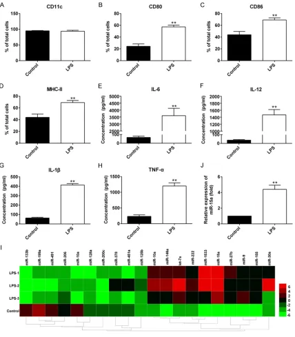
LPS induces the DCs maturation
BMDCs were cultured and treated with 1 μg/
mL LPS for 24 h, then cell surface markers of mature DCs (CD11c, CD80, CD86 and MHC-II)
and cytokines secreted by mature DCs (IL-1β, IL-6, IL-12 and TNF-α) were detected using flow
cytometry and ELISA respectively. Immature DCs and LPS-induced DCs were showed the typical characteristics of DCs that expression
of CD11c (purified > 90%) (Figure 1A). CD80, CD86, MHC-II and cytokines tested were all displayed an increased expression in LPS-induced DCs (P < 0.01) (Figure 1B-H).
MiR-15a is highly expressed in LPS-induced DCs
Total RNA were isolated from immature DCs and LPS-induced DCs to analyze the micro-RNA expression using micro-RNA array. MiR-15a
was elevated significantly in three LPS-induced
DCs group than that in immature DCs (Figure 1I) as well as the qRT-PCR result (P < 0.01) (Figure 1J).
MiR-15a inhibitor suppress the DCs matura-tion induced by LPS
In DCs treated with miR-15a inhibitor in the presence of LPS, surface markers expression (CD80, CD86 and MHC-II) in mature DCs cell were all declined considerably and lower than were used to validate the miR-15a target gene.
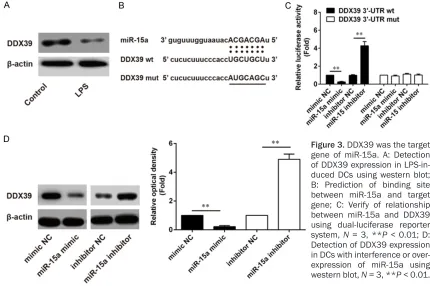
293T cells were transfected with mimic NC, miR-15a mimic, inhibitor NC and miR-15a inhib-itor for 6 h respectively, then cotransfected with the mixture of pGL3.5XDDX39wt-lucifer-ase (pGL3.5XDDX39mut-luciferpGL3.5XDDX39wt-lucifer-ase) and pRL-TK-Renilla-luciferase for 24 h. DDX39wt lucifer-ase and DDX39mut luciferlucifer-ase activities were
measured using microplate reader (Infinite
M1000, TECAN, Switzerland) according to the instruction. Immature DCs were cultured with mimic NC, miR-15a mimic, inhibitor NC and miR-15a inhibitor for 48 h respectively, and then total proteins were isolated to analyze the expression of DDX39 using western blot. Mimic NC, miR-15a mimic, inhibitor NC and miR-15a inhibitor were obtained from GenePharma (Shanghai, China). Antibody against DDX39 and
β-actin were purchased from R&D systems and
Dual-Luciferase Reporter Assay System was obtained from Promega (Madison, WI, USA).
The effect of miR-15a on LPS-induced DCs maturation
To estimate the relationship between miR-15a and DCs maturation, LPS-induced DCs were cultured with inhibitor NC (100 nM) and miR-15a inhibitor (100 nM) for 48 h respectively, and immature DCs were cultured as control. Then the surface markers (CD80, CD86, and
MHC-II), cytokines (IL-1β, IL-6, IL-12 and TNF-α),
endocytic activity and allostimulatory activity were detected as described respectively.
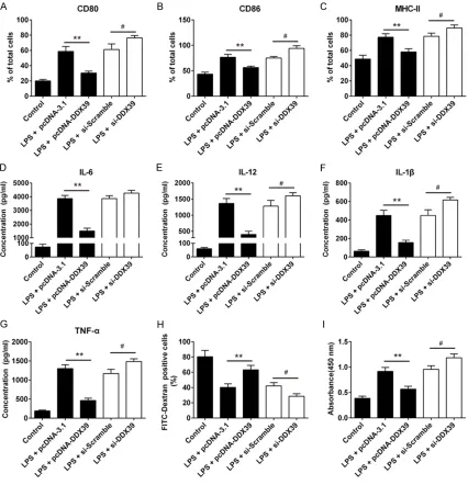
The effect of DDX39 on LPS-induced DCs maturation
To study the relationship between DDX39 and DCs maturation, LPS-induced DCs were transfected with pcDNA-3.1, pcDNA-DDX39, si-scramble and si-DDX39 for 48 h respectively, and immature DCs were cultured as control. The surface markers (CD80, CD86, and
MHC-II), cytokines (IL-1β, IL-6, IL-12 and TNF-α),
endocytic activity and allostimulatory activity were detectedas described respectively. The sequences of si-Scramble and si-DDX39 were
GAAAGACATCAAGGGATCCTACGTT-3’ and 5’-GAACATAACGAGGTACCATCGAGTT-3’.
Combined effect of miR-15a inhibitor and si-DDX39 on LPS-induced DCs maturation
in DCs treated with inhibitor NC in presence of LPS (P < 0.01); These markers in DCs treated with inhibitor NC in presence of LPS were obvi-ously higher than those in control group (P <
0.01) (Figure 2A-C). Cytokines in the cell cul-ture supernatants were also detected by ELISA,
and cytokines in DCs treated with inhibitor NC and miR-15a inhibitor in presence of LPS were all much higher than those in control DCs, but cytokines in DCs treated with miR-15a inhibitor
[image:4.612.94.522.72.561.2]were significantly lower than those in DCs treat -ed with inhibitor NC (P < 0.01) (Figure 2D-G). Figure 1. Expression of maturation markers in BMDCs induced by LPS and screened microRNA with differential ex -pression in mature DCs. A-D: Detection of surface markers (CD11c, CD80, CD86 and MHC-II) of mature DCs using
flow cytometry, N = 3, **P < 0.01 versus control; E-H: Detection of cytokines (IL-6, IL-12, IL-1β, TNF-α) in cell culture
supernatants using ELISA, N = 3, **P < 0.01 versus control; I: Part of heat map of microRNA array in LPS-induced
DCs and control DCs; J: Confirmation of miR-15a expression using qRT-PCR in LPS-induced DCs and control DCs. N
Furthermore, DCs treated with inhibitor NC
sig-nificantly decreased the endocytic activity of
capturing FITC-dextran (P < 0.01), which would be rescued by treating with miR-15a inhibitor (P < 0.01) even if still lower than control DCs (P <
0.05) (Figure 2H); the results of CCK8 assay
indicated that treatment of LPS and inhibitor NC could increase the allostimulatory capacity and it was the strongest when the ratio of DCs and T cells was 1:10, and allostimulatory
capacity would significantly drop when DCs
[image:5.612.93.521.70.525.2]treated with LPS and miR-15a inhibitor (P <
Figure 2. Downregulation of miR-15a suppressed LPS-induced DCs maturation. To evaluate the role of miR-15a in LPS-induced DCs maturation, miR-15a inhibitor and NC inhibitor were respectively transfected into LPS-induced
DCs, and immature DCs were cultured as control. A-C: Detection of surface markers of mature DCs using flow
cytometry, N = 3, *P < 0.05 and **P < 0.01 versus control, ##P < 0.01; D-G: Detection of cytokines secreted by mature DCs in cell culture supernatants using ELISA, N = 3, *P < 0.05 and **P < 0.01 versus control, ##P < 0.01;
H: The endocytic activity of the DCs was detected using flow cytometer and the comparisons were did between
0.01) (Figure 2I). These indicate that miR-15a inhibitor can suppress phenotype and function-al maturation of LPS-induced DCs.
MiR-15a inhibits the DDX39 expression in DCs
According to the result of target gene predic-tion, DDX39 might be the target gene of miR-15a, so the binding site in DDX39 mRNA sequence was mutated as a mutant (Figure 3B). The relationship between miR-15a and
DDX39 3’-UTRwt/DDX39 3’-UTRmut were veri
-fied by luciferase reporter gene assay, and
luciferase activity was increased dramatically
in DDX393’-UTRwt group when treated with
miR-15a inhibitor, that was lowest when treat-ed with miR-15a mimic; but the luciferase
activities in DDX39 3’-UTRmut with different treatments were low and no significant differ -ences between them (Figure 3C). Western blot results showed that DDX39 expression in LPS-induced DCs was lower than that in immature DCs (Figure 3A), and DDX39 expression was changing with different treatments, DCs showed a highest level of DDX39 when treated with miR-15a inhibitor and a lowest level of DDX39 when treated with miR-15a mimic (Figure 3D).
Overexpression of DDX39 inhibits the DCs maturation induced by LPS
[image:6.612.90.520.73.359.2]Cell surface markers and cytokines were increased when DCs were treated with pcDNA-3.1 in presence of LPS, and these increases would be dipped in LPS-induced DCs trans- fected with pcDNA-DDX39 (P < 0.01); on the contrary, cell surface markers expressions and cytokines levels in LPS-induced DCs treated with si-DDX39 were higher than LPS-induced DCs transfected with pcDNA-DDX39 and si-Scramble (P < 0.05) (Figure 4A-G). Endocytic activity of LPS-induced DCs trans-fected with pcDNA-DDX39 was higher than that of LPS-induced DCs transfected with pcDNA-3.1; and endocytic activity of LPS-induced DCs transfected with si-Scramble was higher than that of LPS-induced DCs treated with si-DDX39 (Figure 4H). The changes of allostimulatory capacity of LPS-induced DCs with these different treatments were contrary to the changes of endocytic activity (Figure 4I). These indicate that overexpre- ssion of DDX39 can suppress phenotype and functional maturation of LPS-induced DCs.
Figure 3. DDX39 was the target gene of miR-15a. A: Detection of DDX39 expression in LPS-in-duced DCs using western blot;
B: Prediction of binding site
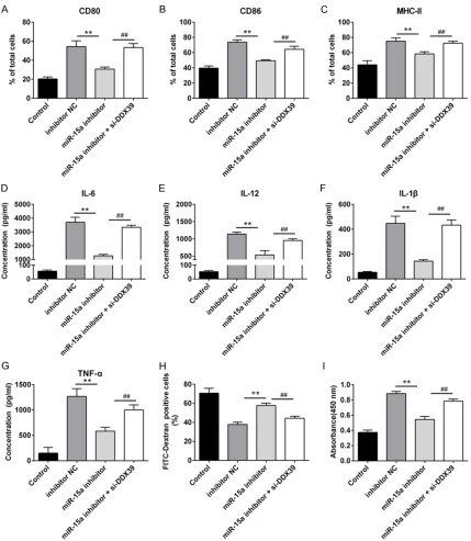
Interference of DDX39 abolishes the inhibition of miR-15a on LPS-induced DCs maturation
Cell surface markers expressions and cyto-kines levels of LPS-induced DCs were declined considerably when transfected with miR-15a inhibitor than transfected with inhibitor NC (P <
0.01), LPS-induced DCs transfected with
[image:7.612.94.520.73.515.2]miR-15a inhibitor and si-DDX39 simultaneously would increase cell surface markers expres-sions and cytokines levels (Figure 5A-G). In the same time, compared with LPS-induced DCs transfected with miR-15a inhibitor, LPS-induced DCs transfected with miR-15a inhi- bitor and si-DDX39 simultaneously would decrease the endocytic activity and increase Figure 4. Interference of DDX39 promoted DCs maturation but overexpression of DDX39 inhibited DCs maturation. To evaluate the role of DDX39 in LPS-induced DCs maturation, recombinant plasmid pcDNA-DDX39 and si-DDX39 were respectively transfected into DCs in the presence of LPS, besides, pcDNA-3.1 and si-Scramble were also respectively transfected into DCs in the presence of LPS as negative control, and immature DCs were cultured as
blank control. A-C: Detection of surface markers of mature DCs using flow cytometry, N = 3, **P < 0.01, #P < 0.05; D-G: Detection of cytokines in cell culture supernatants using ELISA, N = 3, **P < 0.01, #P < 0.05; H: The endocytic
activity of the DCs was detected using flow cytometer and the comparisons were did between the proportion of
the allostimulatory capacity (P < 0.01) (Figure 5H, 5I). These indicate that DDX39 is the target gene of miR-15a involved in DCs maturation.
Discussion
Heart transplantation is the only effective means for treatment of end-stage heart dis-Figure 5. Interference of DDX39 removed the inhibition of miR-15a on DCs maturation. To evaluate the relationship between DDX39 and miR-15a in LPS-induced DCs maturation, miR-15a inhibitor and si-DDX39 were cotransfected into DCs in the presence of LPS, miR-15a inhibitor and inhibitor NC was respectively transfected into LPS-induced DCs as control, immature DCs were cultured as blank control. A-C: Detection of surface markers of mature DCs
[image:8.612.93.522.71.564.2]ease [16]. Although the prognosis of heart
transplantation is improving, but the long-term survival rate is still not satisfactory; and the incidences of chronic homograft rejection in
the first year, the fifth year and the eighth year
after the surgery for patients with heart trans-plantation were 8%, 32% and 43% respectively
[17], which was the most important reason of death [18, 19]. A typical manifestation of chron -ic rejection is CAV, and its severity is far higher than that of other organs for transplant
vascu-lopathy, but there is no effective treatment [20,
21]. At present, immunosuppressant therapy is the main treatment of CAV, but this method
doesn’t controls rejection occurring from the
source and has wide side effects. So, exploring the mechanism of chronic rejection and its intervention strategy has an important
theoreti-cal and practitheoreti-cal significance. DCs are in the
central part of the adaptive immunity and play
a key role in chronic rejection [12, 22]. In the indirect identification process of allogeneic
antigens, DCs activate the T cell response by uptaking, processing and presenting the
alloge-neic antigens to T cells [23-25]. Activation and
mature state of DCs decide the type and degree of subsequent immune response, and imma-ture DCs will mediate the immune tolerance if DCs secrete regulatory cytokines (like IL-10) too much and induce the activation of regulatory T cells; but mature DCs can secrete a lot of
proin-flammatory cytokines and stimulate effect T cell response [26, 27]. In this study, we indicat -ed the effects of overexpress-ed miR-15a and lower expressed DDX39 on the phenotypic and functional maturation of DCs induced by LPS through analyzing the expression of cell sur-face markers (CD80, CD86 and MHC-II) and
cytokines (IL-1β, IL-6, IL-12 and TNF-α), and
endocytosis, stimulatory capacity of DCs for T cell proliferation.
LPS can stimulate the maturation of DCs and
trigger the production of proinflamatory cyto
-kines (IL-1β, IL-6, IL-12, and TNF-α) in vitro [28],
furthermore, mature DCs highly express MHC-II
and co-stimulators (CD80 and CD86) [29], which is essential for an antigen-specific T cell response [30]. In addition, CD11c is a marker
of DCs and involves in phagocytosis, cell
migra-tion, cytokine production and inflammation [31]. So we detected these cytokines and sur -face markers to evaluate LPS induced DCs mat-uration. The results showed that expression
rate of CD11c in all cells was above 90% and indicated DCs culturing was successful. High expression of cytokines and surface markers detected in LPS-induced DCs indicated that DCs were fully mature with treatment of LPS. MicroRNA possesses a wide range of
expres-sion profile and regulatory function, there have
reports about expression and function of
microRNA almost in every system [32, 33], including immune system [34, 35], but its func -tions in immune system are still poorly under-stood. And more studies about microRNA func-tions in DCs maturation and activation should be done to further understand the mechanism of DCs maturation and activation. The results of microRNA array showed difference about microRNAs expression in immature DCs and LPS-induced DCs and high level of miR-15a in LPS-induced DCs. Furthermore, miR-15a inhibi-tor depressed the maturation of LPS-induced DCs which concluded from downregulation of the expressions of cell surface markers and cytokines, increased endocytic activity, and decreased stimulatory capacity for T cell prolif-eration. These turned out that miR-15a partici-pates in the process of LPS-induced DCs maturation.
DExD/H-box helicases participate in the metab-olism process of RNA from transcription to
deg-radation [36], DDX58 (RIG-I) and DDX60 are
sensors of viral RNA molecules and several viruses and elicit antiviral interferon responses
[37]. In this study, DDX39 was predicted as the
target gene of miR-15a and was low expressed in LPS-induced DCs. Overexpression or lower
expression of DDX39 all influenced the expres -sions of cell surface markers and cytokines, endocytic activity and stimulatory capacity on T cell activation of LPS-induced DCs, suggested
results indicated that overexpression of miR-15a promote the LPS-induced DCs maturation may directly via blocking DDX39 expression. In summary, miR-15a is highly expressed in LPS-induced DCs, and miR-15a inhibitor can suppress the phenotype and functional matu-ration of LPS-induced DCs. Furthermore, DDX39 is the downstream target gene of miR-15a and the role of DDX39 in the phenotype and functional maturation of LPS-induced DCs is opposite to that of miR-15a. These results help understanding the mechanism of DCs maturation, which is important for T cell response in innate and adaptive immunity, including chronic rejection after heart trans-plantation. So miR-15a and DDX39 may be potential intervention targets for chronic rejec-tion after heart transplantarejec-tion.
Disclosure of conflict of interest
None.
Address correspondence to: Dr. Demin Xu, De- partment of Cardiac Surgery, Zhongshan Hospital, Fudan University, No. 180 Fenglin Road, Shanghai 200032, China. Tel: 21-64041990; Fax: +86-21-64223006; E-mail: [email protected]
References
[1] Gao SZ, Alderman EL, Schroeder JS, Silverman JF and Hunt SA. Accelerated coronary vascular disease in the heart transplant patient:
coro-nary arteriographic findings. J Am Coll Cardiol
1988; 12: 334-340.
[2] Aziz T, Hasleton P, Hann AW, Yonan N, Deiraniya A and Hutchinson IV. Transforming growth fac-tor beta in relation to cardiac allograft vascu-lopathy after heart transplantation. J Thorac Cardiovasc Surg 2000; 119: 700-708.
[3] Stewart S, Winters GL, Fishbein MC, Tazelaar
HD, Kobashigawa J, Abrams J, Andersen CB, Angelini A, Berry GJ, Burke MM, Demetris AJ,
Hammond E, Itescu S, Marboe CC, McManus
B, Reed EF, Reinsmoen NL, Rodriguez ER,
Rose AG, Rose M, Suciu-Focia N, Zeevi A and
Billingham ME. Revision of the 1990 working
formulation for the standardization of nomen-clature in the diagnosis of heart rejection. J Heart Lung Transplant 2005; 24: 1710-1720.
[4] Kalache S, Dinavahi R, Pinney S, Mehrotra A, Cunningham MW and Heeger PS. Anticardiac myosin immunity and chronic allograft vascu-lopathy in heart transplant recipients. J Immunol 2011; 187: 1023-1030.
[5] Song G, Zhao X, Xu J and Song H. Increased expression of intercellular adhesion mole-cule-1 and vascular cell adhesion molemole-cule-1 in rat cardiac allografts. Transplant Proc 2008; 40: 2720-2723.
[6] Alexis JD, Pyo RT, Chereshnev I, Katz J, Rollins
BJ, Charo IF and Taubman MB. Inhibition of
MCP-1/CCR2 signaling does not inhibit intimal proliferation in a mouse aortic transplant mod-el. J Vasc Res 2008; 45: 538-546.
[7] RH S. A cell culture mode for T lymphocyte co-lonial energy. Science 1990; 4961: 1349-1356.
[8] Heath WR and Carbone FR. Dendritic cell sub-sets in primary and secondary T cell responses at body surfaces. Nat Immunol 2009; 10: 1237-1244.
[9] Segura E and Villadangos JA. Antigen presen-tation by dendritic cells in vivo. Curr Opin Immunol 2009; 21: 105-110.
[10] Sallusto F and Lanzavecchia A. Efficient pre -sentation of soluble antigen by cultured hu-man dendritic cells is maintained by granulo-cyte/macrophage colony-stimulating factor plus interleukin 4 and downregulated by tumor necrosis factor alpha. J Exp Med 1994; 179: 1109-1118.
[11] Sallusto F, Cella M, Danieli C and Lanzavecchia A. Dendritic cells use macropinocytosis and the mannose receptor to concentrate macro-molecules in the major histocompatibility com-plex class II compartment: downregulation by cytokines and bacterial products. J Exp Med 1995; 182: 389-400.
[12] Banchereau J, Briere F, Caux C, Davoust J, Lebecque S, Liu YJ, Pulendran B and Palucka
K. Immunobiology of dendritic cells. Annu Rev Immunol 2000; 18: 767-811.
[13] Jonuleit H, Kuhn U, Muller G, Steinbrink K, Paragnik L, Schmitt E, Knop J and Enk AH.
Pro-inflammatory cytokines and prostaglandins in -duce maturation of potent immunostimulatory dendritic cells under fetal calf serum-free con-ditions. Eur J Immunol 1997; 27: 3135-3142.
[14] Stockwin LH, McGonagle D, Martin IG and Blair
GE. Dendritic cells: immunological sentinels with a central role in health and disease.
Immunol Cell Biol 2000; 78: 91-102.
[15] Inaba K, Inaba M, Romani N, Aya H, Deguchi M, Ikehara S, Muramatsu S and Steinman RM. Generation of large numbers of dendritic cells from mouse bone marrow cultures supple-mented with granulocyte/macrophage colony-stimulating factor. J Exp Med 1992; 176: 1693-1702.
[16] Christie JD, Edwards LB, Aurora P, Dobbels F,
Transplantation: Twenty-sixth Official Adult
Lung and Heart-Lung Transplantation Report- 2009. J Heart Lung Transplant 2009; 28: 1031-1049.
[17] Schmauss D and Weis M. Cardiac allograft vasculopathy: recent developments. Circu- lation 2008; 117: 2131-2141.
[18] Lee MS, Finch W, Weisz G and Kirtane AJ. Cardiac allograft vasculopathy. Rev Cardiovasc Med 2011; 12: 143-152.
[19] Weiss MJ, Madsen JC, Rosengard BR and Allan
JS. Mechanisms of chronic rejection in
cardio-thoracic transplantation. Front Biosci 2008;
13: 2980-2988.
[20] Wehner JR, Morrell CN, Rodriguez ER, Fairchild
RL and Baldwin WM 3rd. Immunological chal -lenges of cardiac transplantation: the need for better animal models to answer current clini-cal questions. J Clin Immunol 2009; 29: 722-729.
[21] Suzuki J, Isobe M, Morishita R and Nagai R. Characteristics of chronic rejection in heart transplantation: important elements of patho-genesis and future treatments. Circ J 2010; 74: 233-239.
[22] Banchereau J and Steinman RM. Dendritic
cells and the control of immunity. Nature 1998; 392: 245-252.
[23] Lechler RI, Sykes M, Thomson AW and Turka LA. Organ transplantation--how much of the promise has been realized? Nat Med 2005; 11: 605-613.
[24] Hornick P. Direct and indirect allorecognition.
Methods Mol Biol 2006; 333: 145-156. [25] Huang YL, Wang YZ, Chen JB, Wang F, Kang XP,
Xia JJ, Lan TS, Xie BY, Ekberg H, Wang XM and
Qi ZQ. Prevention of acute and chronic allograft rejection by combinations of tolerogenic den-dritic cells. Scand J Immunol 2011; 73: 91-101.
[26] Mahnke K, Schmitt E, Bonifaz L, Enk AH and
Jonuleit H. Immature, but not inactive: the tolerogenic function of immature dendritic
cells. Immunol Cell Biol 2002; 80: 477-483. [27] Raker VK, Domogalla MP and Steinbrink K.
Tolerogenic Dendritic Cells for Regulatory T Cell Induction in Man. Front Immunol 2015; 6: 569.
[28] Kelleher M and Beverley PC.
Lipopolysac-charide modulation of dendritic cells is in-
sufficient to mature dendritic cells to generate
CTLs from naive polyclonal CD8+ T cells in vitro, whereas CD40 ligation is essential. J Immunol 2001; 167: 6247-6255.
[29] Quah BJ and O’Neill HC. Maturation of function
in dendritic cells for tolerance and immunity. J Cell Mol Med 2005; 9: 643-654.
[30] Lechmann M, Krooshoop DJ, Dudziak D, Kremmer E, Kuhnt C, Figdor CG, Schuler G and Steinkasserer A. The extracellular domain of CD83 inhibits dendritic cell-mediated T cell stimulation and binds to a ligand on dendritic cells. J Exp Med 2001; 194: 1813-1821.
[31] Georgakopoulos T, Moss ST and Kanaga- sundaram V. Integrin CD11c contributes to monocyte adhesion with CD11b in a differen-tial manner and requires Src family kinase activity. Mol Immunol 2008; 45: 3671-3681.
[32] Ebert MS and Sharp PA. Roles for microRNAs in conferring robustness to biological process-es. Cell 2012; 149: 515-524.
[33] Bushati N and Cohen SM. microRNA functions. Annu Rev Cell Dev Biol 2007; 23: 175-205. [34] O’Connell RM, Rao DS, Chaudhuri AA and
Baltimore D. Physiological and pathological
roles for microRNAs in the immune system. Nat Rev Immunol 2010; 10: 111-122.
[35] Kuchen S, Resch W, Yamane A, Kuo N, Li Z, Chakraborty T, Wei L, Laurence A, Yasuda T, Peng S, Hu-Li J, Lu K, Dubois W, Kitamura Y, Charles N, Sun HW, Muljo S, Schwartzberg PL, Paul WE, O’Shea J, Rajewsky K and Casellas R. Regulation of microRNA expression and abun-dance during lymphopoiesis. Immunity 2010; 32: 828-839.
[36] Fuller-Pace FV. DExD/H box RNA helicases: multifunctional proteins with important roles in transcriptional regulation. Nucleic Acids Res 2006; 34: 4206-4215.