Original Article
HBs, HBc and MHBs promote TRAIL-induced apoptosis
depending on the STAT1 signaling pathway
Da He1,2, Lu Zhang1,2, Dongbo Wu1,2, Xiangjun Chen3, Hong Tang1,2, Taoyou Zhou1,2
1Center of Infectious Diseases, West China Hospital of Sichuan University, Chengdu 610041, Sichuan Province,
China; 2Division of Molecular Biology of Infectious Diseases, State Key Laboratory of Biotherapy, Sichuan
Univer-sity, Chengdu 610041, Sichuan Province, China; 3Department of Medical Quality Control, West China Hospital,
Sichuan University, Chengdu, China
Received April 28, 2016; Accepted August 5, 2016; Epub October 15, 2016; Published October 30, 2016
Abstract: The tumor necrosis factor related apoptosis induced ligand (TRAIL) can selectively induce the apoptosis of virus-infected cells. STAT1 plays an important role in modulating cell apoptosis and anti-virus responses. TRAIL and STAT1 may have a crosstalk in modulating the apoptosis of virue-infected cells. Hepatitis B virus (HBV) has several proteins, such as HBV surface protein (HBs), HBV core protein (HBc), middle hepatitis B surface antigen (MHBs), large hepatitis B surface antigen (LHBs). This study was to investigate the role of different HBV proteins in modulat-ing TRAIL and STAT1 signalmodulat-ing pathway. We found that with TRAIL treatment, the HBV-transfected, HBs-expressmodulat-ing,
HBc-expressing, MHBs-expressing human fibrosarcoma cell line HT1080 (stat1+/+) had higher apoptosis rates while the STAT1 mutant human fibrosarcoma cell line U3A (stat1-/-) did not. These results suggest that HBV may
promote TRAIL-induced apoptosis via HBs, HBc and MHBs relying on the integrity of STAT1 signaling pathway.
Keywords: Tumor necrosis factor related apoptosis inducing ligand, hepatitis B virus protein, STAT1, apoptosis
Introduction
According to the the world health organization (WHO), about 2 billion people has been infect-ed with the hepatitis B virus (HBV) in the world [1]. About 65,000 people infected with HBV died every year for the liver failure, cirrhosis and hepatocellular carcinoma (HCC) [2]. To date, the pathogenic mechanism of HBV is not fully understood. But studies indicate cell apop-tosis may contribute to the development of the disease that can be mediated by Fas ligand (FasL), TNF and TRAIL, etc [3, 4].
TRAIL firstly found by Wiley, is a member of the tumor necrosis factor (TNF) superfamily. Unlike FasL or TNF, TRAIL can specifically induce the apoptosis of virus-infected cells and tumor cells without any effect on the normal ones [5]. It can eliminate the virus by inducing cell apop-tosis through casepase way binding the death receptors DR1 and DR2 which express on the surface of the virus-infected cells.
STAT1 is one member of the signal transducers and activators of transcription (STAT) family. It
hepato-Apoptosis induced by HBV proteins
carcinoma cells to TRAIL-induced apoptosis [10, 11]. However, the regulation machanisms of HBV proteins in cell apoptosis are not fully aware of.
To study the role of HBV proteins in modulating TRAIL-induced apoptosis and whether it was relied on STAT1 signaling pathway, we trans-fected HT1080 and U3A cells with the plasmids pHBV4.1 (containing HBV complete genome), pcDNA3.1/HBs (expressing HBs), pcDNA3.1/ HBc (expressing HBc), pcDNA3.1/MHBs (expre- ssing MHBs) and pcDNA3.1/LHBs (expressing LHBs) and treated them with the low concentra-tion of TRAIL. Cell apoptosis may be observed and the molecular modulation of HBV proteins in TRAIL-induced apoptosis mediated by STAT1 signaling pathway may be revealed.
Materials and methods
Cell culture
Human fibrosarcoma cell line HT1080 (st- at1+/+) was provided by the Laboratory of Transplantation and Immunology, West China Hospital of Sicuan University. STAT1 mutant human fibrosarcoma cell line U3A (stat1-/-) was a gift from Professor Jianhua Tong of the affili-ated Ruijin Hospital of Shanghai Jiao Tong University.
HT1080 cells and U3A cells were cultured in Dulbecco’s Modified Eagle’s Medium (DMEM, Gibco) supplemented with 10% fetal bovine serum (FBS, Gibco) at 37°C in a 5% CO2 incuba-tor. The cells were passaged at a density of 70% to 80%.
Antibodies and reagents
Western blot primary antibody mouse monoclo-nal anti-STAT1 (BD, America); The mouse mono-clonal anti-GAPDH antibody (Zheng Neng, China); the secondary antibody HRP-conjugated anti-mouse IgG (Zheng Neng, China).
Construction and Identification of pcDNA3.1/ HBs
The primers of S gene (expressing HBs) were designed according to the HBV-ayw subtype sequence. S gene forward: 5’-GCGAAGCTTA- TGGAGAGCACAACATCAGGA-3’; S gene reverse: 5’-GCAGCGGCCGCTCAAATGTATACCCAAAG- ACA-3’. S gene fragments were amplified by PCR from the template, pHBV4.1. Both S gene
fragments and pcDNA3.1 were digested with restriction enzymes HindIII and NotI (Thermo, America) and purified using gel extraction kit (Qiagen, German). The recombinant plasmid was successfully constructed after connection, conversion and positive monoclonal selection. pcDNA3.1/HBs was identified by digestion of
HindIII and NotI and was sequence-confirmed. Cell supernatant of pcDNA3.1/HBs-transfected cells was collected and HBs was detected by enzyme-linked immunosorbent assay (ELISA) using HBs ELISA kit (Ke Hua, China), according to the instructions.
Plasmids transfection
Plasmid pHBV4.1 was got from Professor Alan Mclachlan of the American Scripps Institute. Plasmids pcDNA3.1, pcDNA3.1/HBc, pcDNA- 3.1/MHBs and pcDNA3.1/LHBs were provided by the Laboratory of Infectious Disease Center, West China Hospital of Sichuan University.
Cells were seeded into 6-well plates (1×10⁵ cells/well) and were transfected at 50% to 60% density. 2 ug of Plasmids were added into each well and were incubated with Opti-MEM (Gibco, America) and transfection reagent (X-treme GENE HP DNA Transfection Reagent, Roche, Germany) at room temperature for 15 minutes. Then the mixture was added into the wells respectively and mixed gently.
The survival rates of TRAIL-treated cells were detected by MTT method
and their survival rates were detected by MTT assay.
Cells transfected with pHBV4.1, pcDNA3.1/ HBs, pcDNA3.1/HBc, pcDNA3.1/MHB, pcDNA- 3.1/LHBs were treated with low concentration of TRAIL or PBS for 48 h. The cells were col-lected at a concentration of 5×10⁵ to 10×105 cells/mL and were washed by PBS at 4°C, 2000 rpm for twice. The cells were suspended in 150 ul binding buffer, 5 ul Annexin V-FITC and 5 ul propidium iodide (PI). The apoptotic cells were detected by Annexin V-PI kit (Kai Ji, China), according to the instructions.
Detection of STAT1 expression by western blot
The cells with different transfections as before were treated with the low concentration of TRAIL. The cells were collected into the lysis buffer (50 mM Tris-HCl, 1% NP40, 150 mM NaCl, 0.5% sodium deoxycholate, 0.7 ug/ml pepstatin) added with protease inhibitor cock-tail (Roche, German) and phosphatase inhibitor cocktail (Roche, German) for 1 h. Protein super-natant was got from the lysed cells after 12000 rpm centrifugation for 15 min at 4°C. Equal amount of protein was electrophoresed and transferred to a PVDF membrane (Millipore, America) which was then blocked by 5% non-fat milk (BD, America) for 1 h. The membrane was incubated with STAT1 primary antibody over-night at 4°C, then washed by Tris-buffered
saline containing Tween-20 (TBST) and incu-bated with the secondary antibody for an hour. After washing, the membrane was added to the ECL reagent (Millipore, America) to be visual-ized by the gel-imaging instrument (Royal, German). The relative expression of STAT1 was analysed by the Image Lab 5.0 software. Its expression was calculated according to the for-mula: (the value of STAT1 in experimental group/the value of STAT1 in PBS group)/(the value of GAPDH in experimental group/the value of GAPDH in PBS group).
Statistical analysis
Results were statistically analysed by the spss 20.0 software and were presented as means ± standard deviation. The Mann-Whitney U test was used to compare the variables between the rates of two groups. The t test was used to compare the variables between two quantita-tive datas. A P-value <0.05 was considered sta-tistically significant.
Results
PCR amplification of S gene and the identifica-tion of recombinant plasmid pcDNA3.1/HBs
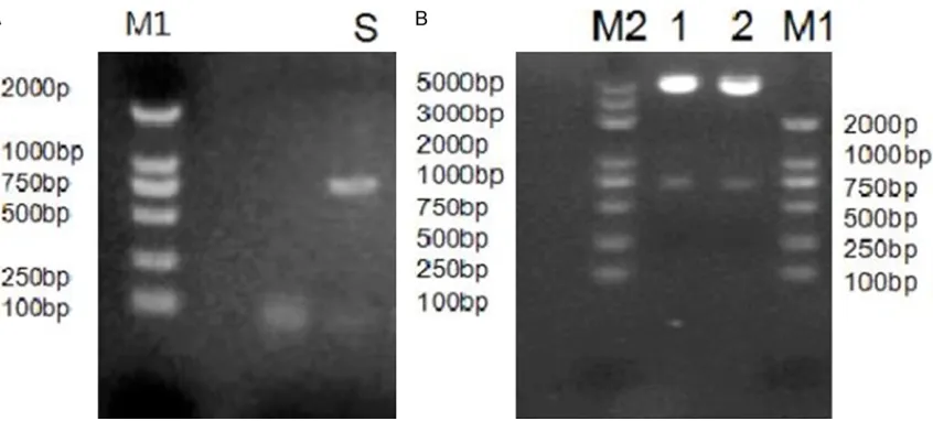
[image:3.612.97.520.72.263.2]S gene was amplified by PCR from the template, pHBV4.1. The PCR products analyzed on 1% agarose gels were consistent with the expected fragment size, about 680 bp (Figure 1A). Figure 1. Electrophoresis of S gene PCR product and pcDNA3.1/HBs digested fragments. Amplification products of
Apoptosis induced by HBV proteins
The recombinant plasmid pcDNA3.1/HBs was digested with restriction enzymes HindIII and
NotI. The nucleic acid fragments were got from 1% agarose gel consistent with the size of S gene (about 680 bp) and pcDNA3.1 (about 5400 bp) (Figure 1B). And the sequencing con-firmed the correct collection of S gene.
The supernatant of HT1080 and U3A cells tran-fected with pcDNA3.1/HBs was detected by ELISA to observe the expression of HBs. It sug-gested that pcDNA3.1/HBs could be success-fully expressed in both the two cells as with the positive ELISA results (S/COV=8.57±0.98).
The effect of different concentration of TRAIL on HT1080 and U3A cells
HT1080 and U3A cells were treated with differ-ent concdiffer-entration of TRAIL (0, 25, 50, 75, 100,
200, 300, 400, 500 ng/mL) for 48 h. The rela-tive survival rates of HT1080 and U3A cells were detected by MTT assay.
The relative survival rates of HT1080 cell had no statistical difference when comparing the 25, 50, 75 ng/mL groups with 0 ng/mL group (P>0.05). However, the relative survival rates of 100, 200, 300, 400, 500 ng/mL groups de- creased comparing with the 0 ng/mL group (P<0.05) (Figure 2A).
[image:4.612.89.519.74.215.2]The relative survival rates of U3A cell had no statistically difference when comparing the 25, 50, 75, 100 ng/mL groups with the group of 0 ng/mL (P>0.05). However, the relative survival rates of 200, 300, 400, 500 ng/mL groups decreased comparing with the 0 ng/mL group (P<0.05) (Figure 2B).
Figure 2. The relative survival rates of cells treated with different concentrations of TRAIL. Cells were treated with
different concentrations of TRAIL for 48 h, and the relative survival rates of HT1080 cells and U3A cells were de -tected by MTT assay (A and B); the datas were obtained from three independent experiments.
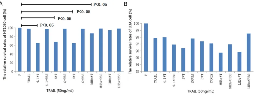
Figure 3. The relative survival rates of transfected cells treated with TRAIL. The relative survival rates of HT1080
cells and U3A cells transfected with HBV, HBs, HBc, MHBs and LHBs were detected by MTT assay after 48 h of TRAIL
[image:4.612.93.523.280.440.2]Apoptosis induced by HBV proteins
Figure 4. The apoptotic cells detected by flow cytometry. HT1080 and U3A cells transfected with HBV, HBs, HBc, MHBs and LHBs were treated with TRAIL (50 ng/ mL) for 48 h. The apoptotic cells were stained by AnnexinV- PI and detected by flow cytometry; the horizontal axis was FITC staining and the vertical axis was PI
TRAIL concentration of 50 ng/mL was consid-ered to have little effect on the survival rates of both cells according to the MTT results and was made to treat cells in the later experi- ment.
The relative survival rates of cells transfected with pHBV4.1, pcDNA3.1/HBs, pcDNA3.1/ HBc, pcDNA3.1/MHBs, pcDNA3.1/LHBs were measured by MTT assay after TRAIL treatment
The transfected HT1080 and U3A cells had been treated with TRAIL (50 ng/mL) for 48 h and MTT assay was performed to detect sur-vival rates.
For HT1080 cells, the relative survival rates of 4.1+T group (65.22±0.75), S+T group (67.60± 1.11), C+T group (65.40±2.21), MHBs+T gro- up (87.19±1.38) decreased significantly com-pared with the PBS group (100.00±0.00) (P< 0.05); however, there were no statistical differ-ences between the transfected groups and the PBS group (P>0.05); nor was there any statisti-cal difference between the TRAIL treated group and the PBS group (P>0.05) (Figure 3A). There was no statistical difference in relative survival rates between the experimental groups and the PBS group of U3A cells (P>0.05) (Figure 3B).
TRAIL could inhibit the relative survival rates of HBV-transfected, HBs-expressing, HBc-expre- ssing, MHBs-expressing HT1080 cells, but not the LHBs-expressing ones. However, HBV, HBs, HBc, MHBs and LHBs could not mediate TRAIL to inhibit the relative survival rates of U3A cells.
The apoptosis rates of cells transfected with pHBV4.1, pcDNA3.1/HBs, pcDNA3.1/HBc, pcDNA3.1/MHBs, pcDNA3.1/LHBs were mea-sured by flow cytometry after TRAIL treatment
The apoptosis of transfected HT1080 and U3A cells treated with TRAIL (50 ng/mL) were dete- cted by flow cytometry.
The apoptosis rates of 4.1+T (22.61±1.75), S+T (24.27±0.30), C+T (19.59±0.53), MHBs+T (22.52±1.88) groups of HT1080 cells increa- sed significantly compared with the PBS group (5.70±0.52) (P<0.05); however, there were no statistical differences between the transfected groups and the PBS group (P>0.05); nor was there any statistical difference between the
TRAIL treated group and the PBS group (P>0.05) (Figures 4A, 5A).
There was no statistical difference in apoptosis rates between the experimental groups and the PBS group of U3A cells (P>0.05) (Figures 4B,
5B).
It suggested that HBV, HBs, HBc, MHBs but not LHBs could promote HT1080 cells to TRAIL-induced apoptosis. However, the HBV proteins could not increase the sensitivity of U3A cells to TRAIL-induced apoptosis.
The relative STAT1 expression of cells trans-fected with pHBV4.1, pcDNA3.1/HBs, pcD-NA3.1/HBc, pcDNA3.1/MHBs,pcDNA3.1/ LHBs were detected by western blot after TRAIL treatment
The relative STAT1 expressions of HT1080 cells treated with TRAIL (50 ng/mL) were analysed by western blot. STAT1 expression of 4.1+T group (16.13±0.86) was higher than that of 4.1+PBS group (11.61±0.55) (P<0.05), S+T gr- oup (13.50±0.57) higher than S+PBS group (8.48±0.51) (P<0.05) and C+T group (12.61± 0.53) higher than C+PBS group (5.75±0.07) (P<0.05); however, no statistical differences of the expression were found between MHBs+T group and MHBs+PBS group, LHBs+T group and LHBs+PBS group (P>0.05) (Figure 6A, 6C). The STAT1 bands were not observed in U3A cells after TRAIL treatment (Figure 6B).
Disccusion
The level of TRAIL in the peripheral blood and liver tissue of patients with HBV is much higher than that of the normal people [12, 13]. Higher TRAIL expression was also found on the CD4+
and CD8+ T cells of patients with chronic
Apoptosis induced by HBV proteins
apoptosis and this might be contributed to HBV proteins as HBs, HBc and MHBs. Because tr- ansfection of HBs, HBc and MHBs could also promote TRAIL-induced apoptosis of HT1080 cells.
The mechanisms that HBV proteins can pro-mote TRAIL-induced apoptosis remain to be clarified, probably related with the high expres-sion of TRAIL death receptors on the infected cells [15]. However, it was reported that preS2 protein was found to increase the sensitivity of HepG2.2.15 to the apoptosis mediated by TRAIL and this was not related with TRAIL re-
ceptor expression [16]. Other studies suggest-ed that HBc can inhibit hepatocyte apoptosis by blocking the TRAIL death receptor, DR5 [17]. However, opposite result that HBc could pro-mote TRAIL-induced apoptosis was found in the HT1080 cells in our study. Except for the ch- ange of TRAIL receptor expression, it may be explained by other mechanisms, for example, the increase of anti-apoptosis protein, Bax [18]. Activation of other signaling pathways may also be involved. It was reported that the truncated middle hepatitis B surface antigen MHBs(t) can increase the TRAIL-mediated apoptosis by acti-vating ERK2 signaling pathway and by the deg-Figure 5. The apoptosis rates of transfected cells treated with TRAIL. The apoptosis rates of HT1080 and U3A cells
[image:8.612.97.519.74.225.2]transfected with HBV, HBs, HBc, MHBs and LHBs after TRAIL treatment (50 ng/mL) for 48 h were compared with the PBS groups (A and B); the datas were obtained from three independent experiments.
Figure 6. STAT1 expression detected by western
blot. STAT1 expressions of HT1080 and U3A
[image:8.612.87.518.296.695.2]radation of procaspases-3 and procaspases-9 [19]. We hypothesized that the modulation of HBV proteins in TRAIL-induced apoptosis may depend on the cellular context of infected cells and the regulation of related signal pathway. We also observed that low concentration of TRAIL could not induce HBV-transfected U3A cell which could not express STAT1. So promo-tion of TRAIL-induced apoptosis by HBV pro-teins may depend on the integrity of STAT1 sig-naling pathway. Further study suggested that when treated with TRAIL, transfection of HBV, HBs, HBc could increase STAT1 expression, indicating that HBV proteins were modulated by STAT1 to promote TRAIL-induced apoptosis. Interferons (IFN) are produced by the cells after the virus infection and exert antiviral effect by activating STAT1 signaling pathway. On the one hand, STAT1 can directly promote casepase expression. On the other hand, IFN is also re- gulated by STAT1 to produce casepase and increase the expression of TRAIL and its recep-tors in order to induce cell apoptosis [6]. He- patitis B viruses infect cells and express HBV proteins which probably activate IFN and STAT1 signaling pathway. As both STAT1 and TRAIL can promote casepase-mediated apoptosis, th- ere may be a crosstalk between STAT1 and TRAIL in apoptotic pathway that makes it pos-sible to promote each other in inducing cell apoptosis. Thus, whether TRAIL-induced apop-tosis promoted by HBV proteins is regulated by STAT1 and IFN signaling pathway needs further study.
In conclusion, this study indicated that HBV can promote TRAIL-induced apoptosis possibly con-tributed to HBs, HBc and MHBs, but not LHBs, and it was depended on the integrity of STAT1 signaling pathway. However, the mechanisms of TRAIL-mediated apoptosis promoted by HBV proteins and modulated by STAT1 remained to be elucidated.
Acknowledgements
This study was financially supported by the National Natural Science Foundation of China (No. 81170371).
Disclosure of conflict of interest
None.
Address correspondence to: Taoyou Zhou, Center of Infectious Diseases, West China Hospital of Sichuan
University, Chengdu 610041, Sichuan Province,
China. E-mail: [email protected]
References
[1] Ott JJ, Stevens GA, Groeger J, Wiersma ST. Global epidemiology of hepatitis B virus
infec-tion: newestimates of age-specific HBsAg sero -prevalence and endemicity. Vaccine 2012; 30: 2212-9.
[2] Lozano R, Naghavi M, Foreman K, Lim S, Shibuya K, Aboyans V, Abraham J, Adair T, Ag-garwal R, Ahn SY, Alvarado M, Anderson HR, Anderson LM, Andrews KG, Atkinson C, Bad-dour LM, Barker-Collo S, Bartels DH, Bell ML , Benjamin EJ , Bennett D , Bhalla K , Bikbov B , Bin Abdulhak A, Birbeck G, Blyth F, Bolliger I, Boufous S , Bucello C Burch M, Burney P, Cara-petis J, Chen H, Chou D, Chugh SS, Coffeng LE, Colan SD, Colquhoun S, Colson KE, Condon J, Connor MD, Cooper LT, Corriere M, Cortinovis M, de Vaccaro KC, Couser W, Cowie BC, Criqui MH, Cross M, Dabhadkar KC, Dahodwala N, De Leo D, Degenhardt L, Delossantos A, Denen-berg J, Desiarlais DC, Dharmaratne SD, Dorsey ER, Driscoll T, Duber H, Ebel B, Erwin PJ, Espin-dola P, Ezzati M, Feigin V, Flaxman AD, Forou-zanfar MH, Fowkes FG, Franklin R ,Fransen M, Freeman MK, Gabriel SE, Gakidou E, Gaspari F, Gillum RF, Gonzalez-Medina D, Halasa YA, Har-ing D, Harrison JE, Havmoeller R, Hay RJ, Hoen B, Hotez PJ, Hoy D, Jacobsen KH, James SL, Jasrasaria R, Jayaraman S, Johns N, Karthikey-an G, Kassebaum N, Keren A, Khoo JP, Knowl-ton LM, Kobusingye O, Koranteng A ,Krish-namurthi R, Lipnick M, Lipshultz SE, Ohno SL, Mabweijano J, MacIntyre MF, Mallinger L, March L, Marks GB, Marks R, Matsumori A, Matzopoulos R, Mayosi BM, McAnulty JH, Mc-Dermott MM, McGrath J, Mensah GA, Merri-man TR, Michaud C, Miller M, Miller TR, Mock C, Mocumbi AO, Mokdad AA, Moran A, Mulhol-land K, Nair MN, Naldi L, Narayan KM, Nasseri K, Norman P, O’Donnell M, Omer SB, Ortblad K, Osborne R, Ozgediz D, Pahari B, Pandian JD, Rivero AP, Padilla RP, Perez-Ruiz F, Perico N, Phillips D, Pierce K, Pope CA, Porrini E, Pour-malek F, Raju M, Ranganathan D, Rehm JT, Rein DB, Remuzzi G, Rivara FP, Roberts T, De León FR, Rosenfeld LC, Rushton L, Sacco RL,
Salomon JA, Sampson U, Sanman E, Schwebel
DC, Segui-Gomez M, Shepard DS, Singh D, Singleton J, Sliwa K, Smith E, Steer A, Taylor JA, Thomas B, Tleyjeh IM, Towbin JA, Truelsen T,
Apoptosis induced by HBV proteins
JD, Woolf AD, Wulf S, Yeh PH, Yip P, Zabetian A, Zheng ZJ, Lopez AD, Murray CJ, AlMazroa MA, Memish ZA . Global and regional mortality from 235 causes of death for 20 age groups in 1990 and 2010: a systematic analysis for the Global Burden of Disease Study 2010. Lancet 2012; 380: 2095-128.
[3] Eichhorst ST. Modulation of apoptosis as a tar-get for liver disease. Expert Opin Ther Tartar-gets 2005; 9: 83-99.
[4] Liu DX. A new hypothesis of pathogenetic mechanism of viral hepatitis B and C. Med Hy-potheses 2001; 56: 405-8.
[5] Wiley SR, Schooley K, Smolak PJ, Din WS, Huang CP, Nicholl JK, Sutherland GR, Smith
TD, Rauch C, Smith CA. Identification and char -acterization of a new member of the TNF fam-ily that induces apoptosis. Immunity 1995; 3: 673-82.
[6] Kim HS, Lee MS. STAT1 as a key modulator of cell death. Cell Signal 2007; 19: 454-65. [7] Shin EC, Ahn JM, Kim CH, Choi Y, Ahn YS, Kim
H, Kim SJ, Park JH. IFN-gamma induces cell death in human hepatoma cells through a TRAIL/death receptor-mediated apoptotic pa- thway. Int J Cancer 2001; 93: 262-8.
[8] Meng RD, El-Deiry WS. p53-independent up-regulation of KILLER/DR5 TRAIL receptor ex-pression by glucocorticoids and interferon-gamma. Exp Cell Res 2001; 262: 154-69. [9] Liu W, Lin YT, Yan XL, Ding YL, Wu YL, Chen WN,
Lin X. Hepatitis B virus core protein inhibits Fas-mediated apoptosis of hepatoma cells via regulation of mFas/FasL and sFas expression. FASEB J 2015; 29: 1113-23.
[10] Wang G, Zhan Y, Wang H, Li W. ABT-263 sensi-tizes TRAIL-resistant hepatocarcinoma cells by downregulating the Bcl-2 family of anti-apop-totic protein. Cancer Chemother Pharmacol 2012; 69: 799-805.
[11] Herr I, Schemmer P, Büchler MW. On the TRAIL to Therapeutic Intervention in Liver Disease. Hepatology 2007; 46: 266-74.
[12] Han LH, Sun WS, Ma CH, Zhang LN, Liu SX, Zhang Q, Gao LF, Chen YH. Detection of soluble TRAIL in HBV infected patients and its clinical implications. World J Gastroenterol 2002; 8: 1077-80.
[13] Liu FW, Wu DB, Chen EQ, Liu C, Liu L, Chen SC, Gong DY, Zhao LS, Tang H, Zhou TY. Expression of TRAIL in liver tissue from patients with differ-ent outcomes of HBV infection. Clin Res Hepa-tol Gastroenterol 2013; 37: 269-74.
[14] Emery JG, McDonnell P, Burke MB, Deen KC, Lyn S, Silverman C, Dul E, Appelbaum ER, Eich-man C, DiPrinzio R, Dodds RA, James IE, Ro- senberg M, Lee JC, Young PR. Osteoprotegerin is a receptor for the cytotoxic ligand TRAIL. J Biol Chem 1998; 273: 14363-7.
[15] Janssen HL, Higuchi H, Abdulkarim A, Gores GJ. Hepatitis B virus enhances tumor necrosis factor-related apoptosis-inducing ligand (TR- AIL) cytotoxicity by increasing TRAIL-R1/death receptor 4 expression. J Hepatol 2003; 39: 414-20.
[16] Liang X, Qu Z, Zhang Z, Du J, Liu Y, Cui M, Liu H, Gao L, Han L, Liu S, Cao L, Zhao P, Sun W. Blockade of preS2 down-regulates the apopto-sis of HepG2.2.15 cells induced by TRAIL. Bio-chem Biophys Res Commun 2008; 369: 456-63.
[17] Du J, Liang X, Liu Y, Qu Z, Gao L, Han L, Liu S, Cui M, Shi Y, Zhang Z, Yu L, Cao L, Ma C, Zhang L, Chen Y, Sun W. Hepatitis B virus core protein inhibits TRAIL-induced apoptosis of hepato-cytes by blocking DR5 expression. Cell Death Differ 2009; 16: 219-29.
[18] Liang X, Liu Y, Zhang Q, Gao L, Han L, Ma C, Zhang L, Chen YH, Sun W. Hepatitis B virus sensitizes hepatocytes to TRAIL-induced apop-tosis through Bax. J Immunol 2007; 178: 503-10.