Original Article
Localization of CD9 on sheep oocytes and
early embryos
Airuungowa1*, Jun Wang2*, Uyhan1*, Uliaas1*, Zhendan Shi1*, Zamgaa1, Oyunnsiqin1, Enkhmaart1, Wanshu Yan1, Liga Wuri3, Yan Cui2, Narankhuar Nasanochir1, Jin Feng1
1Key Laboratory of Animal Genetics, Breeding and Reproduction of The Inner Mongolia Autonomous Region, College of Animal Science, Inner Mongolia Agricultural University, Hohhot 010018, P.R. China; 2College of Animal Science, Inner Mongolia Agricultural University, Hohhot 010018, P.R. China; 3Animal Science Research Center, 920 East Campus Dr, MU, Columbia, Missouri 65211-5300, USA. *Equal contributors.
Received January 30, 2016; Accepted February 24, 2016; Epub May 15, 2016; Published May 30, 2016
Abstract: Gamete cells passing through the genital tracts of both male and female lose some specific structural component to acquire a new one and finish fertilization successfully. It has been identified that some molecules lo
-calized either on gamete cells or in the genital tract tissues involved in particular events of fertilization. Tetraspanin CD9 is considered to be a serious candidate molecule participating in these events. The aim of this study was to
investigate-whether the molecule CD9 is expressed on sheep oocytes during their oogenesis and maturation as
well as on the early embryos and in sheep reproductive organs. The expression of sheep CD9 was examined by im
-munofluorescence and immunoblotting. Sperm binding and penetration of oocytes treated with CD9 antibody were examined by fertilization in vitro. The immunofluorescence studies using an anti-CD9 monoclonal antibody showed
strong staining on the plasma membrane of oocytes at different developmental stages and early embryonic
blas-tomeres. Intensive tissue staining was observed in primary follicles and mature follicles. The Western blot analysis
showed the 24 kDa molecule exists in immature and mature oocytes and early embryos protein obtained from 2-cells, 4-cells, and 8-cells of embryos. Both sperm binding to ooplasma and sperm penetration into oocytes were
significantly reduced in anti-CD9 antibody-treated oocytes (4.4 ± 0.1/oocytes and 41.5% respectively) as compared with oocytes in the controls (6.0 ± 0.3/oocytes and 81.4% respectively) (P < 0.01). The obtained data could be
considered in the interpretation of the role of CD9 in oogenesis , early embryonic growth and that it participates in
sperm-oocyte interactions during fertilization.
Keywords: Tetraspanin CD9, oocyte, embryos, immunofluorescence, tissue
Introduction
Infertility is an emotionally devastating problem for married women at reproductive-age
world-wide [1]. In vitro fertilization (IVF) is the best
chance for reproductive success. Mammalian
fertilization is a very complex process, depend -ing on many events, includ-ing interaction be- tween gametes mediated by multiple proteins on their surface. Successful binding of
sperma-tozoa to zona pellucida followed by fusion is an essential step to zygote formation. Although fertilization process has been described in gen -eral, the molecules and their exact roles are
not fully illustrated. Therefore, every molecule
found in genital system or on the gamete sur-face is considered to be a potential candidate
molecule involved in this process, and signifi
-cant effort has been devoted to characterize and study it closer. The list of molecules known
to be present on gametes is very broad and is still enlarging. Molecules known to be involved in mammalian sperm-egg fusion or a better
interpretation of fertilization process complexi -ty are also reviewed [2, 3]. At present, there are no morphological or physiological features of oocytes that can predict whether conventional
fertilization will be successful. CD9 is a tet -raspanin family membrane protein widely ex- pressed on animal cell membranes [4-6] and implicated in many cellular functions such as adhesion, migration, co-stimulation, signal
transduction, and sperm egg fusion [7, 8]. The
found to be essential for sperm-egg fusion
[9-11]. This function can be restored if CD9 deficient oocytes are injected with CD9 mRNA
[12, 13]. Because CD9 is needed for gamete fusion, measuring the expression of CD9 in women could provide a useful marker for
pre-dicting conventional IVF fertilization in couples
with normal sperm parameters. Potentially, high expression of CD9 could indicate a high
probability of oocyte fertilization using conven
-tional IVF whereas low expression might justify
a need of ICSI instead, although such relation-ship has never been measured. CD9 is also expressed on blastocysts in mice and endome-trium epithelial cells in human and cattle [10, 14, 15]. Given the striking similarities between embryogenesis and the biology of cancer cells, especially in the process of the invasion, CD9 might be involved in embryo-invasive behav-iors. Embryo implantation is an important step in the establishment of pregnancy. Success- ful invasion or migration into the extracellular matrix environment is a fundamental property of embryo implantation. CD9 expression on granulose cells and embryos has been linked with female reproductive function [16-18], how-ever, no study has tested whether CD9 expres-sion on granulosa cells and embryos correlates
with CD9 expression on sheep oocytes. The specific goal of this study, therefore, was to
determine whether CD9 is expressed on sheep oocytes, early embryos and granulosa cells and whether CD9 is involved in sperm-oocyte
inter-action during fertilization and embryo implanta
-tion. It will be used to predict the fertilization success during a conventional IVF cycle and
establishment of pregnancy. Materials and methods
Reagents
Primary antibody anti-CD9 antibody and sec-ondary antibody donkey anti-rabbit IgG H&L were purchased from Abcam Shanghai. Ch- emicals were purchased from Sigma Chemical Co. (St Louis, MO, USA) unless stated
other-wise. The basic medium, designated TCM-199
(pH 7.4), used for the maturation of oocytes,
was tissue culture medium TCM-199 (Gibco;
Grand Island, NY, USA).
Oocyte pick-up solution: TCM199 + 25 mmol/l HEPES + 2.2 mg/ml NaHCO3 + 2% FCS + 100 IU/ml streptomycin + 100 μg/ml penicillin.
Maturation culture medium: TCM199 + 10 mmol/l HEPES + 2.2 mg/ml NaHCO3 + 8 mg/ml BSA + 0.25 mmol/l sodium pyruvate + 2.75 mmol/l lactate + 100 IU/ml penicillin + 100 μg/ ml-streptomycin + 50 ng/ml EGF + 1 μg/ml E2 + 10 μg/ml LH + 10 μg/ml FSH.
SOF working solution: SOF stock solution + 1 mmol/l glutamine + 0.3 mmol/l sodium
py-ruvate.
Oocyte washing solution: SOF working solution
+ 10 mmol/l HEPES + 5 mmol/l NaHCO3 + 0.3% BSA + 100 IU/ml penicillin + 100 μg/ml
strep-tomycin.
Sperm-washing solution: The same
ingredien-ts as oocyte washing solution, with a double amount of double antibody.
Capacitation solution: SOF working solution + 20% estrous sheep serum + 10 mmol/l enicil
-lamine + 10 mmol/l hypotaurine + 10 μg/ml heparin + 0.5 mol/l calcium lactate + 100 IU/ ml penicillin + 100 μg/ml streptomycin.
Embryo culture medium: SOF working solution
+ 10% FCS + 2% essential amino acids (BME-EAA) + 1% nonessential amino acids (MEM-NEAA) + 100 IU/ml penicillin + 100 μg/ml
strep-tomycin.
Oocytes collection and in vitro culture
Sheep ovaries were collected from a local slaughterhouse and were immediately placed in 25-28°C normal saline containing penicillin and streptomycin, then transported to the
labo-ratory within 2-4 h. The ovaries were washed
three times in normal saline and trimmed to
remove fat and the corpus luteum. Then, ova -ries were placed in a petri dish containing oocyte pick-up solution to remove ovarian folli-cle. According to the test needs, cumulus-oocyte complexes (COCs) were sorted out un-
der a stereomicroscope (Olympus SZ40, Tokyo, Japan). The selection criteria were as follows:
complete morphology, dense cytoplasm, uni-form color, at least three layers of granular
cells, and dense encapsulation. Then the
oo-cytes were respectively rinsed three times using maturation culture medium and
trans-ferred into 50 μl of pre-equilibrated maturation
medium droplets (10 oocytes each), overlaid
at 38.5°C in an atmosphere of 5% CO2 under saturated humidity.
Removal of the zona pellucid
The cumulus cells were removed from COCs by vibrating in 0.1% hyaluronidase for 10 minutes and the mature oocytes with a first polar body were collected. Then the zona pellucid (ZP) was
removed by dipping in 0.5 mg/ml pronase
solu-tion for about 1-2 min at 38°C. These ZP-free
oocytes were washed three times in maturation medium and cultured in an incubator for 10
min and then selected for Immunofluorescence
labeling of CD9.
Immunofluorescence staining of CD9 in oo-cytes
Different experimental groups of sheep oocytes
were collected at varying maturity periods. The oocytes were digested in 0.1% hyaluronidase to
completely remove granulosa cells, and the
zona pellucida was removed with phosphate-buffered saline (PBS) (pH 2.5). The digested oocytes were fixed in 4% paraformaldehyde at
room temperature for 20 min and then placed
in 0.2% Triton-X100 (Sigma-Aldrich, MO, USA) for 30 min to osmosis. Thereafter, oocytes were incubated in a blocking agent (PBS + 2% BSA + 10% goat serum + 2% skim milk powder + 0.15
mol/l glycine) at 37°C for 1 h, followed by the
addition of FITC-conjugated Goat Anti-Rabbit IgG (ab150077) with a final concentration of 1 μg/ml and incubation at 37°C for another 45
min. At the end of incubation, oocytes were
thoroughly washed in 0.2% Triton-X100, 5 μg/
ml propidium iodide (Sigma-Aldrich, MO, USA) was added, and then they were placed in a cas-sette for 10 min of nuclide labeling. Finally, oocytes were examined under a confocal
micro-scope (C1/TE2000-U; Nikon, Tokyo, Japan). In
this process, some of ZP-free oocytes without anti-sheep CD9 mAb treatment were used as a control.
In vitro fertilization of oocytes and embryo culture
Sperm capacitation: Fresh semen was washed twice with sperm-washing solution and
centri-fuged at 1500 rpm/min for 5 min. The superna -tant was decanted and sperm at the bottom of the centrifuge tube was added to the pre-equilibrated capacitation solution for 30 min at
38.5°C in an atmosphere of 5% CO2 under sat-urated humidity.
In vitro fertilization: Mature oocytes were
digested with 0.1% hyaluronidase to partially remove granulosa cells. The oocytes were then
washed thrice with the capacitation solution
and transferred into fertilization droplets. Sp-erm was also added into the fertilization drop
-let. Each drop contained 10 μl of sperm with a
density of 5 × 10⁶ sperm/ml. The oocyte-sperm complex was incubated for 18 h at 38.5°C in a
5% CO2 atmosphere under saturated humidity. Embryo culture: After 18 h of oocyte-sperm
co-incubation, zygotes were washed thrice with
oocyte washing solution to remove granulosa cells and sperm. After twice washed with em-
bryo culture medium, zygotes were transferred
into droplet containing monolayer granulose
cell and incubated at 38.5°C in a 5% CO2 atmo-sphere under saturated humidity. Half the me- dium was exchanged with new medium every
other day. The early embryos were collected at
varying maturity periods.
Immunofluorescence staining of CD9 in em-bryos
The early embryos were obtained by IVF and
rinsed three times in Ham’s F-12 medium, then transferred into droplets of preheated Ham’s
F-12 medium supplemented with 0.5% BSA.
For the CD9 block assay, the embryos were incubated with anti-CD9 mAb at concentrations of 10-1000 ng/ml. In control cultures,embryos
were incubated with a purified isotype rat IgG at
the same concentration as the treated
blasto-cysts. To determine embryo attachment, the
plate was shaken for 20 s with one rotation/s. If the blastocyst was found to stay at the same place, this blastocyst was designated as attach-ment; if not, it was designated as non-attach-ment. Each experiment was repeated three times.
Preparation of cryostat sections
Ovaries were taken from a local abattoir as
described above. Then the tissues were fixed in 4% paraformaldehyde overnight (12-24 h) at
4°C until sinking into the bottom of the tube,
sections. All sections were kept in -80°C for fur-ther analysis.
Protein localization by immunofluorescence
The prepared slices were washed with 0.01 M
KPBS (pH 7.4) three times, 10 minutes at a
time on shaker, then fixed with 0.01 M KPBS (+ 1% TritonX-100) for 2 hours at 4°C. After washed another three times, the unspecific staining is blocked by 5% BSA (bovine serum
albumin) in 0.01 M KPBS (pH 7.4) for 30 min at
room temperature. The sections were incubat -ed overnight at 4°C with the primary antibody-Anti-CD9 antibody [EPR2949] diluted at 1:100
in KPBS (+ 0.1% Triton-100) followed by three times washing with KPBS (15 min/time). Then,
the sections were incubated 1 h at room tem-perature with the Goat Anti-Rabbit IgG HandL-Alexa Flour 488 diluted at 1:200 followed by three times washing with KPBS (15 min/time). Finally, the samples were counterstained wi- th DAPI and covered with antifade solution
(DABCO). The staining was observed by confo -Figure 1.Localization of CD9 in sheep ovarian tissue. A. mature follicle (green); D. Tertiary follicle ( green ); G. Pri -mary follicle ( green ); B, E, H. Nuclei of different stage follicle cells were counterstained with DAPI; C, F, I. Merged
[image:4.612.90.524.69.524.2]cal laser scanning microscope (C1/TE2000-U; Nikon, Tokyo, Japan). To check the specificity of
staining, control samples were exposed to the secondary antibody without previous treatment with the primary antibody.
Immunoblotting analysis of CD9 in oocytes and embryos
Total protein was isolated using Tri-Reagent. Proteins were separated in 12% SDS-PAGE
(sodium dodecyl sulfate polyacrylamide gel electrophoresis) and transferred onto a nitro-cellulose membrane (Amersham Biosciences).
The membranes were further kept in blocking
buffer (Beyotime) for 1 h at room temperature, before incubation with an Anti-CD9 antibody [EPR2949] diluted at 1:1000 in primary
anti-body diluent (Beyotime) overnight at 4°C. Then
incubate membrane in horseradish peroxidase conjugated Donkey Anti-Rabbit IgG H&L as sec-ondary antibody (diluted at 1:2000) for 1 h at
room temperature. The membrane was again
cleaned three times with washing buffer (Be- otime) before chemiluminesce assay by using
the ECL western blotting detection system. The
membrane was cut into two pieces; the one
with ß-actin was used as internal control and
for normalization.
In vitro fertilization (IVF)
Before in vitro fertilization,the ZP-free oocytes were treated for 45 min in an incubator filled
with culture medium containing an anti-sheep
CD9 mAb. They were then fertilized in vitro
according to our previous description. In this process, the ZP-free oocytes without anti-sheep CD9 mAb treatment were used as a control.
Assessment of sperm-oocyte binding
At 8 h after insemination, oocytes were re- moved from the microdrops, and the loosely
binding spermatozoa were removed completely
by pipetting. After being washed three to four
times in PBS-0.1% PVA, oocytes were stained with DAPI in PBS-0.1% PVA for 5 min followed by washing 3 times for 10 min in PBS-0.1% PVA. Finally, the oocytes were mounted on
slides and the number of sperm bound to
[image:5.612.88.523.70.337.2]oocyte membrane was counted under a fluo -rescence microscope.
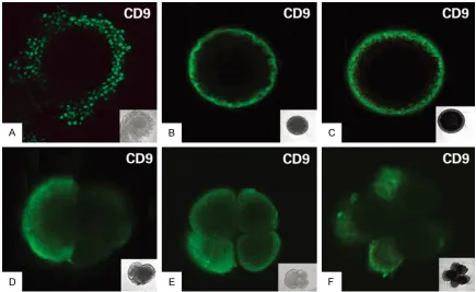
Figure 2. Localization of CD9 protein on cumulus cells, oocytes and early embryos. A. cumulus cells (green); B.
Assessment of sperm penetration
Sperm penetration was assessed 18 h after insemination. Oocytes from each group were
fixed in solution (acetic acid: alcohol 1:3) for 48 h, stained with 1% (w/v) orcein for 5 min and
examined for evidence of sperm penetration under a phase contrast microscope.
Statistical analysis
All experiments were repeated four times ex- ept immunoblotting which was repeated only three times. All percentage data were subject-ed to arc sine transformation before statistical
analysis. Data were analyzed by ANOVA.
Results
Distribution of CD9 in sheep ovarian tissue, oocytes and embryos
As shown in Figure 1, CD9 was present on the membrane at various stages of follicle cells by
immunofluorescent staining. The immunostain -ing was stronger on granulosa cell membrane than that on oocyte plasma membrane in pre-antral follicles (Figure 1G), but the staining on
oocyte plasma membrane was almost the same as that on granulosa cell membrane in the fully grown follicles (Figure 1A and 2A). No staining was observed in the control section (Figure 1B, 1E, 1H).
When the immature oocytes were isolated from antral follicles (3-5 mm in diameter) and cul-tured in vitro for mature, as shown in Figure 2B, 2C, CD9 staining was observed on the mem-brane of oocytes at germinal vesicle(immature)
and metaphase II (mature). The staining was
stronger as the oocyte nuclear stage proceed-ed to MII than at earlier stages.
By using immunofluorescence staining, CD9
antigen was detected to express strongly on the surface of blastomeres of two-, four-, and eight-cell embryos (Figure 2D-F). This expres -sion pattern suggested that CD9 might play a role in sheep embryo development and embryo implantation.
By immunoblotting, a 24 kDa protein was found
in the oocytes at GV and MII stages (Figure 3A), and also found in the embryos (Figure 3B).
[image:6.612.90.524.74.142.2]These results were consistent with those ob-tained by immunofluorescent staining.
Figure 3.The CD9 in the different stages of oocytes and embryos by western blotting. A. Immature and mature
oocytes; B. 2, 4, 8- cells early embryos.
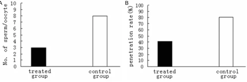
[image:6.612.94.523.194.338.2]Effects of anti-CD9 antibody on sperm-egg binding and sperm penetration
The rate of oocytes reaching the MII stage after 24 h of in vitro maturation was 87.3 ± 1.5%.
When ZP-free oocytes were co-cultured for 45 min with anti-CD9 antibody and then
co-cul-tured with frozen-thawed spermatozoa for 6-8 h, it was found that the number of spermatozoa bound to the oocytes was 4.4 ± 0.1 per oocyte, which was significantly (P < 0.01) fewer than
that in the controls (9.0 ± 1.0 per oocyte). When the oocytes were co-cultured (for IVF) with sper
-matozoa for 20 h, as shown in Figure 4, sperm penetration rate CD9 antibody treatment group
(41.5%) was significantly lower that of control group (81.4%) (P < 0.01).
Discussion
As a member of the tetraspanin family, CD9 is
extensively localized on the membrane of a
variety of cells. CD9 is closely related to other tetraspanin proteins, integrins, IgSF members, glycoproteins, growth factor and other mem-brane proteins [5, 6, 10, 20]. Some proteins in this network participate in many different cel-lular functions, such as adhesion, migration, differentiation, proliferation and signal trans-duction [5, 6]. In the present study, we found that CD9 also located on the plasma mem-brane of ovine oocytes and other cells in prean-tral follicles and fully grown follicles. CD9 was
significantly increased during the final oocyte
maturation, indicating that it is associated with
the competence of the oocyte to be fertilized.
Our data indicated that the presence of CD9 is important for sperm to bind and fuse (penetra-tion) with the oocytes, blocking CD9 by its anti-body inhibits sperm from both binding with and penetrating of oocytes. It has been found that there was a strong CD9 expression on the membrane of oocytes in developing follicles in mouse and the strongest expression was on the membrane of oocytes in fully grown (devel-oped) follicles [21-23]. CD9 was also detected on some cells in the theca layer at the periph-ery of the immature (small) and mature (big) fol-licles, but not in surrounding ovarian tissue [21-24]. Miller et al. [24] reported that there was immunostaining of CD9 on both membrane of oocytes and membrane of cumulus cells but not on ZP in mouse. Houle et al. [25] also found that CD9 expression was in early but not late
corpora lutea in the human ovary. In the pres-ent study, we found that CD9 was extensively expressed in sheep ovarian cells including
oocytes, granulosa cells and theca cells. These
results indicated that CD9 protein was already
synthesized from early follicle development
until oocyte maturation. Most researchers have examined CD9 expression on the membrane of matured mouse oocytes [ 10, 11, 13, 21, 25]. Zhu et al. [13] found that if CD9 mRNA was injected into CD9 knock out mouse oocytes. CD9 could be expressed again on the egg
membrane as revealed by immunofluorescent
staining with anti-mouse CD9 mAb KMC8 or
the anti-human CD9 mAb ALB6. Their results indicated that the localization of CD9 was not
different from that in normal eggs. It has been found that CD9 participates in sperm binding and sperm-egg fusion in mouse, pig and cattle [9-11, 23]. CD9 knockout female mice ovulate normally, and the ovulated oocytes mature to
the MII stage, but they are rarely fertilized
[9-11]. Further studies indicated that sperm were able to adhere to the plasma membrane of ZP-free oocytes from CD9 knockout mouse, but sperm could not fuse with the oocyte
mem-brane [11]. These findings indicate that CD9 on
the membrane of oocytes has an important
effect on fertilization. In the present study, we
found that both sperm binding and
sperm-oocyte fusion were significantly reduced in the
ZP-free sheep oocytes when the CD9 was
blocked by its antibody. These results are the
same as those previously obtained in mice,pig and cattle and they suggest that a similar mechanism may exist for CD9 regulating
fertil-ization in mammals. So far, however, evidence
has only been obtained in mice [10, 11, 13] and pigs [22], cattle [23] and sheep ( present study); whether such a regulation by CD9 during
fertil-ization is present in other mammals remains to be investigated. The mechanisms of CD9 par -ticipates in the sperm-oocyte interaction are not fully understood. Immunoprecipitation and other studies suggest that tetraspanins in the plasma membrane are associated with each other and with several other cell surface mole-cules. In addition, oocytes from CD9 knockout
mice could be fertilized by intracytoplasmic
sperm injection and these embryos developed
to term [11]. These results suggested that CD9
might adjust function through extracellular
may be due to the blocking of sperm-egg
adhe-sion and fuadhe-sion during IVF of sheep oocytes. It
has been reported that another protein integrin
(α6β1) may be the receptor of sperm on the mouse egg surface [26]. The binding of sperm
to egg was achieved by the binding of integrin
α6β1 with the disintegrin domain of fertilin β on
the sperm surface in mice [21, 27]. Several anti-integrin antibodies could inhibit sperm-egg binding in mice, humans and pigs. For example, anti-b1 subunit antibody had a medium inhibi-tory effect on sperm-egg binding during
fertil-ization and it could also inhibit the binding of recombinant fertilin β with mouse oocytes [28]. They might participate in sperm-egg adhesion,
binding and fusion through forming complexes with CD9 or other tetraspanins [29].
Blastocyst adhesion and invasion into uterine endometrium are two important steps of
suc-cessful embryo implantation. To gain an insight
into the cellular and molecular mechanisms that control this process, we have
demonstrat-ed the expression profile of CD9 in pre-implan -tation embryos and the functional role that CD9 plays in embryo implantation in sheep. Results showed that CD9 was expressed on blastomeres of two-, four-, and eight-cell and morula embryos as well as trophoblast cells of blastocysts, and CD9 is involved in the regula-tion of embryo invasion.
Measurement of CD9 expression on sheep granulosa cells and platelets may not be a use-ful indicator for predicting the success of
con-ventional fertilization in couples undergoing IVF. A weak negative relationship between sur -face density of CD9 on granulosa cells and
fer-tilization rate of mature oocytes may reflect a
downregulation of CD9 that accompanies folli-cle maturation at ovulation. More studies are necessary to determine the role of CD9 on granulosa cells during follicle maturity, and to assess if CD9 expression on human granulosa cells could be used as a factor to predict the
success of conventional fertilization during IVF. Fertilization can be blocked by anti-CD9 mAb. These results indicate that CD9 plays an impor -tant role in sheep sperm-oocyte binding, fusion
and fertilization.
Acknowledgements
This work was supported by National Natural
Science Foundation of China (30860189) and
National Natural Science Foundation of China (31360547).
Disclosure of conflict of interest
None.
Address correspondence to: Narankhuar Nasano- chir and Jin Feng, Key Laboratory of Animal Genetics,
Breeding and Reproduction of The Inner Mongolia
Autonomous Region, College of Animal Science, Inner Mongolia Agricultural University, Hohhot 010018, P.R. China. E-mail: 18604716177@163.com (NN); Tel: +86-1363-471 6448, +86-1384-716
4961; E-mail: 13848174696@163.com (JF)
References
[1] Chandra A, Martinez GM, Mosher WD, Abma
JC, Jones J. Fertility, family planning, and repro-ductive health of U.S. women: data from the
2002 National Survey of Family Growth. Vital
Health Stat 2005; 23: 1-160.
[2] Klinovska K, Sebkova, Dvorakova-Hortova K. Sperm-egg fusion: a molecular enigma of mammalian reproduction. Int J Mol Sci 2014; 15: 10652-10668.
[3] Fard Jahromi SS, Shamsir MS. Construction and analysis of the cell surface’s protein net-work for human spermegg interaction. ISRN Bioinformatics 2013; 2013: 1-8.
[4] Berditchevski F. Complexes of tetraspanins with integrins more than meets the eye. J Cell Sci 2001; 114: 4143-4151.
[5] Boucheix C, Rubinstein E. Tetraspanins. Cell
Mol Life Sci 2001; 58: 118-1205.
[6] Hemler ME. Specific tetraspanin function. J
Cell Biol 2001; 155: 1103-1107.
[7] Charrin S, Naour FL, Labas V, Billard M, Le
Caer JP, Emile JF, Petit MA, Boucheix C, Rubinstein E. EWI-2 is a new component of the tetraspanin web in hepatocytes and lymphoid cells. Biochem J 2003; 373: 409-421.
[8] Hemler ME. Tetraspanin proteins mediate cel -lular penetration, invasion, and fusion events
and define a novel type of membrane microdo -main. Annu Rev Cell Dev Biol 2003; 19: 397-422.
[9] Kaji K, Oda S, Shikano T, Ohnuki T, Uematsu Y, Sakagami J, Tada N, Miyazaki S, Kudo A. The
gamete fusion process is defective in eggs of
Cd9-deficient mice. Nat Genet 2000; 24:
279-82.
[10] Le Naour F, Rubinstein E, Jasmin C, Prenant M, Boucheix C. Severely reduced female fertility in
CD9-deficient mice. Science 2000; 287:
319-21.
[11] Miyado K, Yamada G, Yamada S, Hasuwa H,
Ogura A, Okabe M and Mekada E. Requirement of CD9 on the egg plasma membrane for
fertil-ization. Science 2000; 287: 321-324.
[12] Kaji K, Oda S, Miyazaki S, Kudo A. Infertility of CD9-deficient mouse eggs is reversed by
mouse CD9, human CD9, or mouse CD81: polyadenylated mRNA injection developed for molecular analysis of sperm-egg fusion. Dev Biol 2002; 247: 327-334.
[13] GZ Zhu, Miller BJ, Boucheix C. Residues SFQ (173-175) in the large extracellular loop of CD9 are required for gamete fusion. Develop- ment 2002; 129: 1995-2002.
[14] Park KR, Inoue T, Ueda M, Hirano T, Higuchi T,
Maeda M, Konishi I, Fujiwara H, Fujii S. CD9 is expressed on human endometrial epithelial
cells in association with integrins α6, α3 and β1. Molecular Human Reproduction 2000; 6:
252-257.
[15] Xiang W, Maclaren LA. Expression of fertilin
and CD9 in bovine trophoblast and endome-trium during implantation. Biol Reprod 2002; 66: 1790-1796.
[16] Takao Y, Fujiwara H, Yamada S, Hirano T,
Maeda M, Fujii S, Ueda M. CD9 is expressed on the cell surface of human granulosa cells and associated with integrin α6β1. Mol Hum Reprod 1999; 5: 303-310.
[17] Clavero A, Castilla JA, Martinez L, Mendoza N, Fontes J, Maldonado V. Expression of integrin
fraction and adhesion molecules on human granulosa cells and its relation with oocyte ma-turity and follicular steroidogenesis. J Assist Reprod Genet 2004; 21: 187-195.
[18] Patterson KS, RW Ke, MV Jacoski. Expression
of cell surface CD9 in fertile and infertile wom-en. Fertility and Sterility 2004; 82: 283. [19] Hemler ME. Integrin associated proteins. Curr
Opin Cell Biol 1998; 10: 578-585.
[20] Berditchevski F, Odintsova E. Characterization
of integrin-tetraspanin adhesion complexes: role of tetraspanins in integrin signaling. J Cell Biol 1999; 146: 477-492.
[21] Chen MS,Tung KS, Coonrod SA, Takahashi Y,
Bigler D, Chang A, Yamashita Y, Kincade PW, Herr JC, White JM. Role of the integrin-associ-ated protein CD9 in binding between sperm
ADAM 2 and the egg integrin α6β1: implica
-tions for murine fertilization. Proc Natl Acad Sci
1999; 96: 11830-11835.
[22] Li YH, Hou Y, Ma W, Yuan JX, Zhang D, Sun QY, Wang WH. Localazation of CD9 in pig oocytes
and its effects on sperm-egg interaction. Reproduction 2004; 127: 151-157.
[23] Zhou GB, Liu GS, Meng QG, Liu Y, Hou YP,
Wang XX, Li N, Zhu SE. Tetraspanin CD9 in bo
-vine oocytes and its role in fertilization. J
Reprod Dev 2009; 55: 305-8.
[24] Miller BJ, Georges LE, Primakoff P, Myles DG.
Normal fertilization occurs with eggs lacking the integrin α6β1 and is CD9-dependent. J Cell
Biol 2000; 149: 1289-1296.
[25] Houle CD, Ding XY, Foley JF, Afshari CA, Barrett
JC, Davis BJ. Loss of expression and altered
localization of KAI1 and CD9 protein are asso -ciated with epithelial ovarian cancer progres-sion. Gynecol Oncol 2002; 86: 69-78.
[26] Almeida EAC, Huovila APJ, Sutherland AE, Stephens LE, Calarco PG, Shaw LM, Mercuri AM, Sonnenberg A, Primakoff P, Myles DG,
White JM. Mouse egg integrin α6β1 functions
as a sperm receptor. Cell 1995; 81: 1095-1104.
[27] Evans JP. Fertilin beta and other ADAMs as in-tegrin ligands: insights into cell adhesion and
fertilization. Bioessays 2001; 23: 628-639.
[28] Evans JP, Kopf GS, Schultz RM. Characterization
of the binding of recombinant mouse sperm fertilin beta subunit to mouse eggs: evidence for adhesive activity via an egg beta1 integrin-mediated interaction. Dev Biol 1997; 187: 79-93.
[29] Gutierrez-Lopez MD, Ovalle S, Yanez-Mo M, Sanchez-Sanchez N, Rubinstein E, Olmo N, Lizarbe MA, Sanchez-Madrid F, Cabanas C. A