Original Article
Mouse hepatitis virus nucleocapsid protein interacts
receptor for activated C kinase1, and
regulates AKT signal pathway
Pengju Zhang1*, Jun Wang2*, Miao Long3, Xinghong Wu1
1Jilin Academy of Agricultural Sciences, #1363 Shengtai Street, Changchun 130124, China; 2Jilin Agricultural
University, Changchun, China; 3Shenyang Agricultural University, Shenyang, China. *Equal contributors. Received September 26, 2016; Accepted November 2, 2016; Epub March 15, 2017; Published March 30, 2017
Abstract: The nucleocapsid (N) protein of mouse hepatitis virus (MHV-N) has been reported to be a multifunctional protein involved in viral RNA replication and translation. However, how N protein interacts with host protein re-mains largely elusive. To identify cellular proteins that interact with the N protein, a cDNA library of mouse liver was screened using a yeast two-hybrid system assay. We have identified the receptor for activated C kinase 1 (RACK1) as a novel interaction partner of N protein by yeast two-hybrid system. The direct interaction and co-localization of N protein with RACK1 were confirmed by immunoprecipitation and confocal microscopy analysis, respectively. The mapping studies localized the critical N sequences for this interaction to amino acid 150-220 including SR motif by yeast two-hybrid system. Function assay showed that overexpressin N protein significantly inhibited RACK1 expres-sion, thereby leading to suppress Akt pathway activation, which might contribute to induce apoptosis of host cells. To the best of our knowledge, this is the first report that MHV-N protein interacts with the RACK1 within host cells. These findings contribute to enhance our understanding of the pathological mechanisms of MHV.
Keywords: Hepatitis virus, nucleocapsid, receptor for activated C kinase 1, interaction
Introduction
As a member of the family Coronaviridae, mouse hepatitis virus (MHV) is a positive-strand RNA virus representing a significant ubiquitous group of viral pathogens. MHV is a natural pathogen of mice, normally infecting the liver, gastrointestinal tract, and central ner-vous system, causing a wide range of disease, including hepatitis, gastroenteritis, and acute and chronic encephalomyelitis [1-3]. Although MHV had been the wide-studied coronavirus both in vivo and in vitro as well as at the molec-ular level, many scientific questions remain to be answered, such as which specific features of the virus are responsible for its pathogenicity, and which mechanisms are at work during infection. Therefore, understanding the infec-tion mechanism at the molecular level of MHV is crucial need, since it is a foundation for dis-covering and developing anti-MHV drugs or vaccine.
Like other known coronaviruses, MHV is an enveloped virus containing three outer
struc-tural proteins, namely the membrane (M), nucleocapsid (N), and spike (S) proteins [4, 5]. The N protein is the most abundant viral protein in coronaviruses which is produced throughout infection and is an important multifunctional protein. Several functions have been postulat-ed for the coronavirus N protein throughout the virus life cycle, including viral packaging, viral core formation, and signal transduction [6, 7]. Primarily, the N protein of MHV is an extensively phosphorylated, highly basic and vital structur-al protein, the function of which is to form a heli-cal ribonucleoprotein complex with viral RNA; the complex comprises the core structure of the MHV virion. Moreover, the N protein shows intrinsic multimerization and interacts with M protein, suggesting that it is both critical to for-mation of the viral nucleocapsid core and is involved in virion assembly [8, 9].
virus particle assembly and release [10-12]. It has been reported that the N protein of MHV interacts with mouse cellular heterogeneous nuclear ribonucleoprotein A1 (hnRNPA1) [13], which showed that N protein plays a crucial role in viral RNP assembly and replication by inter-action with host proteins. However, how the N protein interacts with other host protein is still unclear. To obtain a more detailed insight into the relationship of the N protein and host cell protein, a yeast two-hybrid system was employed in the present study to identify which proteins can bind to N protein of MHV.
Materials and methods
Strains and general techniques
The strain of Saccharomyces cerevisiae used in this study was AH109 from Clontech (USA). Yeast cells were cultured at 30°C either in a complete YPD medium (1% yeast extract, 1% peptone, 2% glucose) or in a synthetic defined (SD) medium supplemented with required essential nutrients. Plates contained 1-2%
Plasmids and construction of recombinant vectors
[image:2.612.90.371.69.338.2]For bait construction, the full-length N gene of the MHV (MHV-A59) was amplified from a genomic construct of clone by PCR, and cloned into the pMD18-T vector (Takara, Dalian, Japan), and designated as pMD18-T-N. The full-length N gene was subjected to DNA sequenc-ing, and the inserts were verified against the corresponding region of the MHV coronavirus. The N gene was excised from the pMD18-T-N construct using the restriction enzymes EcoR I and BamHI, and ligated into the pGBKT7 vector to generate an N-terminal in frame fusion with the GAL4 activation domain (BD), and the resul-tant plasmid was named as pGBKT7-N. To iden -tify the putative domain of amino acid sequence required for RACK1/N interaction, The truncat -ed mutants N1-149, N150-300, N301-455, N150-220 and N221-300 (constructs and putative functional domains were shown in Figure 1A) were subcloned into pGBKT7. The RACK1 gene was amplified by PCR using the
Figure 1. Analysis of the N and RACK protein interaction by yeast two-hybrid. A. Schematic representation of the MHV-N protein and the truncated mu-tants used in the yeast two-hybrid system, the SR-rich motif is highlighted. B. Mapping the interaction domain of MHV-N. The empty vectors pGBKT7 and pGADT7 co-transformed were used as the negative control and the pG-BKT7-53 and pGADT7-T co-transformed were used as the positive control. Every experiment was repeated for at least three times and the data were obtained by average. The error bars represent standard error of the mean.
agar. Transformation of yeast cells was performed by the lithium acetate. Eherichiacoli KC8 was used for general cloning. DNA manipulation was performed according to established protocol. Beta-galactosidase assays were carried out according to the Clontech Matchmaker manual (PT3024-1, Clontech, USA).
Cell culture
mouse liver cDNA library, and subcloned into the yeast vector pGADT7. For mammalian cell expression, the full-length N gene and RACK1 were subcloned into the pCMV-Myc vector (Clontech, USA) and pCMV-HA, and fluores -cence vector pEGFP-N1 and pDsRed-N1, respectively. siRNA against RACK1 (si-RACK1), siRNA against negative scramble control (si-NC) were purchased from RiboBio (Guangzhou, China). In addition, the N gene was amplified by PCR and inserted into eukaryotic expression vector pCDNA3.0 (Invitrogen, Grand Island, NY, USA) to study its function. All constructs were verified by restriction digestion and sequen-cing.
Yeast two-hybrid screening
Yeast two-hybrid experiments were performed as described in the Clontech manual for the Matchmarker GAL4 two-hybrid system and Clontech yeast protocols handbook (Clontech, USA). Briefly, the mouse liver Matchmaker cDNA library (Clontech, USA) was cloned in frame with the GAL4 activation domain in the PGADT7 vector as a prey. The bait pGBKT7-N was transformed into yeast strain AH109. The bait construct did not show any toxic effect and autonomous transcriptional activation on the host strain. The prey PGADT7-library was then transformed into the bait-transformed AH109 cells, and the cells were incubated on minimal synthetic dropout medium for yeast (SD)/-His/-Leu/-Trp at 30°C. The fresh growing clones were assayed for β-galactosidase activity using
ay, co-immunoprecipitation was performed as previous described [13].
Confocal microscopy
293T cells were grown on coverslips in a 6-well chamber and simultaneously transfected with the recombinant plasmids pEGFP-N and pDsRed-RACK1. After 24 h transfection, the cells were washed with PBS three times and fixed in 4% paraformaldehyde for 20 min at room temperature. The coverslips were then washed with PBS and mounted. Intracellular localization of the N protein and RACK1 was observed under a Leica confocal microscope (Germany).
Western blot assay
[image:3.612.91.374.73.220.2]Total proteins were extracted from cultured cells by using lysis buffer (Beyotime Institute of Biotechnology, Guangzhou, China). The protein concentration was quantified by the bicincho -ninic acid protein assay kit (Beyotime). Equal amounts of protein (30 μg) was insolated using sodium dodecyl sulfate polyacrylamide gel electrophoresis (SDS-PAGE), and then electro-transferred to the polyvinylidene difluoride membranes (PVDF, Millipore, Bedford, MA, USA). The membranes were blocked in 5% non-fat dry milk diluted with TBST (20 mM Tris-HCl, pH 7.5, 150 mM NaCl, 0.1% Tween-20) at room temperature for 2 h and incubated overnight at 4°C with primary antibody against RACK1, Akt, p-Akt (Ser473) and GAPDH (all from Santa Cruz
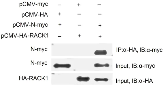
Figure 2. RACK1 protein was immunoprecipitated with the MHV-N protein. Indicated plasmids were simultaneously transfected into 293T cells. Twenty-four hours after transfection, co-immunoprecipitation was performed using anti-HA magnetic microbeads, the proteins immunoprecipitated (IP) were as-sayed with an anti-myc monoclonal antibody. Cell lysates were immunoblot-ted (IB) with anti-Myc to confirm the expression of the object proteins.
the substrate ONPG as desc- ribed standard Protocols Han- dbook (PT3024-1, Clontech, USA). The positive pGADT7-cDNA plasmids were isolated from positive yeast transfor-mants by culture in leucine-deficient medium, and trans -formed into E. coli KC8 for sequence analysis. The result-ing sequence was analyzed in the database of EMBL\Gene Bank by the BLAST program.
In vivo co-immunoprecipita-tion
Biotechnology Inc., California, USA). After the membrane was washed three times with TBST, was further probed with horseradish peroxi-dase (HRP)-conjugated second antibody (Santa Cruz, USA) for 2 h at room temperature. The protein bland was detected with using the enhanced chemiluminescence (ECL) luminol reagent (PerkinElmer Inc.).
Statistical analysis
The data were expressed as the mean ± SD (standard deviation) from at least three inde-pendent experiments. All data are analyzed by Statistical SPSS Version 19.0 (IBM, Chicago, USA). Group differences were compared using two-tailed Student’s T-test or one-way ANOVA. The differences were considered to be statisti-cally significant at P < 0.05.
Results
Yeast two-hybrid screens identified RACK1 as an N interacting protein
A yeast two-hybrid approach was used to iden-tify host proteins that interact with the N pro-tein. Through β-galactosidase assay and tech -nique of segregation analysis. 6 clones were considered to be possible positive clone. One of these clones was identified as receptor for activated C kinase 1 (RACK1), which encodes a 317-amino acid protein. To confirm this protein interaction with N protein, we cloned RACK1 full length by PCR, and subcloned it into pGBK, and then yeast two-hybrid assay and β-galac-tosidase assay were performed. As shown in Figure 1, the protein encoded by the pGAD-RACK1 clones interacted specifically with the N protein and did not interact with the unfused
GAL4-BD protein expressed from the parental pGBKT7 vector.
To map the involved regions of the N protein in the N/RACK1 interaction, the full-length N pro -tein was divided into three domains to study the binding domain of the N protein: the N-terminal domain N1-149, the middle domain N150-300 containing the SR-rich motif and the C-terminal domain N301-454 (Figure 1A). These three truncated proteins were tested for RACK1 binding using yeast two-hybrid assay. The results indicated that N150-300, the mid-dle domain of the N protein, is responsible for the interaction (Figure 1B). To determine fur-ther the region of the N protein involved in RACK1 binding, the middle domain was divided into two parts, N150-220 containing the SR-rich motif and N221-300, In the yeast two-hybrid assay, the β-galactosidase activity of N150-220 fragment is much higher than the activity of N221-300 fragment (Figure 1B). These results implied that the SR-rich motif of the N protein is responsible for the majority of the binding to RACK1.
Co-immunoprecipitation determined the inter-action of the N protein and RACK1
[image:4.612.92.521.72.191.2]To confirm the specific interaction between the N protein and RACK1 protein, co-immunopre -cipitation was performed. The N protein was fused at the amino terminus with a Myc-tag, and RACK1 was fused at the carboxyl terminus with a HA tag. The two plasmids were cotrans-fected into 293T cells and immunoprecipitated. The immunoprecipitated complexes were sepa-rated on SDS-PAGE, and analyzed by Western blot with anti-Myc monoclonal antibodies. As shown in Figure 2, the Myc-fused N protein
immunoprecipitated with HA-RACK1. However, it did not immunoprecipitate with HA alone. These findings indicate that N protein interacts with RACK1 in mammalian cells.
Confocal microscopy assay localized the N pro-tein and RACK1
To further confirm the interaction between N and RACK1 proteins, the localization patterns of the N protein and RACK1 were investigated in 293T cells using confocal microscopy tech-nology. pEGFP-N and pDsRed-RACK1 were transfected simultaneously into 293T cells. As shown in Figure 3, RACK1 and N protein mainly were localized in the cytoplasm. The merged image revealed that the N protein and RACK1 co-localized in the cytoplasm of 293T cells indi-cating that the N protein interacts with RACK1 in cells.
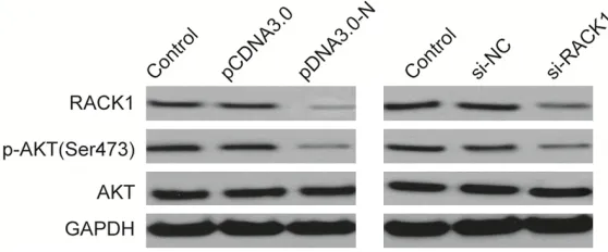
MHV-N downregulated RACK1 expression and inhibited activation AKT signaling pathway
RACK1 (receptor for activated C-kinase1), as a crucial scaffold protein, has been reported to involve in various biological procession includ-ing cell proliferation, cell cycle, metastasis and apoptosis by regulating multiple signaling path-ways, such as PKC pathway, AKT pathway, and IGF1R pathway [14, 15]. Here, we only focus- ed on AKT signal pathway which involved in virus infection, including Coronaviridae [16, 17]. Therefore, we hypothesized that by inter-action with RACK1, N protein might affect the AKT signaling pathway in infected cells. To test this hypothesis, RACK1, AKT and pAKT protein levels were determined in vero cells after
trans-function by interaction with RACK1, and thus indirectly regulating the AKT signaling pathway.
Discussion
In the present study, we have firstly reported that the MHV-N protein has a specific binding to mouse RACK1, and the further yeast two-hybrid assay demonstrated that the fragment amino acid 150-220 of MHV-N probably contribute to the N/RACK1 interaction. RACK1 was originally identified on the basis of its ability to bind the activated form of PKC, stabilize this protein and facilitate its trafficking within the cell [18]. It has been reported that RACK1 played a role in complex protein-protein interactions between signaling molecules, such as integrins, phos-phodiesterase 4D5, Src tyrosine kinase, RhoA small GTPase, as well as PKC [19, 20]. Our results combined with previous study demon-strated that RACK1 may function as a scaffold -ing protein to mediate protein-protein interac-tion and facilitate tight regulainterac-tion of various cellular biological functions, including cell cycle, survival, adhesion, and migration.
[image:5.612.91.370.74.190.2]Accumulating evidence has implied that RACK1 was involved in various biological procession by regulating multiple signal pathways, inculding AKT pathway, IGF1R pathway, and Sonic hedge -hog signaling pathway [14, 15, 18, 21, 22]. In this study, we only focused on AKT signal path -way since this path-way has been confirmed to involve in Coronaviridae infection, and induce apoptosis of host cells [16, 17, 23]. In this study, we found that overexpression of MHV-N or knockdown of RACK1 in vero cells signifi -cantly inhibited RACK1 expression and phos -pho-Akt expression, suggesting that MHV-N
Figure 4. MHV-N downregulated RACK1 expression and inhibited
activa-tion AKT signaling pathway. Western blot analysis of p-AKT, AKT and RACK1
protein levels in U2OS cells transfected with N overexpression plasmid (pCDNA3.0-N) or si-RACK1. GAPDH was used as internal control.
protein targets RACK1, and thus indirectly regu -lating the AKT signaling pathway. Previous a report showed that that SARS coronavirus nucleocapsid protein induced apoptosis of host cells by regulating MAPK pathway and AKT pathway [23]. Our study together with previous study implied that coronavirus nucleocapsid protein could induce apoptosis of host cells by regulating AKT signal pathway.
In addition, the yeast two-hybrid result has clearly indicated that only one fragment of MHV-N (aa 150-220) is required for N/RACK1 interaction. To our knowledge, the fragment aa 150-220 contains SR-rich motif (containing rich serine and arginine) that is multifunctional and conserved in the N protein of coronavirus-es [13]. The SR-related proteins are often involved in protein-RNA and protein-protein interactions, and the SR-rich motif is conserved in the N protein of coronavirus [24]. It has been reported that SR-rich motif is indispensable for SARS N oligomerization and for N protein inter-action with SARS-CoV membrane protein [25]. The SR-rich motif is also responsible for the interaction with hnRNPA1 in SARS [26]. In addi-tion, this motif is also involved in interaction with Hubc9 in SARS [27]. All of these facts indi-cate that the SR-rich motif of the N protein might play a crucial role in MHV infection.
In conclusion, our data have shown for the first time that the nucleocapsid protein of MHV has a high binding affinity to mouse RACK1 and such a protein-protein interaction involves the region aa 150-220 of MHV-N. In addition, our study also revealed that MHV-N overexpression could decrease RACK1 expression and down -regulate its downstream signal pathway, AKT pathway. To the best of our knowledge, this is the first report that MHV-N protein interacts with the receptor for activated C kinase 1 (RACK1) within host cells and our findings con -tribute to enhance our understanding of the pathological mechanisms of MHV.
Disclosure of conflict of interest
None.
Address correspondence to: Xinghong Wu, Jilin Academy of Agricultural Sciences, #1363 Shengtai Street, Changchun 130124, China. E-mail: wuxh@ cjaas.com
References
[1] Rose KM, Elliott R, Martinez-Sobrido L, Garcia-Sastre A and Weiss SR. Murine coronavirus delays expression of a subset of interferon-stimulated genes. J Virol 2010; 84: 5656-5669.
[2] Rose KM and Weiss SR. Murine coronavirus cell type dependent interaction with the type I interferon response. Viruses 2009; 1: 689-712.
[3] Ye Y, Hauns K, Langland JO, Jacobs BL and Hogue BG. Mouse hepatitis coronavirus A59 nucleocapsid protein is a type I interferon an-tagonist. J Virol 2007; 81: 2554-2563. [4] Lai MM and Cavanagh D. The molecular
biolo-gy of coronaviruses. Adv Virus Res 1997; 48: 1-100.
[5] Versteeg GA, Bredenbeek PJ, van den Worm SH and Spaan WJ. Group 2 coronaviruses pre-vent immediate early interferon induction by protection of viral RNA from host cell recogni-tion. Virology 2007; 361: 18-26.
[6] He R, Leeson A, Andonov A, Li Y, Bastien N, Cao J, Osiowy C, Dobie F, Cutts T, Ballantine M and Li X. Activation of AP-1 signal transduction pathway by SARS coronavirus nucleocapsid protein. Biochem Biophys Res Commun 2003; 311: 870-876.
[7] Hiscox JA, Wurm T, Wilson L, Britton P, Cavanagh D and Brooks G. The coronavirus in-fectious bronchitis virus nucleoprotein localiz-es to the nucleolus. J Virol 2001; 75: 506-512. [8] He R, Dobie F, Ballantine M, Leeson A, Li Y,
Bastien N, Cutts T, Andonov A, Cao J, Booth TF, Plummer FA, Tyler S, Baker L and Li X. Analysis of multimerization of the SARS coronavirus nu-cleocapsid protein. Biochem Biophys Res Commun 2004; 316: 476-483.
[9] He R, Leeson A, Ballantine M, Andonov A, Baker L, Dobie F, Li Y, Bastien N, Feldmann H, Strocher U, Theriault S, Cutts T, Cao J, Booth TF, Plummer FA, Tyler S and Li X. Charact- erization of protein-protein interactions be-tween the nucleocapsid protein and mem-brane protein of the SARS coronavirus. Virus Res 2004; 105: 121-125.
[10] Hogue BG. Bovine coronavirus nucleocapsid protein processing and assembly. Adv Exp Med Biol 1995; 380: 259-263.
[11] Narayanan K, Chen CJ, Maeda J and Makino S. Nucleocapsid-independent specific viral RNA packaging via viral envelope protein and viral RNA signal. J Virol 2003; 77: 2922-2927. [12] Narayanan K, Maeda A, Maeda J and Makino
[13] Wang Y and Zhang X. The nucleocapsid protein of coronavirus mouse hepatitis virus interacts with the cellular heterogeneous nuclear ribo-nucleoprotein A1 in vitro and in vivo. Virology 1999; 265: 96-109.
[14] Battaini F, Pascale A, Paoletti R and Govoni S. The role of anchoring protein RACK1 in PKC ac-tivation in the ageing rat brain. Trends Neurosci 1997; 20: 410-415.
[15] Wang F, Yamauchi M, Muramatsu M, Osawa T, Tsuchida R and Shibuya M. RACK1 regulates VEGF/Flt1-mediated cell migration via activa-tion of a PI3K/Akt pathway. J Biol Chem 2011; 286: 9097-9106.
[16] Tsoi H, Li L, Chen ZS, Lau KF, Tsui SK and Chan HY. The SARS-coronavirus membrane protein induces apoptosis via interfering with PDK1-PKB/Akt signalling. Biochem J 2014; 464: 439-447.
[17] Chan CM, Ma CW, Chan WY and Chan HY. The SARS-Coronavirus Membrane protein induces apoptosis through modulating the Akt survival pathway. Arch Biochem Biophys 2007; 459: 197-207.
[18] Ron D, Chen CH, Caldwell J, Jamieson L, Orr E and Mochly-Rosen D. Cloning of an intracellu-lar receptor for protein kinase C: a homolog of the beta subunit of G proteins. Proc Natl Acad Sci U S A 1994; 91: 839-843.
[19] McCahill A, Warwicker J, Bolger GB, Houslay MD and Yarwood SJ. The RACK1 scaffold pro-tein: a dynamic cog in cell response mecha-nisms. Mol Pharmacol 2002; 62: 1261-1273. [20] Chiba Y, Tanabe M, Sakai H, Kimura S and
Misawa M. A functional interaction between CPI-17 and RACK1 proteins in bronchial smooth muscle cells. Biochem Biophys Res Commun 2010; 401: 487-490.
[21] Shi S, Deng YZ, Zhao JS, Ji XD, Shi J, Feng YX, Li G, Li JJ, Zhu D, Koeffler HP, Zhao Y and Xie D. RACK1 promotes non-small-cell lung cancer tumorigenicity through activating sonic hedge-hog signaling pathway. J Biol Chem 2012; 287: 7845-7858.
[22] Peng R, Jiang B, Ma J, Ma Z, Wan X, Liu H, Chen Z, Cheng Q and Chen R. Forced downregula-tion of RACK1 inhibits glioma development by suppressing Src/Akt signaling activity. Oncol Rep 2013; 30: 2195-2202.
[23] Surjit M, Liu B, Jameel S, Chow VT and Lal SK. The SARS coronavirus nucleocapsid protein in-duces actin reorganization and apoptosis in COS-1 cells in the absence of growth factors. Biochem J 2004; 383: 13-18.
[24] Toney JH, Navas-Martin S, Weiss SR and Koeller A. Sabadinine: a potential non-peptide anti-severe acute-respiratory-syndrome agent identified using structure-aided design. J Med Chem 2004; 47: 1079-1080.
[25] Blencowe BJ, Bowman JA, McCracken S and Rosonina E. SR-related proteins and the pro-cessing of messenger RNA precursors. Bio- chem Cell Biol 1999; 77: 277-291.
[26] Luo H, Chen Q, Chen J, Chen K, Shen X and Jiang H. The nucleocapsid protein of SARS coronavirus has a high binding affinity to the human cellular heterogeneous nuclear ribonu-cleoprotein A1. FEBS Lett 2005; 579: 2623-2628.