Original Article
Expression and function of TAK1 in osteosarcoma tissue
Yuxi Wei1*, Ruiming Zhou1*, Quanbing Wang1, Beibei Fu2, Weili Jing3, Honglin Wang1
Departments of 1Orthopedics, 2Neurology, Renmin Hospital, Hubei University of Medicine, Shiyan 442000, Hubei, P. R. China; 3Taihe Hospital, Hubei University of Medicine, No. 39 Chaoyang Road, Maojian District, Shiyan 442000, Hubei, P. R. China. *Co-first authors.
Received January 12, 2016; Accepted May 19, 2016; Epub June 15, 2016; Published June 30, 2016
Abstract: Objective: To investigate the expression and function of transforming growth factor β-activated kinase 1 (TAK1) in osteosarcoma (OS) tissue. Methods: TAK1 protein expression in OS and adjacent normal tissues was detected by immunohistochemistry. The association of TAK1 expression with clinicopathological features and prog-nosis in OS patients was analyzed. In vitro cell culture experiments were performed using a selective TAK1 inhibitor, 5Z-7-oxozeaenol, to determine the effects of TAK1 on proliferation and apoptosis in human U2OS cells. Results: The positive expression rate of TAK1 was higher in OS tissue (73.6%) than in adjacent normal bone tissue (19.8%, P < 0.05). TAK1 expression was unrelated to age, tumor site, gender, or subtype of the OS patients (P > 0.05). However, TAK1 expression was closely associated with the presence or absence of OS metastasis and Enneking stage (P < 0.05). TAK1-positive patients had shorter 5-year survival times than TAK1-negative patients (P < 0.05). A mechanis-tic study revealed that the TAK1 inhibitor 5Z-7-oxozeaenol inhibited proliferation and promoted apoptosis in an OS cell line (P < 0.05). Western blotting analysis showed that 5Z-7-oxozeaenol suppressed p-TAK1 (Thr87), cyclin D1, and intranuclear p65 expression levels, whereas it increased cleaved-caspase 3 protein levels in OS cells (P < 0.05). Conclusion: TAK1 expression was significantly associated with clinical stage and prognosis in OS patients, and this protein may represent a new target for the treatment of OS.
Keywords: Osteosarcoma, TAK1
Introduction
Osteosarcoma (OS) is the most common type of malignant bone tumor, and it primarily occurs in adolescents, with a high mortality. However, the molecular mechanisms underlying OS de- velopment and progression have not been fully
elucidated, leading to a lack of specific thera -peutic clinical drugs [1]. Therefore, in-depth studies of the pathological mechanisms under-lying OS development and progression could lead to the development of targeted therapeu-tic drugs, potentially improving the prognosis of OS patients. Recent studies have found that
transforming growth factor β-activated kinase
1 (TAK1) plays an essential role in the develop-ment and progression of various tumor tissues, including esophageal cancer [2], breast cancer [3], ovarian cancer [4], liver cancer [5], pancre-atic cancer [3], and colon cancer [6]. However,
little is known about the expression and role of TAK1 in OS. In the present study, we first ana
-lyzed the expression and clinical significance of
TAK1 in OS tissue. Then, we used in vitro cell culture experiments and a selective TAK1 inhib-itor, 5Z-7-oxozeaenol (5Z7O), to assess the role of TAK1 in OS development and progression. Our results reveal novel insights into the role of TAK1 in OS and point to new approaches for the treatment of this disease.
Materials and methods
General data
In total, tumor and peritumoral normal bone tissue samples were selected from 106 patients with OS. These patients underwent surgical treatment in our hospital between August 2000 and August 2010, and OS was
postoper-ative auxiliary examination results, pathological stage, and therapeutic conditions. The study excluded patients who had received radiothera-py or chemotheraradiothera-py prior to surgery. Patients were followed up with every three months, either by telephone or hospital visit. The
follow-up duration was five years.
Immunohistochemistry and hematoxylin & eo-sin staining
Surgical specimens of OS and adjacent normal
tissues were fixed in 10% formalin and embed
-ded in paraffin. Specimens were then sliced into 4-µm-thick sections using a microtome and baked in an oven at 60°C for 8 h prior to
use. Immunohistochemical (IHC) detection was
performed as follows: paraffin sections were deparaffinized and rehydrated. After washing
with phosphate-buffered saline (PBS), speci-men antigens were retrieved by treatspeci-ment with
an EDTA buffer at 100°C for 20 min, followed by
natural cooling to room temperature. The sam-ples were washed with distilled water, and
per-oxidase was added dropwise to the block solu -tion. Specimens were incubated at room tem-perature, and after washing the specimens with PBS, non-immune animal serum was added dropwise. The specimens in solution were incubated at room temperature for 10 min and then decanted. Anti-TAK1 primary antibody (Santa Cruz Biotechnology, Santa Cruz, CA, USA; 1:50 dilution) was added dropwise, and the specimens were incubated at room tem-perature for 60 min. PBS was substituted for the primary antibody as a negative control. After washing the specimens with PBS, a biotin-labeled goat anti-rabbit IgG secondary antibody (ZsBio, Beijing, China) was added, and the specimens were incubated at room tempera-ture for 10 min. Following a PBS wash, a strep-tavidin-biotin-peroxidase solution was added dropwise, and the specimens were incubated at room temperature for 10 min. After washing the specimens with PBS, a colorimetric reac-tion was performed using the DAB chromogenic reagent. After washing the specimens with tap water, they were counter-stained with hema-toxylin and then washed with tap water, dehy-drated with alcohol, cleared with xylene, and mounted with neutral gum. Hematoxylin & eosin (HE) staining was performed according to standard procedures.
Three experienced pathologists independently interpreted the IHC results, as previously des-
cribed [7]. Briefly, the double-blind method was used to count cells within 15 high-magnifica
-tion (× 400) fields of view for each sec-tion spec -imen. The average number of TAK1-positive
cells per 100 cells was defined as the positive
rate for TAK1. Positive IHC data were interpret-ed using guidelines in the literature, which were
based on two criteria. The first criterion was the
intensity of positively stained cells: negative was scored as 0 points, pale yellow as 1 point,
yellow as 2 points, and brown as 3 points (final
interpretation: 0-2 points as negative and 3-7 points as positive). The second criterion was the percentage of positive cells: 0%-5% was
scored as negative, 6%-25% as weakly positive,
26%-75% as moderately positive, and > 76% as
strongly positive (final interpretation: weakly
positive, moderately positive, and strongly
posi-tive were defined as a posiposi-tive result).
Cell culturing and grouping
The human OS cell line U2OS was provided
by the Cell Bank of Shanghai Institutes for
Biological Sciences of Chinese Academy of Sciences. After they were thawed, the cells
were seeded into culture flasks and incubated at 37°C under 5% CO2. Logarithmic phase U2OS cells were divided into four groups for
the experiments: blank, control (solvent),
low-dose 5Z7O treatment (500 nM), and high-low-dose 5Z7O treatment (1000 nM). Thirty-nine repli-cate wells were set for each group. 5Z7O was purchased from Sigma-Aldrich (St. Louis, MO, USA).
MTT cell proliferation assay
After 5Z7O treatment, culture plates were pl-
aced in a cell incubator at 37°C under 5% CO2
for 24, 48, or 72 h. Four hours before the end of the incubation, 20 µL of an MTT solution was added to each well. At the end of the incubation period, the supernatant from each well was
aspirated, and 150 μL of DMSO was added to
each well. The plates were oscillated for 10 min, and the absorbance was measured at 570 nm (A570) using an ELISA analyzer. The results are expressed as the mean value from six repli-cate wells. The cell proliferation inhibition rate for each group was calculated as follows: inhibi-tion rate = (1-OD of the experimental group/OD
Flow cytometry assay for measuring apoptosis
Apoptosis in human U2OS cells was detected
using an Annexin V-FITC kit (Biovision, San
Francisco, CA, USA), according to the manufac-turer’s instructions. Cells harvested at 24, 48, or 72 h were digested with trypsin and then resuspended in complete medium. After cen-trifugation, the supernatant was discarded. Following a PBS wash, 1 × binding buffer was added dropwise to the cells. Each sample was then treated with 5 µL Annexin V-FITC and 10 µL PI. The samples were thoroughly mixed before analysis.
Western blotting assay of cell proliferation
Human OS cells were harvested after 48 h of treatment. Protein was extracted using a cell
total protein and nuclear protein extraction kit
(Beyotime, Nantong, China), according to the manufacturer’s instructions. The BCA method was used for protein quantitation. Thirty micro-grams of each protein sample was separated
by electrophoresis and transferred to a
mem-brane. The membranes was blocked with block -ing solution (Beyotime) for 1 h and then incu-bated with the diluted primary antibodies
against β-actin, H3, p65, p-TAK1 (Thr187), and
cleaved-caspase-3 (CST, Beverly, MA, USA) at
4°C overnight. After they were washed, the
membranes were incubated with horseradish peroxidase-conjugated goat anti-rabbit
second-ary antibody diluted in 5% milk. After the mem -branes were washed, a luminescence solution was added dropwise to the membrane. Images were acquired using a gel imager, and gray scale data analysis was performed using Image J.
Statistical analysis
Data were processed using the SPSS 19.0 Statistics software program (IBM SPSS, So- mers, NY, USA). Count data were analyzed using
[image:3.612.92.523.73.399.2]the χ2 test. Means were compared among mul-tiple groups using one-way ANOVA with post-hoc multiple comparisons by LSD-t test. The
survival rate of OS patients with different char-acteristics was estimated using the Kaplan-Meier method, and univariate analysis was
per-formed using the log-rank test. Multivariate
analysis of prognostic factors for OS patients was conducted using the Cox proportional hazards regression model. Statistical tests
were conducted with a significance level of α =
0.05, and a value of P < 0.05 was considered
significant.
Results
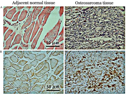
HE staining of OS and adjacent normal tissues
All clinical OS specimens used in the study were
confirmed by postoperative pathological diag -nosis. Typical HE staining results from the OS samples are shown in Figure 1A. The OS cells were interspersed with the trabecular or
sheet-like structure of the osteoid tissue.
Morpholo-gically, the OS cells had clear spindle or polygo-nal shapes. The nuclei of these cells varied in shape, although they were large and intensely stained with prominent nucleoli.
Expression and significance of TAK1 protein in OS tissue
In the OS tissue, TAK1 staining was primarily cytoplasmic and appeared as pale yellow, yel-low, or brown staining. TAK1-positive cells were
markedly less common in the adjacent normal
tissue (Figure 1B) compared with the OS tis-sue. The positive expression rate of TAK1 was 73.6% in the OS tissue, whereas it was only 19.8% in the adjacent normal bone tissue,
which was significantly different.
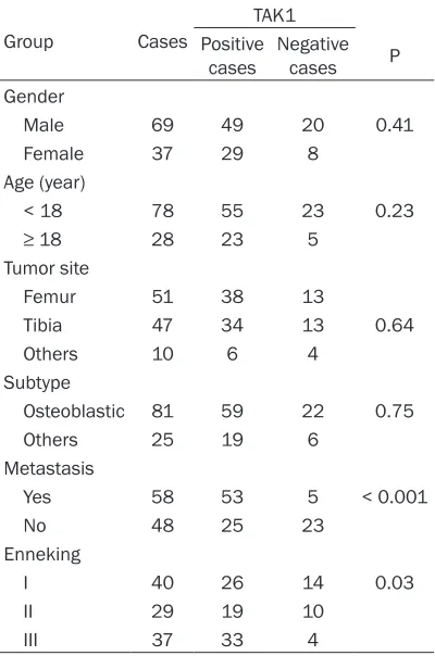
Association of TAK1 protein expression with the pathological stage of OS
The positive expression rate of TAK1 was unre-lated to the gender, age, tumor site, and sub-type of the OS patients (P > 0.05). However, TAK1 expression was strongly associated with the presence or absence of metastasis and
Enneking stage (P < 0.05, Table 1).
Association of TAK1 protein expression with clinical prognosis
Statistical analysis using the Kaplan-Meier method revealed that (Figure 2) among the
TAK1-positive OS patients, 37 died within five
years, with a median survival time of 30 months. By contrast, among the TAK1-negative
OS patients, seven died within five years, with a
median survival time of 51 months. Therefore,
TAK1-negative OS patients had a significantly
higher 5-year survival rate than TAK1-positive OS patients (P = 0.009).
Effects of the TAK1 inhibitor 5Z7O on OS cell proliferation and apoptosis
[image:4.612.89.291.87.565.2]Tables 2 and 3 show that the 5Z7O-treated
Table 1. Association of TAK1 with osteosar-coma clinicopathological stage
Group Cases
TAK1 Positive
cases Negative cases P Gender
Male 69 49 20 0.41
Female 37 29 8
Age (year)
< 18 78 55 23 0.23
≥ 18 28 23 5
Tumor site
Femur 51 38 13
Tibia 47 34 13 0.64
Others 10 6 4
Subtype
Osteoblastic 81 59 22 0.75
Others 25 19 6
Metastasis
Yes 58 53 5 < 0.001
No 48 25 23
Enneking
I 40 26 14 0.03
II 29 19 10
III 37 33 4
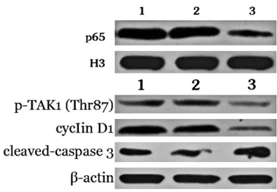
[image:4.612.91.291.97.399.2]significantly lower intranuclear p65, p-TAK1
(Thr187), and cyclin D1 expression (P < 0.05). By contrast, cleaved-caspase 3 protein expres-sion was strongly enhanced in the 5Z7O group compared with the control group (P < 0.05).
Univariate analysis of prognostic factors in OS patients
The results of univariate analysis by log-rank test showed significant differences in the sur -vival of OS patients with regard to age,
metas-tasis, Enneking stage, and TAK1 expression (P
< 0.05). However, no significant differences
were found in the survival of OS patients with regard to gender, tumor site, and subtype (P > 0.05) (Table 4).
Multivariate analysis of prognostic factors in OS patients
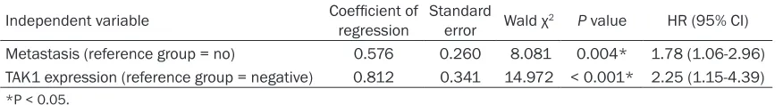
Multivariate Cox regression analysis revealed that metastasis and TAK1 expression were
independent risk factors for a poor prognosis of
OS patients (P < 0.05) (Table 5).
Discussion
[image:5.612.90.391.86.154.2]Recent studies have observed abnormally high TAK1 expression in a variety of tumor tissues, and TAK1 has been shown to play an essential role in the development and progression of dif-ferent tumors. Inhibiting the TAK1 activity can
Table 2. Cell proliferation in different groups
Group n 24 h 48 h 72 h
Blank 10 0 0 0
Control 10 3.1±1.12 4.9±1.38 5.8±1.92
Low-dose 5Z-7-oxozeaenol 10 20±3.12* 24±3.79* 29±4.68* High-dose 5Z-7-oxozeaenol 10 35±4.59#,* 39±6.78#,* 41±6.21#,*
[image:5.612.90.391.211.292.2]*P < 0.05, compared with the control group; #P < 0.05, compared with the low-dose group.
Table 3. Cell apoptosis in different groups
Group n 24 h 48 h 72 h
Blank 10 2.78±1.22 4.01±2.11 4.45±2.09
Control 10 3.2±1.12 4.1±2.01 4.5±1.96
Low-dose 5Z-7-oxozeaenol 10 31.1±4.56* 36.8±5.03* 41.1±6.12* High-dose 5Z-7-oxozeaenol 10 41.6±7.12#,* 47.8±8.89#,* 54.1±9.81#,*
[image:5.612.91.299.349.693.2]*P < 0.05, compared with the control group; #P < 0.05, compared with the low-dose group.
Table 4. Univariate analysis of prognostic factors in patients with osteosarcoma (n = 106)
Factor Case survival time Median (months) χ
2 P
Gender
Male 69 48.0 2.285 0.131
Female 37 54.5
Age (years)
< 60 78 52.0 5.219 0.022*
≥ 60 28 35.0
Tumor site
Femur 51 48.0 2.048 0.359
Tibia 47 46.0
Other 10 53.5
Subtype
Osteoblastic 81 52.0 1.669 0.196
Other 25 48.0
Metastasis
Yes 58 22.5 9.591 0.002*
No 48 57.0
Enneking stage
I 40 52.0 8.252 0.016*
II 29 31.0
III 37 23.5
TAK1 expression
Positive 78 30.0 6.606 0.009* Negative 28 51.0
*P< 0.05.
tion and higher apoptosis in OS cells, which occurred in a dose-dependent man-ner (P < 0.05).
Effects of the TAK1 in-hibitor 5Z7O on p-TAK1 (Thr87), intranuclear p65, cyclin D1, and cleaved-caspase 3 expression
As the high-dose 5Z7O group showed the stron-gest effects, we used this dose in further experime- nts to determine the mech-anisms underlying TAK1 activity in OS. Table 4 and
significantly suppress proliferation and pro -mote apoptosis in tumor cells, suggesting that TAK1 could represent a novel target for tumor treatment [8]. TAK1 is located at the intersec-tion of multiple signal transducintersec-tion pathways. For example, while the activation of TAK1 can trigger the MAPK pathway, it can also lead to the activation of IKK, which causes the
degra-dation of IκBα; NF-κBp65, now freed from inhi
-bition by IκBα, can move from the cytoplasm to the nucleus to bind to specific target gene
sequences and regulate the transcription of
rel-evant genes [9]. MAPK expression is signifi -cantly elevated in OS tissue, and inhibiting
MAPK expression can markedly suppress pro -liferation and induce apoptosis in OS [10-12].
Similarly, NF-κB shows abnormal activation in OS tissue, and the inhibition of NF-κB activity
can strongly suppress proliferation and
facili-cantly associated with the presence or absence
of metastasis and Enneking stage, suggesting
that TAK1 may be important for tumor progres-sion in OS patients. A Kaplan-Meier survival analysis showed that TAK1-positive patients
had a significantly shorter 5-year survival time
compared with TAK1-negative patients, again
suggesting that TAK1 is a key factor affecting
OS patient prognosis. Therefore, postoperative detection of TAK1 expression in OS tissue could
have prognostic significance for OS patients.
Our results demonstrate that TAK1 expression
is significantly higher in OS tissue and is associ -ated with prognosis in OS patients. Thus, using in vitro cell culture experiments, we treated an
OS cell line with a specific TAK1 inhibitor, 5Z7O,
to further elucidate the role of TAK1 in OS cell proliferation and apoptosis. Numerous
experi-ments have shown that 5Z7O can significantly
inhibit proliferation and promote apoptosis in a variety of tumor cells [14]. The drug dose used in the present study was based on previous studies, and the results showed that 5Z7O
sig-nificantly inhibited proliferation and promoted
apoptosis of the OS cell line in a
dose-depen-dent manner. Thr187 is a key site for TAK1 acti -vation [15], and 5Z7O can strongly inhibit Thr187 phosphorylation, leading to TAK1 inhibi-tion [16]. Indeed, we found that the 5Z7O group
showed markedly lower p-TAK1 (Thr187), intra -nuclear p65, and cyclin D1 expression levels than the control group. By contrast,
cleaved-caspase 3 protein expression was markedly
increased in the 5Z7O group compared with the control group. Therefore, this may be the
[image:6.612.91.372.72.231.2]mech-Figure 3. Cell apoptosis in the different groups. *P < 0.05, compared to the blank group; #P < 0.05, compared to the control group.
Figure 4. Western blotting analysis of P65, p-TAK1 (Thr187), cyclin D1, and cleaved-caspase 3 expres-sion in the different treatment groups (representa-tive images are presented).
tate apoptosis in OS [13].
These findings suggest that
TAK1 plays an essential role in OS development and pro-gression. In the present study,
we first analyzed TAK1 expres -sion in tumor tissue samples from clinical OS patients us- ing IHC. The results clearly showed that TAK1 expression
was significantly higher in the
OS tissue samples than in the adjacent normal tissue sam-ples. Further analysis found
no link between TAK1 expres -sion and the age, gender, or tumor site of OS patients.
[image:6.612.90.287.283.420.2]-anism through which 5Z7O inhibits proliferation and promotes apoptosis in OS cells. We do note that the present study shows only that the selective TAK1 inhibitor 5Z7O can suppress proliferation and enhance apoptosis in OS ce- lls in an in vitro setting. Further experiments should be performed using in vivo animal ex- periments to determine the best methods for treating OS by targeting TAK1.
In summary, TAK1 expression was significantly
elevated in OS tissue and was closely associ-ated with clinical stage and prognosis in OS
patients. We showed that a specific TAK1 inhib -itor can inhibit proliferation and promote apop-tosis in OS cells, suggesting that TAK1 may be a promising new target for the treatment of OS.
Disclosure of conflict of interest
None.
Address correspondence to: Dr. Quanbing Wang, Department of Orthopedics, Renmin Hospital, Hubei University of Medicine, No. 39 Chaoyang Road, Maojian District, Shiyan 442000, Hubei, P. R. China. Tel: +86-15897824431; Fax: +0086-07198637852; E-mail: [email protected]; Beibei Fu, De- partment of Neurology, Renmin hospital, Hubei University of Medicine, No. 39 Chaoyang Road, Maojian District, Shiyan 442000, Hubei, P. R. China. Tel: +86-18071352388; Fax: +0086-07198637852; E-mail: [email protected]
References
[1] Isakoff MS, Bielack SS, Meltzer Gorlick R. Osteosarcoma: Current Treatment and a Co- llaborative Pathway to Success. J Clin Oncol 2015; 33: 3029-35.
[2] Qiao F, Qiang T. The expression and clinical sig-nificance of TAK1 in esophageal cancer. J Pract Med 2014; 3084-3086, 3087.
[3] Wu X, Zhang W, Font-Burgada J, Palmer T, Hamil AS, Biswas SK, Poidinger M, Borcherding N, Xie Q, Ellies LG, Lytle NK, Wu LW, Fox RG,Yang J, Dowdy SF, Reya T, Karin M.
Ubiquitin-conjugating enzyme Ubc13 controls breast cancer metastasis through a TAK1-p38 MAP kinase cascade. Proc Natl Acad Sci U S A 2014; 111: 13870-5.
[4] Ying L, Chunxia Y, Wei L. Inhibition of ovarian cancer cell growth by a novel TAK1 inhibitor LYTAK1. Cancer Chemother Pharmacol 2015; 76: 641-50.
[5] Roh YS, Song J, Seki E. TAK1 regulates hepatic cell survival and carcinogenesis. J Gastroen- terol 2014; 49: 185-94.
[6] Hrabe JE, O’Leary BR, Fath MA, Rodman SN, Button AM, Domann FE, Spitz DR, Mezhir JJ. Disruption of thioredoxin metabolism enhanc-es the toxicity of transforming growth factor beta-activated kinase 1 (TAK1) inhibition in KRAS-mutated colon cancer cells. Redox Biol 2015; 5: 319-327.
[7] Brown RS, Goodman TM, Zasadny KR, Greenson JK, Wahl RL. Expression of hexoki -nase II and Glut-1 in untreated human breast cancer. Nucl Med Biol 2002; 29: 443-53. [8] Kilty I, Jones LH. TAK1 selective inhibition:
state of the art and future opportunities. Future Med Chem 2015; 7: 23-33.
[9] Mihaly SR, Ninomiya-Tsuji J, Morioka S. TAK1 control of cell death. Cell Death Differ 2014; 21: 1667-76.
[10] Hu KH, Li WX, Sun MY, Zhang SB, Fan CX, Wu Q, Zhu W, Xu X. Cadmium Induced Apoptosis in MG63 Cells by Increasing ROS, Activation of p38 MAPK and Inhibition of ERK 1/2 Pathways. Cell Physiol Biochem 2015; 36: 642-54. [11] Niu NK, Wang ZL, Pan ST, Ding HQ, Au GH, He
ZX, Zhou ZW, Xiao G, Yang YX, Zhang X, Yang T, Chen XW, Qiu JX, Zhou SF. Pro-apoptotic and pro-autophagic effects of the Aurora kinase A inhibitor alisertib (MLN8237) on human osteo-sarcoma U-2 OS and MG-63 cells through the activation of mitochondria-mediated pathway and inhibition of p38 MAPK/PI3K/Akt/mTOR signaling pathway. Drug Des Devel Ther 2015; 9: 1555-84.
[image:7.612.90.528.98.157.2][12] Wang F, Ke ZF, Wang R, Wang YF, Huang LL, Wang LT. Astrocyte elevated gene-1 (AEG-1) promotes osteosarcoma cell invasion through the JNK/c-Jun/MMP-2 pathway. Biochem Bio- phys Res Commun 2014; 452: 933-9.
Table 5. Multivariate Cox regression analysis of prognostic factors for patients with osteosarcoma (n = 106)
Independent variable Coefficient of regression Standard error Wald χ2 P value HR (95% CI)
Metastasis (reference group = no) 0.576 0.260 8.081 0.004* 1.78 (1.06-2.96) TAK1 expression (reference group = negative) 0.812 0.341 14.972 < 0.001* 2.25 (1.15-4.39)
[13] Golden D, Saria EA, Hansen MF. Regulation of Osteoblast Migration Involving Receptor Acti- vator of Nuclear Factor-kappa B (RANK) Sig-naling. J Cell Physiol 2015; 230: 2951-60. [14] Zhang J, Li B, Wu H, Ou J, Wei R, Liu J, Cai W,
Liu X, Zhao S, Yang J, Zhou L, Liu S, Liang A. Synergistic action of 5Z-7-oxozeaenol and bortezomib in inducing apoptosis of Burkitt lymphoma cell line Daudi. Tumour Biol 2016; 37: 531-9.
[15] Sakurai H. Targeting of TAK1 in inflammatory disorders and cancer. Trends Pharmacol Sci 2012; 33: 522-30.